. 2022 Jul 29;15(15):5240.
doi: 10.3390/ma15155240.

Evaluation of the Biocompatibility and Osteogenic Properties of Metal Oxide Coatings Applied by Magnetron Sputtering as Potential Biofunctional Surface Modifications for Orthopedic Implants

Affiliations

Evaluation of the Biocompatibility and Osteogenic Properties of Metal Oxide Coatings Applied by Magnetron Sputtering as Potential Biofunctional Surface Modifications for Orthopedic Implants

Mariana Fernández-Lizárraga et al. Materials (Basel). .

Abstract

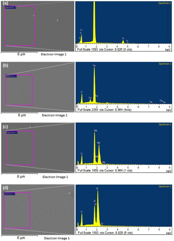
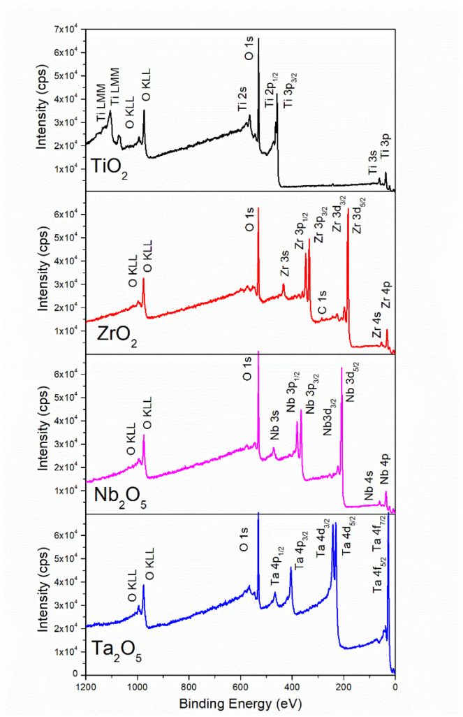
Biomaterials with adequate properties to direct a biological response are essential for orthopedic and dental implants. The surface properties are responsible for the biological response; thus, coatings with biologically relevant properties such as osteoinduction are exciting options to tailor the surface of different bulk materials. Metal oxide coatings such as TiO2, ZrO2, Nb2O5 and Ta2O5 have been suggested as promising for orthopedic and dental implants. However, a comparative study among them is still missing to select the most promising for bone-growth-related applications. In this work, using magnetron sputtering, TiO2, ZrO2, Ta2O5, and Nb2O5 thin films were deposited on Si (100) substrates. The coatings were characterized by Optical Profilometry, Scanning Electron Microscopy, Energy-Dispersive X-ray Spectroscopy, X-ray Photoelectron Spectroscopy, X-ray Diffraction, Water Contact Angle measurements, and Surface Free Energy calculations. The cell adhesion, viability, proliferation, and differentiation toward the osteoblastic phenotype of mesenchymal stem cells plated on the coatings were measured to define the biological response. Results confirmed that all coatings were biocompatible. However, a more significant number of cells and proliferative cells were observed on Nb2O5 and Ta2O5 compared to TiO2 and ZrO2. Nevertheless, Nb2O5 and Ta2O5 seemed to induce cell differentiation toward the osteoblastic phenotype in a longer cell culture time than TiO2 and ZrO2.

Keywords: magnetron sputtering; mesenchymal stem cells; metal oxide coatings; osteogenesis.


Conflict of interest statement

The authors declare no conflict of interest.

Figures

Figure 1
Figure 1
Representative pictures of the macroscopic appearance of Si (100) wafers coated with metal oxide thin films by magnetron sputtering. Coated samples were named as TiOx, TaOx, NbOx, ZrOx, for titanium, tantalum, niobium and zirconium oxide coatings, while Si samples correspond to uncoated Si (100) wafers.
Figure 2
Figure 2
Representative SEM images of the areas analyzed (pink rectangles marked on the micrographs) for EDS elemental composition studies and of the corresponding EDS spectra obtained for the (a) TiOx; (b) TaOx; (c) NbOx; and (d) ZrOx coated samples.
Figure 3
Figure 3
Example low resolution XPS spectra of TiOX (TiO2), ZrOX (ZrO2), NbOX (Nb2O5) and TaOX (Ta2O5).
Figure 4
Figure 4
Representative grazing incidence XRD patterns of (a) TiOx, (b) ZrOx, (c) NbOx, and (d) TaOx coatings. In Figure 3a, the A and R stand for the expected XRD pattern for Anatase and Rutile crystalline phases of TiO2, respectively. In Figure 3b, the B stands for the expected XRD pattern for the Baddeleyite crystalline phase of ZrO2.
Figure 5
Figure 5
Representative micrographs, obtained by an optical profilometer, of BM-MSC cell cultured on uncoated Si (ac) and coated, TiOx (df), TaOx (gi), NbOx (jl), and ZrOx (mo), samples. Three incubation times were handled, 3, 7, and 14 days. Micrographs were acquired at two different magnifications, 5× for main micrographs and 50× for insert magnifications.
Figure 6
Figure 6
Qualitative evaluation of BM-MSC viability at 3, 7, and 14 days of culture on uncoated Si (df) and TiOx (gi), TaOx (jl), NbOx (mo), and ZrOx (pr) coated samples. Ctrl (ac) corresponds to BM-MSC culture on the surface of standard tissue culture plates. Micrographs were acquired at two different magnifications, 5× and 20×. The fluorescent LIVE/DEADTM Viability/Cytotoxicity Kit (Invitrogen®, Waltham, MA, USA) for mammalian cells was used to mark viable (green) and dead (red nuclei) cells.
Figure 7
Figure 7
Quantitative evaluation of BM-MSC viability cultured on the coatings at (a) 3 days, (b) 7days, (c) 14 days of culture. Thermo Fisher Alamar BlueTM cell viability reagent kit was used. Ctrl corresponds to BM-MSC cultured on the surface of standard tissue culture plates. Data are presented as the mean ± standard error using three independent cultures per variable and analyzed by one-way ANOVA using a Dunnett’s post hoc test. * p ≤ 0.05 vs. Ctrl; ** p ≤ 0.001 vs. Ctrl; **** p ≤ 0.0001; & p ≤ 0.05 vs. TiOx; && p ≤ 0.01 vs. TiOx.
Figure 8
Figure 8
Evaluation of cell proliferation for BM-MSC cultured on the coatings studied. (a) 3 days and (b) 5 days of culture. The Roche Cell Proliferation ELISA, BrdU (colorimetric) kit was used. Ctrl corresponds to BM-MSC cultured on standard TCP. Data are presented as the mean ± standard error using three independent cultures per variable and analyzed by one-way ANOVA using a Dunnett’s post hoc test. * p ≤ 0.05 vs. Ctrl; *** p ≤ 0.005 and **** p ≤ 0.0005; &&& p ≤ 0.001 vs. TiOx; &&&& p ≤ 0.0001 vs. TiOx; $$ p ≤ 0.01 vs. TaOx; $$$$ p ≤ 0.001 vs. TaOx; % p ≤ 0.05 vs. NbOx.
Figure 9
Figure 9
RUNX2 (ae), OP (fj) and OC (ko) cellular expression characterized by IF assays in BM-MSC cultured on the coatings. In green, positive expression of the corresponding osteogenic marker is observed, while in blue, cell nuclei stained with Hoechst are observed. Ctrl corresponds to cells cultured on standard tissue culture plates.
Figure 10
Figure 10
Quantification of proteins involved in osteogenic differentiation. Quantification of ALP activity, at (a) 7 days, and (b) 14 days of cells culture. Quantification of (c) osteopontin (OP) expression, (d) osteoprotegerin (OPG), and (e) osteocalcin (OC) expression after 14 days of cells culture. Ctrl- and Ctrl correspond to BM-MSC cultured on TCP surfaces with supplemented Mesenchymal Stem Basal Medium (Ctrl−), or DMEM/F-12 supplemented with 10% v/v FBS (Ctrl). Data are presented as the mean ± standard error using three independent cultures per variable and analyzed by one-way ANOVA using a Dunnett’s post hoc test. * p ≤ 0.05 vs. Ctrl; ** p ≤ 0.01 vs. Ctrl; *** p ≤ 0.001; and **** p ≤ 0.0001 vs. Ctrl vs Ctrl.

Similar articles

Cited by

References

    1. Grün N.G., Holweg P.L., Donohue N., Klestil T., Weinberg A.-M. Resorbable Implants in Pediatric Fracture Treatment. Innov. Surg. Sci. 2018;3:119–125. doi: 10.1515/iss-2018-0006. - DOI - PMC - PubMed
    1. Chandra G., Pandey A. Design Approaches and Challenges for Biodegradable Bone Implants: A Review. Expert Rev. Med. Devices. 2021;18:629–647. doi: 10.1080/17434440.2021.1935875. - DOI - PubMed
    1. Siti Nur Hazwani M.R., Lim L.X., Lockman Z., Zuhailawati H. Fabrication of Titanium-Based Alloys with Bioactive Surface Oxide Layer as Biomedical Implants: Opportunity and Challenges. Trans. Nonferrous Met. Soc. China Engl. Ed. 2022;32:1–44. doi: 10.1016/S1003-6326(21)65776-X. - DOI
    1. Liu Y., Rath B., Tingart M., Eschweiler J. Role of Implants Surface Modification in Osseointegration: A Systematic Review. J. Biomed. Mater. Res. Part A. 2020;108:470–484. doi: 10.1002/jbm.a.36829. - DOI - PubMed
    1. Rizzo P. A Review on the Latest Advancements in the Non-Invasive Evaluation/Monitoring of Dental and Trans-Femoral Implants. Biomed. Eng. Lett. 2020;10:83–102. doi: 10.1007/s13534-019-00126-8. - DOI - PMC - PubMed

LinkOut - more resources