

doi: 10.3390/ma15155355.
Polymorphic Biological and Inorganic Functional Nanomaterials
Affiliations
- PMID: 35955287
- PMCID: PMC9369650
- DOI: 10.3390/ma15155355
Item in Clipboard
Polymorphic Biological and Inorganic Functional Nanomaterials
Materials (Basel).
.
Abstract
This perspective involves two types of functional nanomaterials, amyloid fibrils and metal oxide nanowires and nanogrids. Both the protein and the inorganic nanomaterials rely on their polymorphism to exhibit diverse properties that are important to sensing and catalysis. Several examples of novel functionalities are provided from biomarker sensing and filtration applications to smart scaffolds for energy and sustainability applications.
Keywords: amyloids; electrospinning; nanomaterials; polymorphism.
Conflict of interest statement
Authors declare no conflict of interest.
Figures
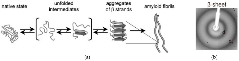
(a) Amyloid fibril formation stages [4]; (b) XRD pattern typical of β-sheet structures with equatorial (d1) and meridional (d2) reflections (from [8]).

(a) Process map of amyloid fibrils from wheat flour; (b) confocal image of amyloid fibrils; (c) TEM image of amyloid fibrils. Images from Gouma et al. [4].
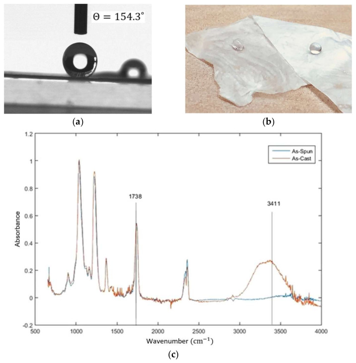
(a) Close image of water droplet on super water repellent CA fibrous mats; (b) water droplets on super water repellent CA fibrous mats; (c) FTIR analysis illustrating the intensity of the hydroxyl band at 3450 cm−1 that decreased in the as-spun fiber [25].
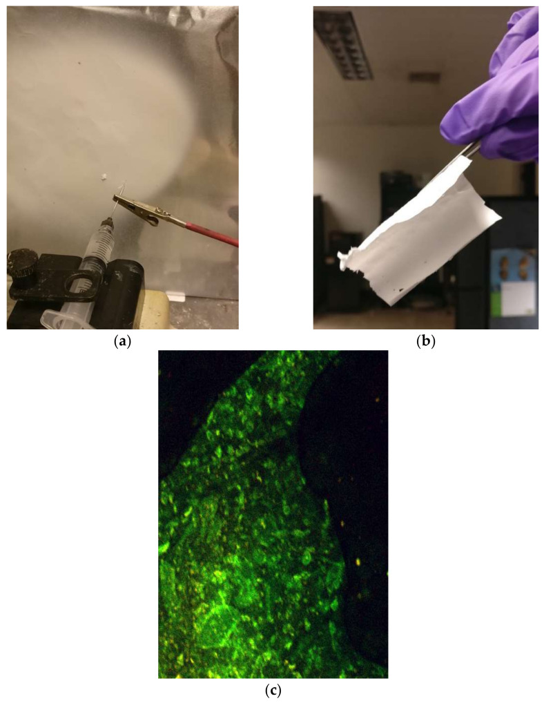
(a) Electrospinning of the amyloid and CA solution; (b) the electrospun, fibrous mat; (c) confocal microscopy of the mat, confirming a uniform dispersion of amyloid fibers in the CA mat.
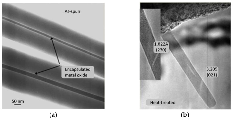
(a) TEM images of PVP-MoO3 nanocomposite before heat treatment [28]; (b) HRTEM image of a MoO3 nanowire after heat treatment [28].
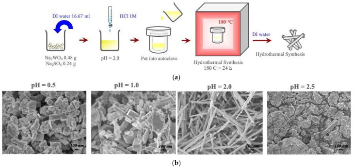
(a) Process map for the formation of the metastable hexagonal WO3 polymorph via hydrothermal treatment [32]; (b) SEM images of the microstructure at varying pH [32]. Nanowires are formed at a pH of 2 with lengths of 10 microns.

(a) TEM image of γ-WO3 nanowires grown on silicon nitride grid [33]; (b) TEM image of γ-WO3 nanowires grown on copper mesh grid [33].
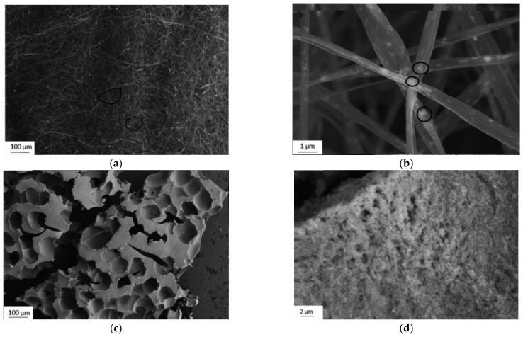
(a) SEM images of as-spun fibers, highlighting the formation of a 3D fiber-woven foam (circled) [34]; (b) high magnification SEM image of fibers highlighting the sol-phase particles in the fibers (circled) [34]; (c) high magnification SEM image of foam surface showing the agglomerated sol particles and nano porosity [34]; (d) SEM image of foam structures in the fibers [34].
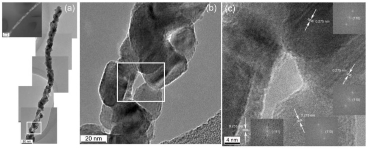
CuO nanowires forming “links in a chain” architecture of nanocrystals with low angle boundaries; this pseudo-monocrystalline porous architecture is called “nanogrids”; (a) collage of five HRTEM images of a single chain [40]; (b,c) englarged views of particle arrangement within the nanowires [40]. The white squares highlight the area captured of the following image.
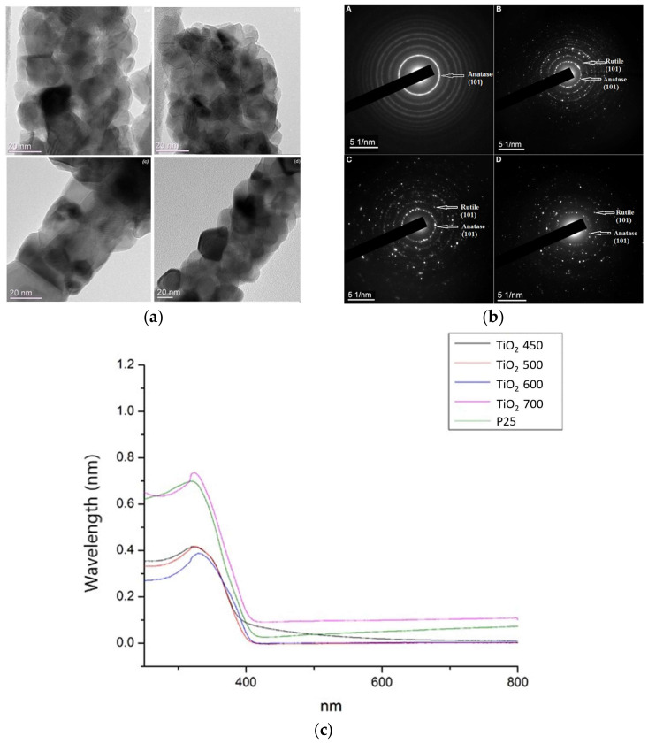
(a) TEM images of TiO2 nanogrids at 450 °C, 500 °C, 600 °C, and 700 °C; (b) SAED patterns of TiO2 nanofibers annealed at 450 °C, 500 °C, 600 °C, and 700 °C [43]; (c) UV-Vis spectra of P25 and TiO2 nanofibers annealed at 450 °C, 500 °C, 600 °C, and 700 °C [43].
Similar articles
-
Inorganic Hydrogel Based on Low-Dimensional Nanomaterials.ACS Nano. 2024 Jan 30;18(4):2730-2749. doi: 10.1021/acsnano.3c11262. Epub 2024 Jan 14. ACS Nano. 2024. PMID: 38221737 Review.
-
Functional micro/nanostructures: simple synthesis and application in sensors, fuel cells, and gene delivery.Acc Chem Res. 2011 Jul 19;44(7):491-500. doi: 10.1021/ar200001m. Epub 2011 May 25. Acc Chem Res. 2011. PMID: 21612197
-
Nanomaterials: amyloids reflect their brighter side.Nano Rev. 2011;2. doi: 10.3402/nano.v2i0.6032. Epub 2011 May 31. Nano Rev. 2011. PMID: 22110868 Free PMC article.
-
Catalytically Active Amyloids as Future Bionanomaterials.Nanomaterials (Basel). 2022 Oct 28;12(21):3802. doi: 10.3390/nano12213802. Nanomaterials (Basel). 2022. PMID: 36364578 Free PMC article. Review.
-
Material Nanoarchitectonics of Functional Polymers and Inorganic Nanomaterials for Smart Supercapacitors.Small. 2022 Feb;18(7):e2102397. doi: 10.1002/smll.202102397. Epub 2021 Dec 3. Small. 2022. PMID: 34862722 Review.
References
-
- Schultz M. Rudolf Virchow. Emerg. Infect. Dis. 2008;14:1480. doi: 10.3201/eid1409.086672. - DOI
Grants and funding
LinkOut - more resources
Full Text Sources
