Impact of Gamma Irradiation on the Properties of Magnesium-Doped Hydroxyapatite in Chitosan Matrix
- PMID: 35955308
- PMCID: PMC9369862
- DOI: 10.3390/ma15155372
Impact of Gamma Irradiation on the Properties of Magnesium-Doped Hydroxyapatite in Chitosan Matrix
Abstract
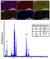
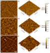
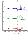
This is the first report regarding the effect of gamma irradiation on chitosan-coated magnesium-doped hydroxyapatite (xMg = 0.1; 10 MgHApCh) layers prepared by the spin-coating process. The stability of the resulting 10 MgHApCh gel suspension used to obtain the layers has been shown by ultrasound measurements. The presence of magnesium and the effect of the irradiation process on the studied samples were shown by X-ray photoelectron spectroscopy (XPS). The XPS results obtained for irradiated 10 MgHApCh layers suggested that the magnesium and calcium contained in the surface layer are from tricalcium phosphate (TCP; Ca3(PO4)2) and hydroxyapatite (HAp). The XPS analysis has also highlighted that the amount of TCP in the surface layer increased with the irradiation dose. The energy-dispersive X-ray spectroscopy (EDX) evaluation showed that the calcium decreases with the increase in the irradiation dose. In addition, a decrease in crystallinity and crystallite size was highlighted after irradiation. By atomic force microscopy (AFM) we have obtained images suggesting a good homogeneity of the surface of the non-irradiated and irradiated layers. The AFM results were also sustained by the scanning electron microscopy (SEM) images obtained for the studied samples. The effect of gamma-ray doses on the Fourier transform infrared spectroscopy (ATR-FTIR) spectra of 10 MgHApCh composite layers was also evaluated. The in vitro antifungal assays proved that 10 MgHApCh composite layers presented a strong antifungal effect, correlated with the irradiation dose and incubation time. The study of the stability of the 10 MgHApCh gel allowed us to achieve uniform and homogeneous layers that could be used in different biomedical applications.
Keywords: antifungal activity; chitosan; gamma irradiation; hydroxyapatite; magnesium.
Conflict of interest statement
The authors declare no conflict of interest. The funders had no role in the design of the study; in the collection, analyses, or interpretation of data; in the writing of the manuscript; or in the decision to publish the results.
Figures


Similar articles
-
Biological and Physico-Chemical Properties of Composite Layers Based on Magnesium-Doped Hydroxyapatite in Chitosan Matrix.Micromachines (Basel). 2022 Sep 22;13(10):1574. doi: 10.3390/mi13101574. Micromachines (Basel). 2022. PMID: 36295927 Free PMC article.
-
Exploring the physicochemical traits, antifungal capabilities, and 3D spatial complexity of hydroxyapatite with Ag+Mg2+ substitution in the biocomposite thin films.Micron. 2024 Sep;184:103661. doi: 10.1016/j.micron.2024.103661. Epub 2024 May 22. Micron. 2024. PMID: 38833994
-
Biocomposite Coatings Doped with Magnesium and Zinc Ions in Chitosan Matrix for Antimicrobial Applications.Materials (Basel). 2023 Jun 15;16(12):4412. doi: 10.3390/ma16124412. Materials (Basel). 2023. PMID: 37374594 Free PMC article.
-
The Effects of Electron Beam Irradiation on the Morphological and Physicochemical Properties of Magnesium-Doped Hydroxyapatite/Chitosan Composite Coatings.Polymers (Basel). 2022 Jan 31;14(3):582. doi: 10.3390/polym14030582. Polymers (Basel). 2022. PMID: 35160570 Free PMC article.
-
Morphological and fractal features of cancer cells anchored on composite layers based on magnesium-doped hydroxyapatite loaded in chitosan matrix.Micron. 2024 Jan;176:103548. doi: 10.1016/j.micron.2023.103548. Epub 2023 Oct 4. Micron. 2024. PMID: 37813055
Cited by
-
Biological and Physico-Chemical Properties of Composite Layers Based on Magnesium-Doped Hydroxyapatite in Chitosan Matrix.Micromachines (Basel). 2022 Sep 22;13(10):1574. doi: 10.3390/mi13101574. Micromachines (Basel). 2022. PMID: 36295927 Free PMC article.
-
Bioactivity and Thermal Stability of Collagen-Chitosan Containing Lemongrass Essential Oil for Potential Medical Applications.Polymers (Basel). 2022 Sep 17;14(18):3884. doi: 10.3390/polym14183884. Polymers (Basel). 2022. PMID: 36146031 Free PMC article.
-
Complex Evaluation of Nanocomposite-Based Hydroxyapatite for Biomedical Applications.Biomimetics (Basel). 2023 Nov 6;8(7):528. doi: 10.3390/biomimetics8070528. Biomimetics (Basel). 2023. PMID: 37999169 Free PMC article.
-
Influence of Electron Beam Irradiation and RPMI Immersion on the Development of Magnesium-Doped Hydroxyapatite/Chitosan Composite Bioactive Layers for Biomedical Applications.Polymers (Basel). 2025 Feb 18;17(4):533. doi: 10.3390/polym17040533. Polymers (Basel). 2025. PMID: 40006195 Free PMC article.
-
Development of Chrome-Doped Hydroxyapatite in a PVA Matrix Enriched with Amoxicillin for Biomedical Applications.Antibiotics (Basel). 2025 Apr 30;14(5):455. doi: 10.3390/antibiotics14050455. Antibiotics (Basel). 2025. PMID: 40426522 Free PMC article.
References
-
- Mrázová H., Koller J., Kubišová K., Fujeríková G., Klincová E., Babál P. Comparison of structural changes in skin and amnion tissue grafts for transplantation induced by gamma and electron beam irradiation for sterilization. Cell Tissue Bank. 2016;17:255–260. doi: 10.1007/s10561-015-9536-3. - DOI - PubMed
Grants and funding
LinkOut - more resources
Full Text Sources
Miscellaneous

