

The Vascular Endothelium and Coagulation: Homeostasis, Disease, and Treatment, with a Focus on the Von Willebrand Factor and Factors VIII and V
- PMID: 35955419
- PMCID: PMC9425441
- DOI: 10.3390/ijms23158283
The Vascular Endothelium and Coagulation: Homeostasis, Disease, and Treatment, with a Focus on the Von Willebrand Factor and Factors VIII and V
Abstract
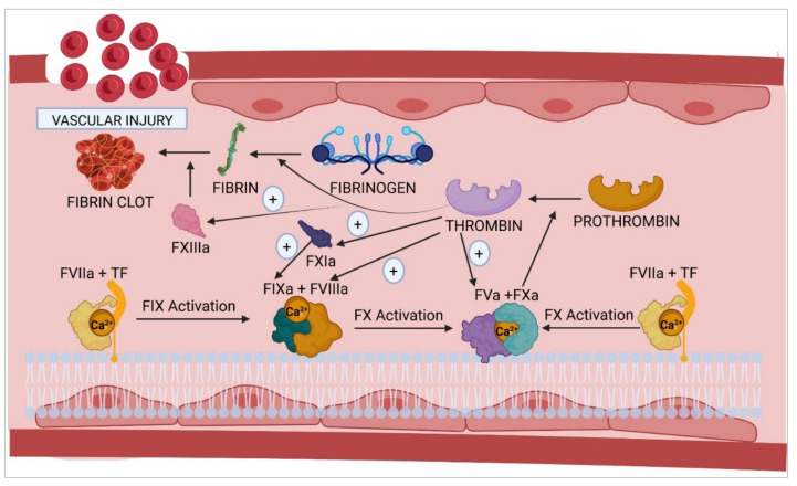
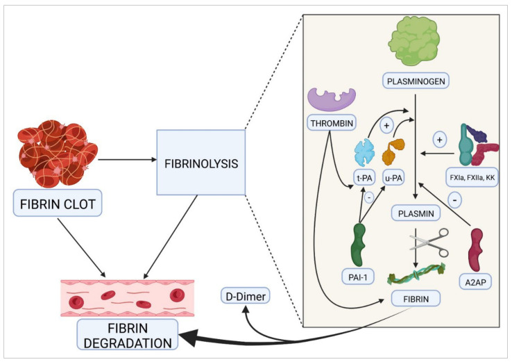
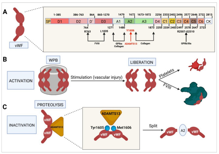
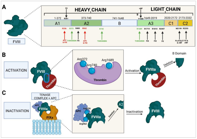
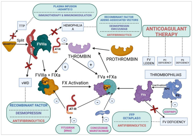
The vascular endothelium has several important functions, including hemostasis. The homeostasis of hemostasis is based on a fine balance between procoagulant and anticoagulant proteins and between fibrinolytic and antifibrinolytic ones. Coagulopathies are characterized by a mutation-induced alteration of the function of certain coagulation factors or by a disturbed balance between the mechanisms responsible for regulating coagulation. Homeostatic therapies consist in replacement and nonreplacement treatments or in the administration of antifibrinolytic agents. Rebalancing products reestablish hemostasis by inhibiting natural anticoagulant pathways. These agents include monoclonal antibodies, such as concizumab and marstacimab, which target the tissue factor pathway inhibitor; interfering RNA therapies, such as fitusiran, which targets antithrombin III; and protease inhibitors, such as serpinPC, which targets active protein C. In cases of thrombophilia (deficiency of protein C, protein S, or factor V Leiden), treatment may consist in direct oral anticoagulants, replacement therapy (plasma or recombinant ADAMTS13) in cases of a congenital deficiency of ADAMTS13, or immunomodulators (prednisone) if the thrombophilia is autoimmune. Monoclonal-antibody-based anti-vWF immunotherapy (caplacizumab) is used in the context of severe thrombophilia, regardless of the cause of the disorder. In cases of disseminated intravascular coagulation, the treatment of choice consists in administration of antifibrinolytics, all-trans-retinoic acid, and recombinant soluble human thrombomodulin.
Keywords: coagulation; coagulopathies; embryo; factor V; factor VIII; homeostasis; treatment; vascular endothelium; von Willebrand factor.
Conflict of interest statement
The authors declare no conflict of interest.
Figures
Similar articles
-
Spinal dural arteriovenous fistulas are not associated with prothrombotic factors.Stroke. 2004 Sep;35(9):2069-71. doi: 10.1161/01.STR.0000135766.22285.dc. Epub 2004 Jul 1. Stroke. 2004. PMID: 15232118 Review.
-
Hypercoagulability in adolescent girls on oral contraceptives-global coagulation profile and estrogen receptor polymorphisms.Am J Hematol. 2015 Aug;90(8):725-31. doi: 10.1002/ajh.24064. Am J Hematol. 2015. PMID: 26014094
-
Influence of factor VIII/von Willebrand complex on the activated protein C-resistance phenotype and on the risk for venous thromboembolism in heterozygous carriers of the factor V Leiden mutation.Blood Coagul Fibrinolysis. 1999 Oct;10(7):409-16. Blood Coagul Fibrinolysis. 1999. PMID: 10695766
-
Orthostatic hypercoagulability: a novel physiological mechanism to activate the coagulation system.Hypertension. 2008 Jun;51(6):1545-51. doi: 10.1161/HYPERTENSIONAHA.108.112003. Epub 2008 Apr 14. Hypertension. 2008. PMID: 18413485
-
Protein C.Prog Hemost Thromb. 1984;7:25-54. Prog Hemost Thromb. 1984. PMID: 6099583 Review.
Cited by
-
Cardiotoxicity of Electronic Cigarettes and Heat-Not-Burn Tobacco Products-A Problem for the Modern Pediatric Cardiologist.Healthcare (Basel). 2023 Feb 8;11(4):491. doi: 10.3390/healthcare11040491. Healthcare (Basel). 2023. PMID: 36833024 Free PMC article. Review.
-
The advent of RNA-based therapeutics for metabolic syndrome and associated conditions: a comprehensive review of the literature.Mol Biol Rep. 2024 Apr 5;51(1):493. doi: 10.1007/s11033-024-09457-x. Mol Biol Rep. 2024. PMID: 38580818 Review.
-
A Review of the Mechanisms of Astragaloside IV and Berberine in Vascular Dysfunction Associated with Obesity and Diabetes.Drug Des Devel Ther. 2025 Jun 7;19:4911-4932. doi: 10.2147/DDDT.S520323. eCollection 2025. Drug Des Devel Ther. 2025. PMID: 40502549 Free PMC article. Review.
-
Knee replacement surgery in a patient with acquired von Willebrand disease: a case study with recommendations for patient management.Ann Med Surg (Lond). 2024 Jan 8;86(3):1681-1686. doi: 10.1097/MS9.0000000000001690. eCollection 2024 Mar. Ann Med Surg (Lond). 2024. PMID: 38463081 Free PMC article.
-
Diagnostic Value of Protein C Depletion in Pathologies Associated with the Activation of the Blood Coagulation System.Int J Mol Sci. 2025 Jun 25;26(13):6122. doi: 10.3390/ijms26136122. Int J Mol Sci. 2025. PMID: 40649900 Free PMC article.
References
Publication types
MeSH terms
Substances
Grants and funding
LinkOut - more resources
Full Text Sources
Miscellaneous
