. 2022 Jul 27;23(15):8315.
doi: 10.3390/ijms23158315.

Hypoxia Aggravates Inhibition of Alveolar Epithelial Na-Transport by Lipopolysaccharide-Stimulation of Alveolar Macrophages

Affiliations

Hypoxia Aggravates Inhibition of Alveolar Epithelial Na-Transport by Lipopolysaccharide-Stimulation of Alveolar Macrophages

Emel Baloglu et al. Int J Mol Sci. .

Abstract

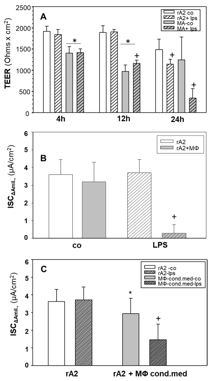
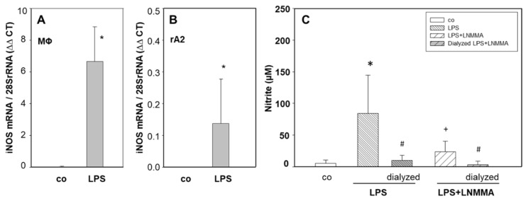
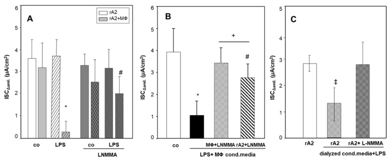
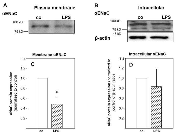
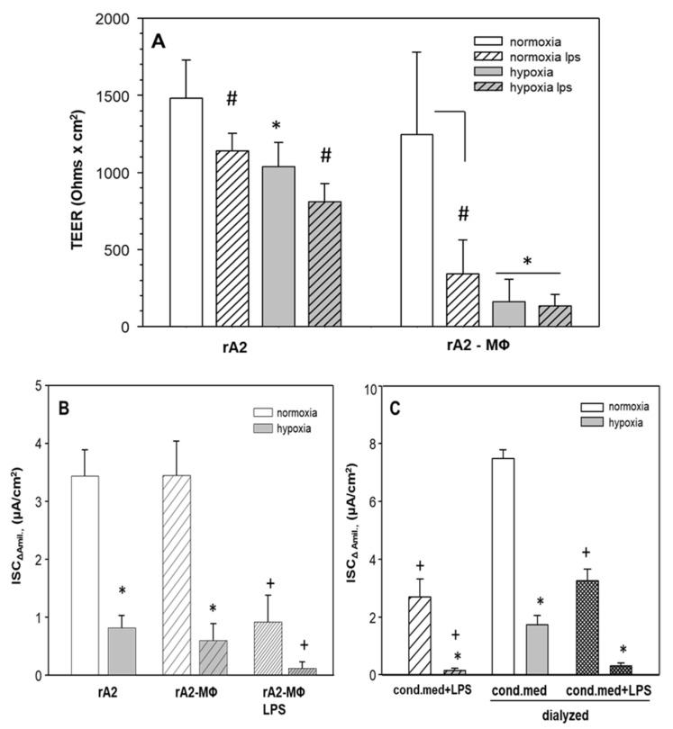
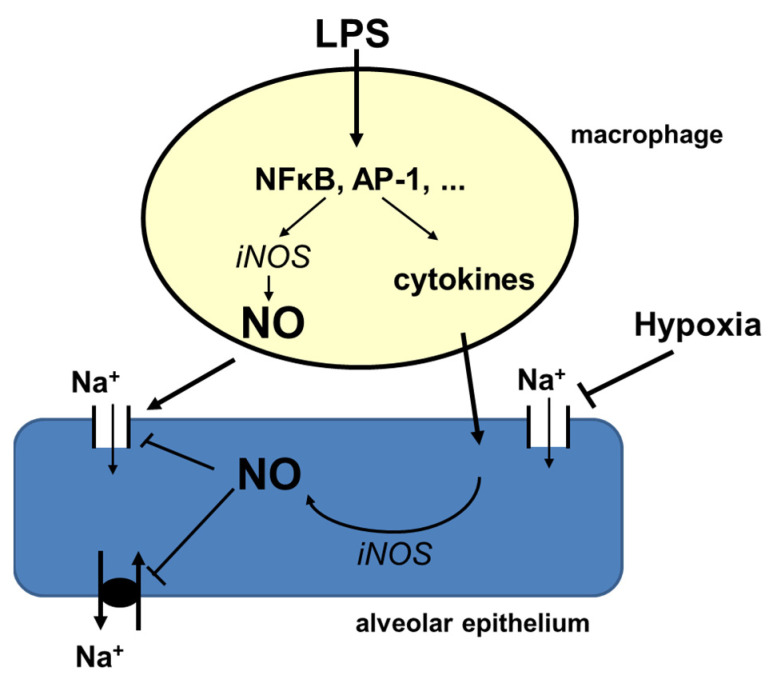
Inflammation and hypoxia impair alveolar barrier tightness, inhibit Na- and fluid reabsorption, and cause edema. We tested whether stimulated alveolar macrophages affect alveolar Na-transport and whether hypoxia aggravates the effects of inflammation, and tested for involved signaling pathways. Primary rat alveolar type II cells (rA2) were co-cultured with rat alveolar macrophages (NR8383) or treated with NR8383-conditioned media after stimulation with lipopolysaccharide (LPS; 1 µg/mL) and exposed to normoxia and hypoxia (1.5% O2). LPS caused a fast, transient increase in TNFα and IL-6 mRNA in macrophages and a sustained increase in inducible nitric oxide synthase (NOS2) mRNA in macrophages and in rA2 cells resulting in elevated nitrite levels and secretion of TNF-α and IL-6 into culture media. In normoxia, 24 h of LPS treated NR8383 decreased the transepithelial electrical resistance (TEER) of co-cultures, of amiloride-sensitive short circuit current (ISCΔamil); whereas Na/K-ATPase activity was not affected. Inhibition was also seen with conditioned media from LPS-stimulated NR8383 on rA2, but was less pronounced after dialysis to remove small molecules and nitrite. The effect of LPS-stimulated macrophages on TEER and Na-transport was fully prevented by the iNOS-inhibitor L-NMMA applied to co-cultures and to rA2 mono-cultures. Hypoxia in combination with LPS-stimulated NR8383 totally abolished TEER and ISCΔamil. These results indicate that the LPS-stimulation of alveolar macrophages impairs alveolar epithelial Na-transport by NO-dependent mechanisms, where part of the NO is produced by rA2 induced by signals from LPS stimulated alveolar macrophages.

Keywords: Na/K-ATPase; alveolar epithelial Na-channels; alveolar macrophages; hypoxia; iNOS; inflammation; pulmonary edema.


Conflict of interest statement

The authors declare no conflict of interest.

Figures

Figure 1
Figure 1
Effects of LPS on the transepithelial resistance and ion transport of rA2 cells in mono- and co-culture with alveolar macrophages and of conditioned medium obtained from LPS stimulated macrophages. Mono and co-cultures of rA2 cells with alveolar macrophages were prepared as described in the Methods section and treated with LPS (1 μg/mL) at indicated time points. Transepithelial electrical resistance (TEER) was measured by EVOM (Ohms × cm2) (A). Short circuit currents were measured in Ussing chambers in mono and co-cultures treated with LPS (1 μg/mL) for 24 h (B). Conditioned macrophage media was prepared by exposing mono-cultures of macrophages to LPS (1 µg/mL) for 24 h and kept on rA2 monolayers for 24 h. ENaC activity was measured as the amiloride (10 µM) sensitive component of Ieq in Ussing chambers (ISCΔamil). (C). Mean values ± SD of 5–11 experiments from at least 3 independent preparations of primary rA2 cells. * p < 0.001 effect of culturing macrophages or conditioned media with rA2 cells, + p < 0.001 effect of LPS treatment compared to relevant controls. MΦ: rat alveolar macrophages; rA2: rat alveolar epithelial cells alone; rA2 co: control rat alveolar epithelial cells that did not receive MΦ, MΦ conditioned medium or LPS; MA: MΦ and rA2 co-culture.
Figure 2
Figure 2
Effect of LPS treatment on the mRNA expression of iNOS in macrophages and rA2 mono-layers and nitrite levels in dialyzed macrophage conditioned media. Macrophage (A) and rA2 cells (B) were treated with LPS (1 µg/mL) for 24 h. mRNA expression was normalized to 28S-rRNA. Mean values ± SD of 4 to 5 independent cell preparations. Conditioned macrophage media were prepared by exposing mono-cultures of macrophages to LPS (1 µg/mL) and L-NMMA (1 mM) for 24 h (C). Five ml of conditioned medium was dialyzed and nitrite production was measured. Mean values ± SD of 3–6 independent preparations. Level of significance was p < 0.05: * effect of LPS treatment, # effect of dialysis, + effect of L-NMMA treatment. MΦ: rat alveolar macrophages; rA2: rat alveolar epithelial cells.
Figure 3
Figure 3
Effect of inhibiting of iNOS activity with LNMMA on ion transport activity in co-cultures and cells exposed to LPS stimulated conditioned media. Mono and co-cultures of rA2 cells with alveolar macrophages were pretreated with L-NMMA (1 mM) for 30 min prior to LPS (1 μg/mL) stimulation. ENaC activity was measured as the amiloride (10 µM) sensitive component of Ieq in Ussing chambers (ISCΔamil) (A). Conditioned macrophage media from mono-cultures of macrophages treated with LPS (1 µg/mL) and L-NMMA (1 mM) was kept on rA2 cell monolayers and on macrophages for 24 h. (B). Conditioned macrophage media from mono-cultures of macrophages treated with LPS (1 µg/mL) were dialyzed and kept on rA2 monolayers for 24 h, in parallel rA2 cells treated with L-NMMA (1 mM) 30 min prior receiving LPS treated conditioned media (C). Mean values ± SD of 4–6 experiments from at least 3 independent preparations of primary rA2 cells. * effect of LPS treatment of co-cultures and conditioned media p < 0.001; + effect of L-NMMA, # effect of L-NMMA treatment of rA2 cells compared to MΦ + L-NMMA, ‡ effect of dialysis p < 0.001. MΦ: rat alveolar macrophages; rA2: rat alveolar epithelial cells.
Figure 4
Figure 4
Plasma membrane and intracellular αENaC protein expression of rA2 cells treated with LPS stimulated macrophage conditioned media (Figure S6). Conditioned media from LPS stimulated macrophages were kept on primary rA2 cell monolayers for 24 h. Biotinylated proteins represent the apical membrane fraction, non-biotinylated is considered intracellular. Representative immunoblots showing αENaC (A,C) surface expression (biotinylated). (B,D) Intracellular αENaC (non-biotinylated). β-actin was not detected in the surface (biotinylated) membrane fraction but only in the intracellular pool. * effect of LPS treated macrophage conditioned media, p = 0.03.
Figure 5
Figure 5
Effects of hypoxia on transepithelial resistance and ion transport of rA2 cells in mono- and co-culture with alveolar macrophages and of dialysis of conditioned medium obtained from LPS stimulated macrophages. Mono and co-cultures of rA2 cells with alveolar macrophages were prepared as described in Methods section and treated with LPS (1 μg/mL) and kept in hypoxia for 24 h. Transepithelial electrical resistance (TEER) was measured by EVOM (Ohms × cm2) (A,B). Dialyzed and non-dialyzed conditioned macrophage media treated with LPS (1 µg/mL) was prepared as described in Methods section and kept on rA2 monolayers for 24 h in normoxia or hypoxia (C). ENaC activity was measured as the amiloride (10 µM) sensitive component of Ieq in Ussing chambers (ISCΔamil). Mean values ± SD of 4–8 experiments from at least 3 independent preparations of primary rA2 cells. # p < 0.001 effect of LPS in (A), * p < 0.001 effect of hypoxia, + p < 0.001 effect of LPS treatment compared to relevant controls. MΦ: rat alveolar macrophages; rA2: rat alveolar epithelial cells.
Figure 6
Figure 6
Alveolar macrophages release cytokines, chemokines, nitrite and related products upon stimulation with LPS which impair alveolar epithelial Na-transport by NO-dependent mechanisms that can be prevented by blocking iNOS activity in macrophages. iNOS is also activated in alveolar epithelial cells in response to cytokine and nitrite related products released from activated macrophages. Hypoxia by its direct effects on the alveolar epithelium additively inhibits alveolar epithelial barrier tightness and ion transport in lung inflammation.

Similar articles

Cited by

References

    1. Delclaux C., Azoulay E. Inflammatory response to infectious pulmonary injury. Eur. Respir. J. 2003;22:10s–14s. doi: 10.1183/09031936.03.00420203. - DOI - PubMed
    1. Bode J.G., Ehlting C., Haussinger D. The macrophage response towards LPS and its control through the p38(MAPK)-STAT3 axis. Cell. Signal. 2012;24:1185–1194. doi: 10.1016/j.cellsig.2012.01.018. - DOI - PubMed
    1. Chen Z.T., Li S.L., Cai E.Q., Wu W.L., Jin J.S., Zhu B. LPS induces pulmonary intravascular macrophages producing inflammatory mediators via activating NF-kappaB. J. Cell. Biochem. 2003;89:1206–1214. doi: 10.1002/jcb.10590. - DOI - PubMed
    1. Wang W., Ye L., Ye L., Li B., Gao B., Zeng Y., Kong L., Fang X., Zheng H., Wu Z., et al. Up-regulation of IL-6 and TNF-alpha induced by SARS-coronavirus spike protein in murine macrophages via NF-kappaB pathway. Virus Res. 2007;128:1–8. doi: 10.1016/j.virusres.2007.02.007. - DOI - PMC - PubMed
    1. Haddad I.Y., Zhu S., Crow J., Barefield E., Gadilhe T., Matalon S. Inhibition of alveolar type II cell ATP and surfactant synthesis by nitric oxide. Am. J. Physiol. 1996;14:L898–L906. doi: 10.1152/ajplung.1996.270.6.L898. - DOI - PubMed

LinkOut - more resources