. 2022 Jul 27;23(15):8313.
doi: 10.3390/ijms23158313.

A Preclinical Study on Brugada Syndrome with a CACNB2 Variant Using Human Cardiomyocytes from Induced Pluripotent Stem Cells

Affiliations

A Preclinical Study on Brugada Syndrome with a CACNB2 Variant Using Human Cardiomyocytes from Induced Pluripotent Stem Cells

Rujia Zhong et al. Int J Mol Sci. .

Abstract

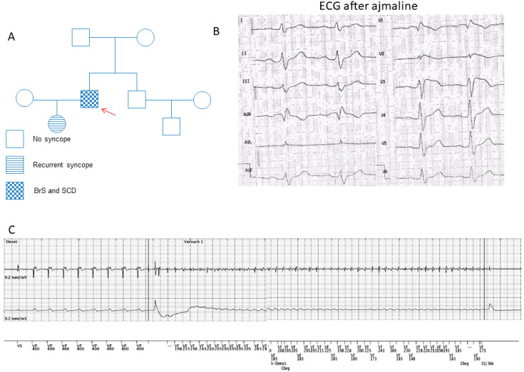
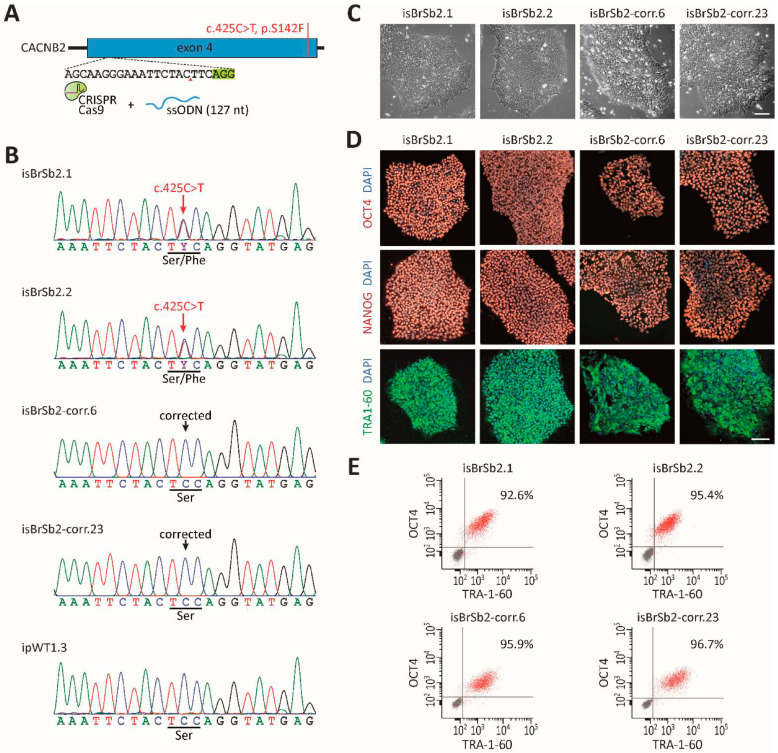
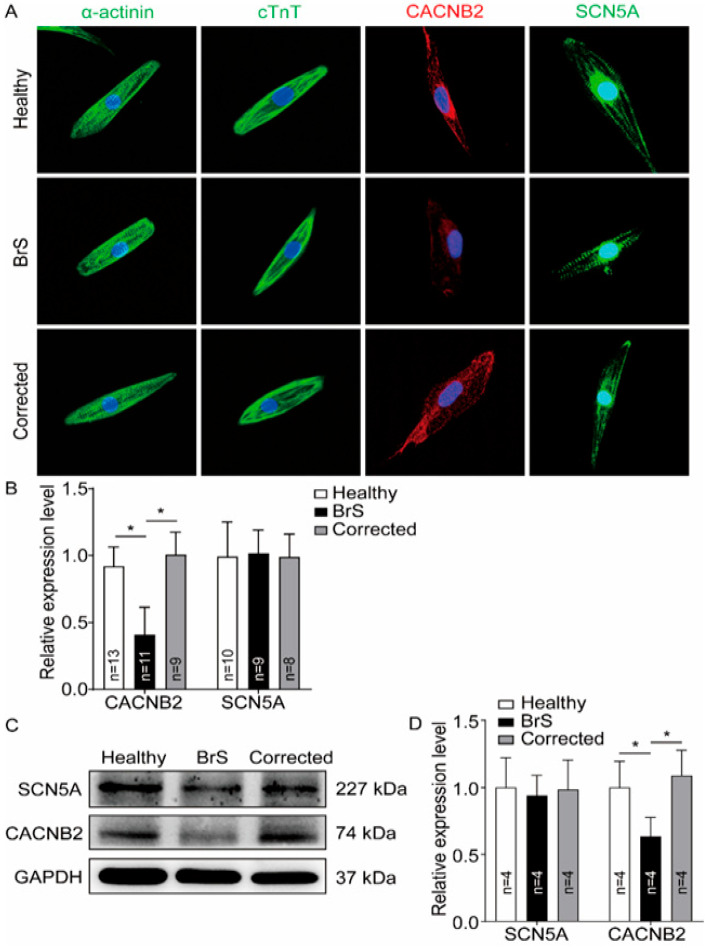
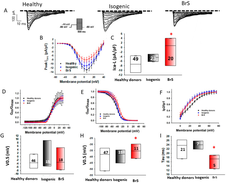
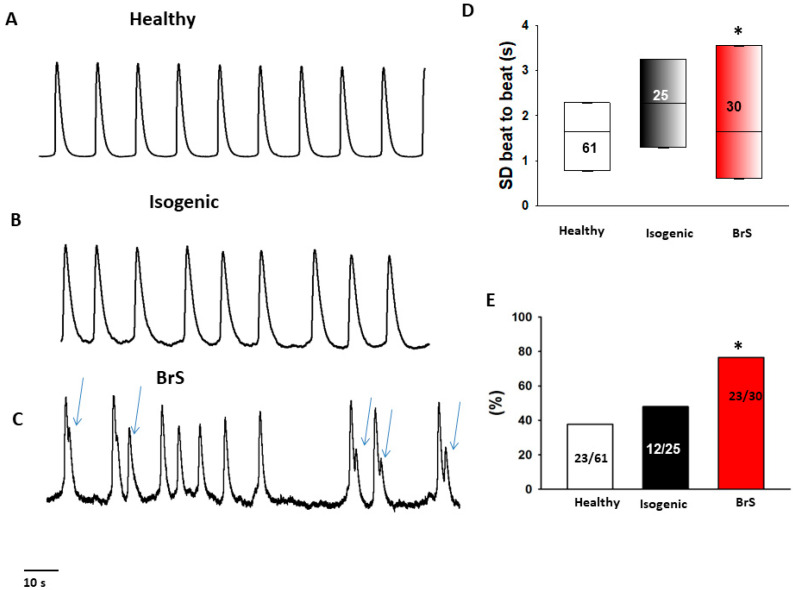
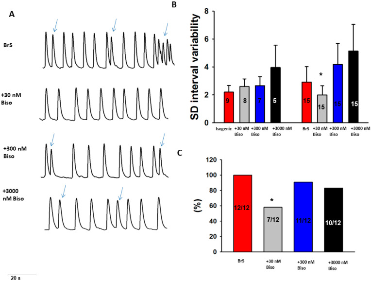
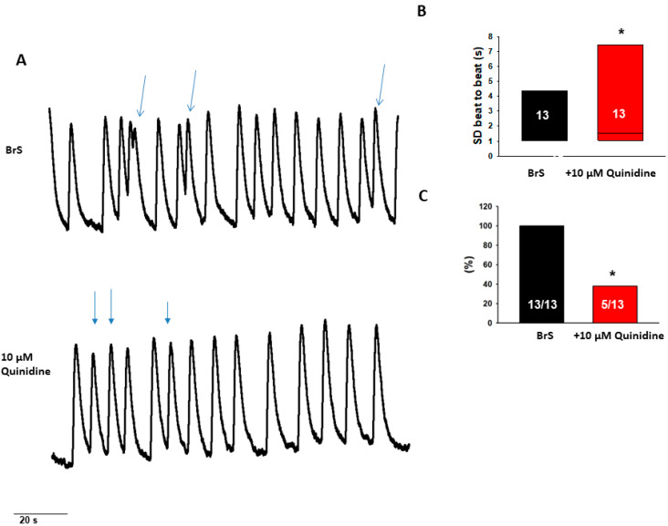
Aims: Some gene variants in the sodium channels, as well as calcium channels, have been associated with Brugada syndrome (BrS). However, the investigation of the human cellular phenotype and the use of drugs for BrS in presence of variant in the calcium channel subunit is still lacking. Objectives: The objective of this study was to establish a cellular model of BrS in the presence of a CACNB2 variant of uncertain significance (c.425C > T/p.S142F) using human-induced pluripotent stem cell-derived cardiomyocytes (hiPSC-CMs) and test drug effects using this model. Methods and results: This study recruited cells from a patient with Brugada syndrome (BrS) and recurrent ventricular fibrillation carrying a missense variant in CACNB2 as well as from three healthy independent persons. These cells (hiPSC-CMs) generated from skin biopsies of healthy persons and the BrS patient (BrS-hiPSC-CMs) as well as CRISPR/Cas9 corrected cells (isogenic control, site-variant corrected) were used for this study. The hiPSC-CMs from the BrS patient showed a significantly reduced L-type calcium channel current (ICa-L) compared with the healthy control hiPSC-CMs. The inactivation curve was shifted to a more positive potential and the recovery from inactivation was accelerated. The protein expression of CACNB2 of the hiPSC-CMs from the BrS-patient was significantly decreased compared with healthy hiPSC-CMs. Moreover, the correction of the CACNB2 site-variant rescued the changes seen in the hiPSC-CMs of the BrS patient to the normal state. These data indicate that the CACNB2 gene variant led to loss-of-function of L-type calcium channels in hiPSC-CMs from the BrS patient. Strikingly, arrhythmia events were more frequently detected in BrS-hiPSC-CMs. Bisoprolol (beta-blockers) at low concentration and quinidine decreased arrhythmic events. Conclusions: The CACNB2 variant (c.425C > T/p.S142F) causes a loss-of-function of L-type calcium channels and is pathogenic for this type of BrS. Bisoprolol and quinidine may be effective for treating BrS with this variant.

Keywords: Brugada syndrome; CACNB gene; arrhythmias; human-induced pluripotent stem cell-derived cardiomyocytes.


Conflict of interest statement

The authors declare no conflict of interest.

Figures

Figure 1
Figure 1
Clinical characteristics of the BrS patient. (A) The pedigree of the BrS patient’s family. The patient recruited for this study is marked by the arrow. He has had sudden cardiac death. (B) The electrocardiogram (ECG) from the BrS patient shows a classic BrS–ECG pattern after infusion of ajmaline. (C) Electrocardiogram of the ICD showing ventricular fibrillation terminated with an appropriate ICD shock after stopping the bisoprolol treatment.
Figure 2
Figure 2
Genetic correction of BrS-hiPSCs by CRISPR/Cas9 genome editing. (A) Corrected hiPSCs were generated with a CRISPR guide RNA targeting the CACNB2 exon 4 and a single-stranded oligonucleotide (ssODN) for homology-directed repair. (B) Confirmation of genetic correction, assessed by Sanger sequencing of genomic DNA. (C) Patients’ and CRISPR-corrected hiPSC lines exhibited a typical human stem cell-like morphology. Scale bar: 100 μm. (D) Immunofluorescence staining for key pluripotency markers OCT4, NANOG and TRA1–60 in patients’ and corrected hiPSC lines. Nuclei were counter-stained with DAPI. Scale bar: 100 μm. (E) Purity of patient-specific and CRISPR-corrected hiPSC lines was evaluated by the flow cytometry analysis of pluripotency markers OCT4 and TRA1–60. Gray dots represent the negative controls.
Figure 3
Figure 3
Reduction of CACNB2 protein expression level in hiPSC-CMs from the BrS-patient. Western blot and immunostaining analyses were performed to examine the protein levels of CACNB2 in hiPSC-CMs from healthy donor (Healthy), the patient (BrS) and isogenic control cell line (Corrected). (A) Representative immunostaining images. (B) Statistical analyses of fluorescence intensity from immunostained cells. (C) Representative examples of Western blots of cell lysates from healthy donor cell line (healthy), the patient (BrS) and the CRISPR/Cas9-corrected (Corrected) hiPSC-CMs. (D) Statistical analyses of relative expression levels of SCN5A and CACNB2 from Western blot experiments. Data were shown as mean ± standard deviation (SD). Numbers given represent the number of cells (B) or experiments (D). * p < 0.05 versus healthy analyzed by one-way analysis of variance (ANOVA) with Holm–Sidak post-test.
Figure 4
Figure 4
Reduction of L-type calcium channel currents in hiPSC-CMs from the BrS patient. Peak L-type calcium (ICa-L) and sodium channel (INa) currents were analyzed in hiPSC-CMs from healthy donors (Healthy), the BrS patient (BrS) and variant-corrected cells (Isogenic). (A) Representative traces of ICa-L in hiPSC-CMs from healthy donors (Healthy), the BrS patient (BrS) and variant-corrected cells (Isogenic). (B) I-V curves of ICa-L from healthy donors (Healthy), the BrS patient (BrS) and variant-corrected cells (Isogenic). (C) Median values of peak ICa-L at −10 mV from healthy donors (Healthy), the BrS patient (BrS) and variant-corrected cells (Isogenic). (D) Activation curves of ICa-L in hiPSC-CMs from each cell line. (E) Inactivation curves of ICa-L in hiPSC-CMs from each cell line. (F) Recovery curves of ICa-L in hiPSC-CMs from each cell line. (G) Median values of the potential at 50% activation (V0.5) of ICa-L in hiPSC-CMs from each cell line. (H) Median values of the potential at 50% inactivation (V0.5) of ICa-L in hiPSC-CMs from each cell line. (I) Median values of the time constants (Tau) of recovery from the inactivation of ICa-L in hiPSC-CMs from each cell line. Numbers given in (C) represent the number of cells for (B,C). Numbers given in (F) represent the number of cells for (E,F). * p < 0.05 versus Healthy according to the analysis of one-way ANOVA with Holm–Sidak post-test.
Figure 5
Figure 5
Increased beating variability and arrhythmic events in BrS-hiPSC-CMs. Spontaneous calcium transients were recorded in spontaneously beating hiPSC-CMs from the BrS patient, the healthy donor (healthy) and the CRISPR-corrected cells (isogenic). The beating variability (standard deviation (SD) of cell beating intervals) and the occurrence of arrhythmic events (irregular or triggered beats or EAD-like events) were compared among the three cell groups. (AC) Representative traces of calcium transients in cells from each line. Arrhythmic events are marked by arrows. (D) Median values of standard deviation (SD) of cell beating intervals. (E) Percentage of cells showing arrhythmic events. The numbers given represent number of cells showing arrhythmic events versus total cell number. The numbers given represent number of cells. * p < 0.05 versus healthy according to the one-way ANOVA (D) or Fisher’s exact test (E).
Figure 6
Figure 6
Effects of beta-blocker on the beating variability and arrhythmic events in BrS-hiPSC-CMs. Spontaneous calcium transients were recorded in regularly beating hiPSC-CMs from the BrS patient and the isogenic control(isogenic) in the absence (baseline) and presence of 30 nM, 300 nM and 3000 nM bisoprolol (Biso). The beating variability (standard deviation (SD) of cell beating intervals) and the occurrence of arrhythmic events (irregular or triggered beats or EAD-like events) were compared between with and without drug application. (A) Representative trace of BrS (at baseline) and after the administration of different concentrations of bisoprolol. (B) Mean values of standard deviation (SD) of cell beating intervals. (C) Percentage of cells showing arrhythmic events. * p < 0.05 versus isogenic according to Fisher’s exact test (C) or versus baseline according to t-test (B).
Figure 7
Figure 7
Effects of quinidine on the beating variability and arrhythmic events in BrS-hiPSC-CMs. (A) Representative traces of calcium transient before and after administration of 10 µM quinidine in BrS-hiPSC-CMs. Only cells with arrhythmic events were used. (B) Median values of standard deviation (SD) of cell beating intervals. (C) Percentage of cells showing arrhythmic events. * p < 0.05 using Fisher’s exact test (C) or versus baseline according to t-test (B).

References

    1. Milman A., Andorin A., Gourraud J.-B., Postema P.G., Sacher F., Mabo P., Kim S.-H., Juang J.J., Maeda S., Takahashi Y., et al. Profile of patients with Brugada syndrome presenting with their first documented arrhythmic event: Data from the Survey on Arrhythmic Events in BRUgada Syndrome (SABRUS) Heart Rhythm. 2018;15:716–724. doi: 10.1016/j.hrthm.2018.01.014. - DOI - PubMed
    1. Casado-Arroyo R., Berne P., Rao J.Y., Rodriguez-Mañero M., Levinstein M., Conte G., Sieira J., Namdar M., Ricciardi D., Chierchia G.B., et al. Long-Term Trends in Newly Diagnosed Brugada Syndrome: Implications for Risk Stratification. J. Am. Coll. Cardiol. 2016;68:614–623. doi: 10.1016/j.jacc.2016.05.073. - DOI - PubMed
    1. Mascia G., Della Bona R., Ameri P., Canepa M., Porto I., Parati G., Crotti L., Brignole M. Brugada syndrome and syncope: A practical approach for diagnosis and treatment. Europace. 2021;23:996–1002. doi: 10.1093/europace/euaa370. - DOI - PubMed
    1. Mascia G., Della Bona R., Ameri P., Canepa M., Porto I., Brignole M. Brugada syndrome and syncope: A systematic review. J. Cardiovasc. Electrophysiol. 2020;31:3334–3338. doi: 10.1111/jce.14787. - DOI - PubMed
    1. Brugada J., Campuzano O., Arbelo E., Sarquella-Brugada G., Brugada R. Present Status of Brugada Syndrome: JACC State-of-the-Art Review. J. Am. Coll. Cardiol. 2018;72:1046–1059. doi: 10.1016/j.jacc.2018.06.037. - DOI - PubMed

MeSH terms

LinkOut - more resources