

Microbeam Irradiation as a Simultaneously Integrated Boost in a Conventional Whole-Brain Radiotherapy Protocol
- PMID: 35955454
- PMCID: PMC9368396
- DOI: 10.3390/ijms23158319
Microbeam Irradiation as a Simultaneously Integrated Boost in a Conventional Whole-Brain Radiotherapy Protocol
Abstract
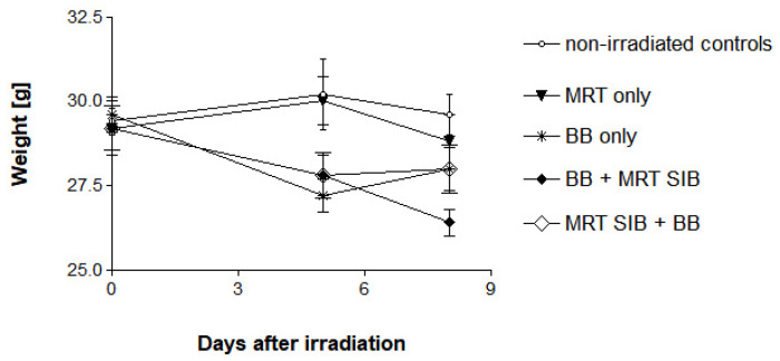
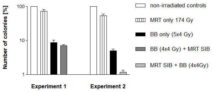
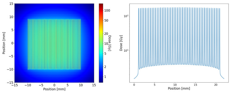
Microbeam radiotherapy (MRT), an experimental high-dose rate concept with spatial fractionation at the micrometre range, has shown a high therapeutic potential as well as good preservation of normal tissue function in pre-clinical studies. We investigated the suitability of MRT as a simultaneously integrated boost (SIB) in conventional whole-brain irradiation (WBRT). A 174 Gy MRT SIB was administered with an array of quasi-parallel, 50 µm wide microbeams spaced at a centre-to-centre distance of 400 µm either on the first or last day of a 5 × 4 Gy radiotherapy schedule in healthy adult C57 BL/6J mice and in F98 glioma cell cultures. The animals were observed for signs of intracranial pressure and focal neurologic signs. Colony counts were conducted in F98 glioma cell cultures. No signs of acute adverse effects were observed in any of the irradiated animals within 3 days after the last irradiation fraction. The tumoricidal effect on F98 cell in vitro was higher when the MRT boost was delivered on the first day of the irradiation course, as opposed to the last day. Therefore, the MRT SIB should be integrated into a clinical radiotherapy schedule as early as possible.
Keywords: F98 glioma cells; brain tissue tolerance; microbeam radiotherapy (MRT); simultaneously integrated boost (SIB).
Conflict of interest statement
The authors declare no conflict of interest.
Figures
Similar articles
-
MRT-boost as the last fraction may be the most efficient irradiation schedule for increased survival times in a rat glioma model.J Synchrotron Radiat. 2023 May 1;30(Pt 3):591-595. doi: 10.1107/S1600577523002606. Epub 2023 Apr 17. J Synchrotron Radiat. 2023. PMID: 37067258 Free PMC article.
-
Synchrotron X-Ray Boost Delivered by Microbeam Radiation Therapy After Conventional X-Ray Therapy Fractionated in Time Improves F98 Glioma Control.Int J Radiat Oncol Biol Phys. 2020 Jun 1;107(2):360-369. doi: 10.1016/j.ijrobp.2020.02.023. Epub 2020 Feb 21. Int J Radiat Oncol Biol Phys. 2020. PMID: 32088292
-
Good Timing Matters: The Spatially Fractionated High Dose Rate Boost Should Come First.Cancers (Basel). 2022 Dec 2;14(23):5964. doi: 10.3390/cancers14235964. Cancers (Basel). 2022. PMID: 36497446 Free PMC article.
-
Effects of pulsed, spatially fractionated, microscopic synchrotron X-ray beams on normal and tumoral brain tissue.Mutat Res. 2010 Apr-Jun;704(1-3):160-6. doi: 10.1016/j.mrrev.2009.12.003. Epub 2009 Dec 23. Mutat Res. 2010. PMID: 20034592 Review.
-
Microbeam radiation therapy: Clinical perspectives.Phys Med. 2015 Sep;31(6):564-7. doi: 10.1016/j.ejmp.2015.02.011. Epub 2015 Mar 13. Phys Med. 2015. PMID: 25773883 Review.
Cited by
-
The Impact of Synchrotron Microbeam Radiation Therapy Combined With Broad Beam in a Preclinical Breast Cancer Model.Adv Radiat Oncol. 2024 Nov 13;10(1):101680. doi: 10.1016/j.adro.2024.101680. eCollection 2025 Jan. Adv Radiat Oncol. 2024. PMID: 39687472 Free PMC article.
-
Application of Synchrotron Radiation in Fundamental Research and Clinical Medicine.Biomedicines. 2025 Jun 10;13(6):1419. doi: 10.3390/biomedicines13061419. Biomedicines. 2025. PMID: 40564139 Free PMC article. Review.
-
Effects of Microbeam Irradiation on Rodent Esophageal Smooth Muscle Contraction.Cells. 2022 Dec 31;12(1):176. doi: 10.3390/cells12010176. Cells. 2022. PMID: 36611969 Free PMC article.
-
The Spinal Cord as Organ of Risk: Assessment for Acute and Subacute Neurological Adverse Effects after Microbeam Radiotherapy in a Rodent Model.Cancers (Basel). 2023 Apr 26;15(9):2470. doi: 10.3390/cancers15092470. Cancers (Basel). 2023. PMID: 37173938 Free PMC article.
-
From Basic Radiobiology to Translational Radiotherapy.Int J Mol Sci. 2022 Dec 14;23(24):15902. doi: 10.3390/ijms232415902. Int J Mol Sci. 2022. PMID: 36555542 Free PMC article.
References
-
- Scherer H.J. Structural development in gliomas. Am. J. Cancer. 1938;34:333–351.
-
- Stupp R., Mason W.P., van den Bent M.J., Weller M., Fisher B., Taphoorn M.J., Belanger K., Brandes A.A., Marosi C., Bogdahn U., et al. European Organisation for Research and Treatment of Cancer Brain Tumor and Radiotherapy Groups; National Cancer Institute of Canada Clinical Trials Group. Radiotherapy plus concomitant and adjuvant temozolomide for glioblastoma. N. Engl. J. Med. 2005;352:987–996. doi: 10.1056/NEJMoa043330. - DOI - PubMed
MeSH terms
Grants and funding
LinkOut - more resources
Full Text Sources
Medical
