. 2022 Jul 28;23(15):8335.
doi: 10.3390/ijms23158335.

Ciliary Neurotrophic Factor (CNTF) and Its Receptors Signal Regulate Cementoblasts Apoptosis through a Mechanism of ERK1/2 and Caspases Signaling

Affiliations

Ciliary Neurotrophic Factor (CNTF) and Its Receptors Signal Regulate Cementoblasts Apoptosis through a Mechanism of ERK1/2 and Caspases Signaling

Jiawen Yong et al. Int J Mol Sci. .

Abstract

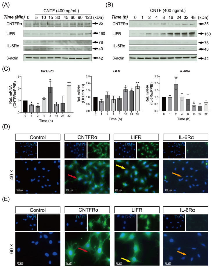
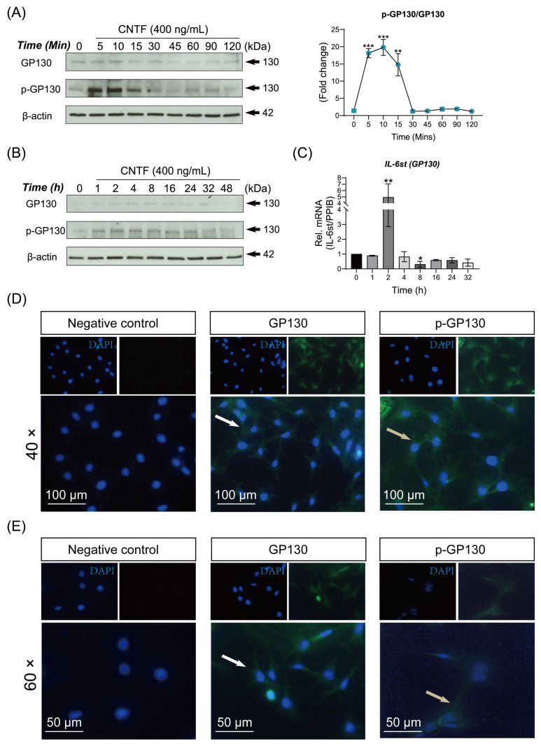
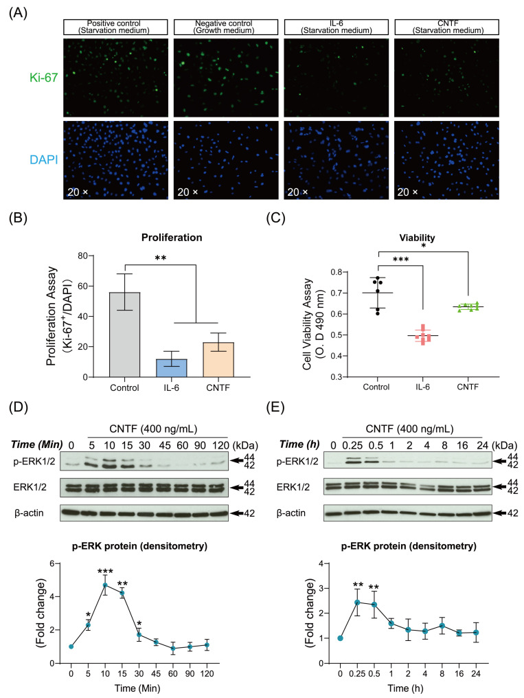
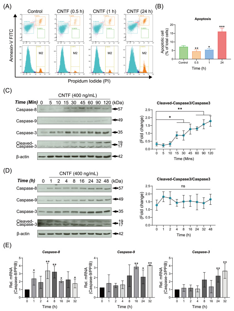
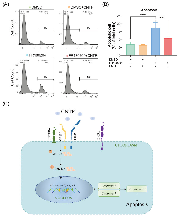
Ciliary neurotrophic factor (CNTF) was identified as a survival factor in various types of peripheral and central neurons, glia and non-neural cells. At present, there is no available data on the expression and localization of CNTF-receptors in cementoblasts as well as on the role of exogenous CNTF on this cell line. The purpose of this study was to determine if cementoblasts express CNTF-receptors and analyze the mechanism of its apoptotic regulation effects on cementoblasts. OCCM-30 cementoblasts were cultivated and stimulated kinetically using CNTF protein (NBP2-35168, Novus Biologicals). Quantified transcriptional (RT-qPCR) and translational (WB) products of CNTFRα, IL-6Rα (CD126), LIFR, p-GP130, GP130, p-ERK1/2, ERK1/2, Caspase-8, -9, -3 and cleaved-caspase-3 were evaluated. Immunofluorescence (IF) staining was applied to visualize the localization of the CNTF-receptors within cells. The apoptosis ratio was measured with an Annexin-V FITC/PI kit. The ERK1/2 antagonist (FR180204, Calbiochem) was added for further investigation by flow cytometry analysis. The CNTF-receptor complex (CNTFRα, LIFR, GP130) was functionally up-regulated in cementoblasts while cultivated with exogenous CNTF. CNTF significantly attenuated cell viability and proliferation for long-term stimulation. Flow cytometry analysis shows that CNTF enhanced the apoptosis after prolonged duration. However, after only a short-term period, CNTF halts the apoptosis of cementoblasts. Further studies revealed that CNTF activated phosphorylated GP130 and the anti-apoptotic molecule ERK1/2 signaling to participate in the regulation of the apoptosis ratio of cementoblasts. In conclusion, CNTF elicited the cellular functions through a notable induction of its receptor complex in cementoblasts. CNTF has an inhibitory effect on the cementoblast homeostasis. These data also elucidate a cellular mechanism for an exogenous CNTF-triggered apoptosis regulation in a mechanism of ERK1/2 and caspase signaling and provides insight into the complex cellular responses induced by CNTF in cementoblasts.

Keywords: CNTF; CNTF-receptors; ERK1/2; apoptosis; caspase; cementoblasts.


Conflict of interest statement

The authors declare no conflict of interest.

Figures

Figure 1
Figure 1
The tripartite CNTF-receptor complex is up-regulated by ciliary neurotrophic factor in cementoblasts. (A,B) WB showed protein expression of the CNTF-receptors (CNTFRα, LIFR and IL-6Rα) in OCCM-30 cells induced by CNTF protein (400 ng/mL) for various periods. Internal β-actin serves as loading control. (C) The expression of mRNAs encoding the CNTF-receptors were quantified by RT-qPCR. The relative mRNA expression of each gene was obtained through normalizing to internal PPIB. The statistical significance was determined by student t-test (n = 3 for each group). (D,E) The CNTF-receptors immunofluorescent localization showed the expression of CNTFRα (red arrow), LIFR (yellow arrow) and IL-6Rα (orange arrow) in CNTF-treated OCCM-30 cells. Nuclei are stained with DAPI (blue). Scale bar: 100 μm (image magnification: 40×); 50 μm (image magnification: 60×). Bar indicates values ± standard deviation (SD) which represent three independent experiments. Statistically significant differences (indicated by asterisks) are shown as follows (* p < 0.05; ** p < 0.005).
Figure 2
Figure 2
Ciliary neurotrophic factor triggers GP130 protein expression and phosphorylated GP130 in cementoblasts. (A,B) The protein expression of GP130 and phosphorylated GP130 were determined by WB. Internal β-actin serve as loading control. The line chart shows the densitometric analysis of p-GP130 expression related to total GP-130 expression. (C) RT-qPCR quantification of GP130 (IL-6st) gene expression in OCCM-30 cells when treated with CNTF (400 ng/mL) for indicated time. The relative mRNA expression was obtained through normalizing to internal PPIB. (D,E) IF staining of subcellular localization of GP130 (white arrow) as well as p-GP130 (grey arrow) in non-stimulated cells (negative control) and CNTF-stimulated OCCM-30 cells. Nuclei are stained with DAPI (blue). Individual and merged images of GP130 and p-GP130 are shown. Scale bar: 100 μm (image magnification: 40×); 50 μm (image magnification: 60×). Bar indicates values ± standard deviation (SD) which represent three independent experiments. Statistically significant differences (indicated by asterisks) are shown as follows (* p < 0.05; ** p < 0.005; *** p < 0.0005).
Figure 3
Figure 3
Ciliary neurotrophic factor impairs OCCM-30 homeostasis and activates the expression of ERK1/2 MAPK signaling. (A,B) Immunofluorescence microscopy images show representative proliferation markers Ki-67 expression. OCCM-30 cells were exposed to CNTF (400 ng/mL) and then underwent immunofluorescence staining for Ki-67 to visualize cells in the proliferation stage. The proportion of proliferating cells for each group was quantified according to Ki-67 positive cells (Ki-67+)/total cell counting (DAPI). (C) Cell viability assay was performed by MTS assay. IL-6 cytokine served as positive control. (D,E) Representative immunoblot of p-ERK1/2 protein expression in the presence of CNTF (400 ng/mL) at different time points. Internal β-actin serves as loading control. Densitometric immunoblot analysis of bands indicated the enhanced p-ERK1/2 expression relative to that of the control group. Densitometric results are showed as fold change. Bar indicates values ± standard deviation (SD) which represent three independent experiments. Statistically significant differences (indicated by asterisks) are shown as follows (* p < 0.05; ** p < 0.005; *** p < 0.0005).
Figure 4
Figure 4
Ciliary neurotrophic factor regulates apoptosis rate and triggers the caspases signaling. (A,B) Representative plots from Annexin-V FITC and PI staining by flow cytometry analysis performed in triplicate are shown. Apoptotic cells (Annexin-V FITC+/PI+) are shown in the upper right quadrant. Graphics show the percentages of apoptotic cells exposed to CNTF (400 ng/mL) at different time periods. (C,D) Representative immunoblot showed that the protein expression of Caspase-8, -9 and -3 as well as cleaved-caspase-3 in response to CNTF (400 ng/mL) in a time-dependent manner. β-actin was loaded as an internal control. (E) mRNA expression of Caspase-8, -9 and -3 in response to CNTF (400 ng/mL) stimulation at indicated time period. Bar indicates values ± standard deviation (SD) which represent three independent experiments. Statistically significant differences (indicated by asterisks) are shown as follows (ns, no significant difference; * p < 0.05; ** p < 0.005; *** p < 0.0005).
Figure 5
Figure 5
ERK1/2 signal is involved in the regulation of apoptosis of cementoblasts and the caspases pathway. (A,B) Graphics show the percentages of apoptotic cells exposed to ERK1/2 inhibitor (1.0 μg/mL, FR180204) as well as co-stimulation with CNTF (400 ng/mL). (C) The scheme summarizes the mode of CNTF action in cementoblasts: CNTF activated the tripartite CNTF-receptor complex targets and phosphorylated GP130 protein, which recruits ERK1/2 signaling and caspases signaling expression. FR180204 promotes apoptosis in OCCM-30 cells and CNTF addition suppressed the ERK1/2 inhibitor-induced apoptosis within a short-term period. Bar indicates values ± standard deviation (SD) which represent three independent experiments. Statistically significant differences (indicated by asterisks) are shown as follows (** p < 0.005; *** p < 0.0005).

References

    1. Stockli K.A., Lottspeich F., Sendtner M., Masiakowski P., Carroll P., Gotz R., Lindholm D., Thoenen H. Molecular cloning, expression and regional distribution of rat ciliary neurotrophic factor. Nature. 1989;342:920–923. doi: 10.1038/342920a0. - DOI - PubMed
    1. Lin L.F., Mismer D., Lile J.D., Armes L.G., Butler E.T., 3rd, Vannice J.L., Collins F. Purification, cloning, and expression of ciliary neurotrophic factor (CNTF) Science. 1989;246:1023–1025. doi: 10.1126/science.2587985. - DOI - PubMed
    1. Kruttgen A., Grotzinger J., Kurapkat G., Weis J., Simon R., Thier M., Schroder M., Heinrich P., Wollmer A., Comeau M., et al. Human ciliary neurotrophic factor: A structure-function analysis. Pt 1Biochem. J. 1995;309:215–220. doi: 10.1042/bj3090215. - DOI - PMC - PubMed
    1. Schuster B., Kovaleva M., Sun Y., Regenhard P., Matthews V., Grotzinger J., Rose-John S., Kallen K.J. Signaling of human ciliary neurotrophic factor (CNTF) revisited. The interleukin-6 receptor can serve as an alpha-receptor for CTNF. J. Biol. Chem. 2003;278:9528–9535. doi: 10.1074/jbc.M210044200. - DOI - PubMed
    1. Heinrich P.C., Behrmann I., Muller-Newen G., Schaper F., Graeve L. Interleukin-6-type cytokine signalling through the gp130/Jak/STAT pathway. Pt 2Biochem. J. 1998;334:297–314. doi: 10.1042/bj3340297. - DOI - PMC - PubMed

MeSH terms

Substances