. 2022 Jul 28;23(15):8355.
doi: 10.3390/ijms23158355.

GTP Binding Protein Gtr1 Cooperating with ASF1 Regulates Asexual Development in Stemphylium eturmiunum

Affiliations

GTP Binding Protein Gtr1 Cooperating with ASF1 Regulates Asexual Development in Stemphylium eturmiunum

Shi Wang et al. Int J Mol Sci. .

Abstract

The Gtr1 protein was a member of the RagA subfamily of the Ras-like small GTPase superfamily and involved in phosphate acquisition, ribosome biogenesis and epigenetic control of gene expression in yeast. However, Gtr1 regulation sexual or asexual development in filamentous fungi is barely accepted. In the study, SeGtr1, identified from Stemphylium eturmiunum, could manipulate mycelial growth, nuclear distribution of mycelium and the morphology of conidia in Segtr1 silenced strains compared with its overexpression transformants, while the sexual activity of Segtr1 silenced strains were unchanged. SeASF1, a H3/H4 chaperone, participated in nucleosome assembly/disassembly, DNA replication and transcriptional regulation. Our experiments showed that deletion Seasf1 mutants produced the hyphal fusion and abnormal conidia. Notably, we characterized that Segtr1 was down-regulated in Se∆asf1 mutants and Seasf1 was also down-regulated in SiSegtr1 strains. We further confirmed that SeGtr1 interacted with SeASF1 or SeH4 in vivo and vitro, respectively. Thus, SeGtr1 can cooperate with SeASF1 to modulate asexual development in Stemphylium eturmiunum.

Keywords: ASF1; GTP binding protein Gtr1; Stemphylium eturmiunum; asexual development; interaction.


Conflict of interest statement

There are no conflict of interest for all authors and organizations that mentioned in this manuscript.

Figures

Figure 1
Figure 1
Seasf1 regulates asexual developmental characterization in S. eturmiunum. (A) Growth of two Se∆asf1 mutants, two Se∆asf1::Seasf1 transformants, and WT strains on CM medium. The cultures were photographed after 9 days of incubation. (B) Characterizations of hyphal fusion in two Se∆asf1 mutants, two Se∆asf1::Seasf1 transformants, and WT strains. The images were photographed after growing on PDA medium for 8 days. The fusions in the hyphae were marked with red arrows. (C,D) Conidia morphology of four mutants and WT strains were cultured on CM medium for 4 weeks. Bar = 20 μm, 500 μm.
Figure 2
Figure 2
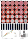
The colonial phenotypes and nuclear distribution of Segtr1 silenced transformants. (A) Colonial growth of two Segtr1 silenced transformants (SiSegtr1-T63 and SiSegtr1-T66) and two Segtr1 overexpression transformants (OESegtr1-T3 and OESegtr1-T8) was observed on PDA medium. WT was S. eturmiunum strain and Control was a negative control strain. The cultures were photographed after 1 day, 3 days, 5 days, 7 days and 9 days. (B) The mycelium of these four transformants were grown on PDA medium for 6 days and examined by DIC and fluorescence microscopy. The nuclei of the mycelia were discovered under the fluorescence microscopy after staining by DAPI. Bar = 20 μm. (C) Colony diameters were measured in each independent biological experiment at 1–9 days of growth on PDA medium. Rates of colonial growth were calculated for each treatment. (D) qRT-PCR was used to measure the expression levels of Segtr1 in silenced transformants, overexpression transformants, Control and WT. The degree of WT was assigned to value 1.0. Actin gene of S. eturmiunum was used as endogenous control. The bars indicated statistically significant differences (ANOVA; ** p < 0.01). Each experiment was repeated at least three times.
Figure 3
Figure 3
SeGtr1 plays a role in asexual development, but the sexual activity was unchanged. (A) For the microscopic investigation of conidiophores and conidia development, two silenced transformants (SiSegtr1-T63 and SiSegtr1-T66), two overexpression transformants (OESegtr1-T3 and OESegtr1-T8), Control and WT strains were grown on CM medium for 35 days, respectively. Control was a negative control strain. Red arrowheads indicated abnormal conidiophores. (B) To further observe the role of Segtr1 during the sexual development of S. eturmiunum, all transformants and WT strains were cultured on PDA medium for inducing perithecia production. At 35 days, all transformant strains produced abundant perithecia. Bar = 20 μm and 500 μm.
Figure 4
Figure 4
The expression patterns of Seasf1, SeH4, and Segtr1 in knockout mutants, silenced lines or overexpression strains. (A) The expression levels of Segtr1 and SeH4 in two Se∆asf1 mutants and two complemented transformants were measured by qRT-PCR. (B) The expression levels of Segtr1 and Seasf1 in two SiSeH4 lines were measured by qRT-PCR. (C) The expression levels of Seasf1 and SeH4 in two SiSegtr1 lines and two OESegtr1 lines were measured by qRT-PCR. The degree of WT was assigned to value 1.0. Two Seasf1 deleted mutants were Se∆asf1-0 and Se∆asf1-5, two complemented transformants were Se∆asf1::Seasf1-1 and Se∆asf1::Seasf1-2. Two SeH4-silenced lines were SiSeH4-T8 and SiSeH4-T20. Two Segtr1-silenced lines were SiSegtr1-T63 and SiSegtr1-T66, two OESegtr1 lines were OESegtr1-T3 and OESegtr1-T8. The Actin in S. eturmiunum was used as endogenous control. The bars indicated statistically significant differences (ANOVA; ** p < 0.01).
Figure 5
Figure 5
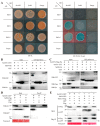
SeASF1 interaction with SeH4, and SeGtr1 interaction with SeASF1 or SeH4. (A) SeASF1 interacted with SeH4, and SeGtr1 interaction with SeASF1 or SeH4 using Y2H. SeASF1 or SeH4 was cloned into plasmid pGBKT7 (BD). SeH4 or SeGtr1 was cloned into plasmid pGADT7 (AD). Yeast transformants were first grown on SD/-Trp/-Leu, and selected on SD/-Trp/-Leu/-His/-Ade/X-α-gal. A positive interaction results in the activation of the lacZ reporter, which turned the blue in the presence of X-α-galactosidase. The images were photographed at 3 days after incubation. (B) SeASF1 was cloned into plasmid pGEX-6P-1. SeH4 was cloned into plasmid pET28a. SeASF1-GST was expressed in E. coli and incubated with SeH4-His, purified (pull-down) by glutathione sepharose beads. Recombinant GST was control. SeH4-His was pulled down by SeASF1-GST. (C) SeGtr1 was cloned into plasmid pGEX-6P-1. Flag-SeASF1 or SeH4 was cloned into plasmid pET28a. SeH4-His and Flag-SeASF1-His were both retained by SeGtr1-GST. (D) SeASF1 was cloned into plasmid pDL2, SeH4 was cloned into plasmid pFL7. Total proteins were extracted from F. graminearum protoplasts expressing SeASF1-GFP and SeH4-Flag. Recombinant GFP or Flag was control. The immune complexes were immunoprecipitated with α-Flag antibody (α-Flag IP). Coprecipitation of SeH4-Flag was detected by immunoblotting. (E) SeH4 was cloned into plasmid pDL2. SeGtr1 was cloned into plasmid pFL7. Total proteins were extracted from F. graminearum protoplasts expressing SeASF1-GFP, SeH4-GFP, and SeGtr1-Flag. Coprecipitation of SeGtr1-Flag was detected by immunoblotting. Membranes were stained with Ponceau S to confirm equal loading. Protein sizes are indicated in kDa. Each experiment was repeated at least three times.
Figure 6
Figure 6
A model for ASF1 binding H4 (ASF1-H4) to interact with GTR1 and then to mediate asexual reproduction in Stemphylium eturmiunum. ASF1 interacts with H4 and then they are translocated into nuclei through the nuclear pore. The dimer of ASF1-H4 regulates DNA replication and damage repair in previous studies. Herein, ASF1-H4 combines with GTR1 to constitute a trimeric complex which in turn is involved in the central regulatory pathway to modulate asexual reproduction.

Similar articles

References

    1. Wu M.Y., Mead M.E., Lee M.K., Neuhaus G.F., Adpressa D.A., Martien J.I., Son Y.E., Moon H., Amador-Noguez D., Han K.H., et al. Transcriptomic, protein-DNA interaction, and metabolomic studies of VosA, VelB, and WetA in Aspergillus nidulans asexual spores. Mol. Biol. 2021;12:e03128-20. doi: 10.1128/mBio.03128-20. - DOI - PMC - PubMed
    1. Dyer P.S., Ingram D.S., Johnstone K. The control of sexual morphogenesis in the Ascomycotina. Biol. Rev. 1992;67:421–458. doi: 10.1111/j.1469-185X.1992.tb01189.x. - DOI
    1. Milgroom M.G. Recombination and the multilocus structure of fungal populations. Annu. Rev. Phytopathol. 1996;34:457–477. doi: 10.1146/annurev.phyto.34.1.457. - DOI - PubMed
    1. Grigsby I.F., Rutledge E.M., Morton C.A., Finger F.P. Functional redundancy of two C. elegans homologs of the histone chaperone Asf1 in germline DNA replication. Dev. Biol. 2009;329:64–79. doi: 10.1016/j.ydbio.2009.02.015. - DOI - PubMed
    1. Taylor J., Jacobson D., Fisher M. The volution of asexual fungi: Reproduction, speciation and classification. Annu. Rev. Phytopathol. 1999;37:197–246. doi: 10.1146/annurev.phyto.37.1.197. - DOI - PubMed

MeSH terms

Supplementary concepts

LinkOut - more resources