. 2022 Jul 29;23(15):8419.
doi: 10.3390/ijms23158419.

Persistent TLR4 Activation Promotes Hepatocellular Carcinoma Growth through Positive Feedback Regulation by LIN28A/Let-7g miRNA

Affiliations

Persistent TLR4 Activation Promotes Hepatocellular Carcinoma Growth through Positive Feedback Regulation by LIN28A/Let-7g miRNA

I-Ting Chen et al. Int J Mol Sci. .

Abstract

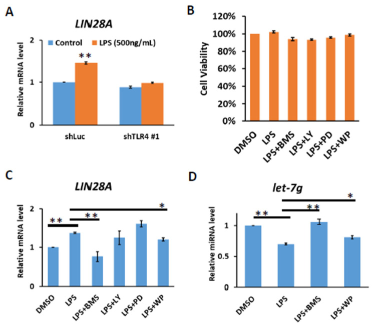
Chronic inflammation caused by liver damage or infection plays an important role in the development and progression of hepatocellular carcinoma (HCC). The activation of Toll-like receptors 4 (TLR4) is involved in HCC tumorigenesis. Moreover, high TLR4 expression in HCC has been linked to poor prognosis. Although the expression of TLR4 in HCC is relatively low compared to hematopoietic cells, it is important to explore the molecular mechanism leading to the elevation of TLR4 in HCC. In this study, we aimed to investigate the positive regulating loop for TLR4 expression in HCC in response to chronic inflammation. Our results confirm that the mRNA expression of TLR4 and proinflammatory cytokines, including interleukin 6 (IL6) and C-C motif chemokine ligand 2 (CCL2), positively correlate in human HCC samples. High TLR4 expression in HCC is more susceptible to lipopolysaccharide (LPS); TLR4 activation in HCC provides growth and survival advantages and thus promotes tumorigenesis. It has been shown that the LIN28/let-7 microRNA (miRNA) axis is a downstream effector of the TLR4 signal pathway, and let-7 miRNA is a potential post-transcriptional regulator for TLR4. Thus, we investigated the correlation between TLR4 and LIN28A mRNA and let-7g miRNA in HCC clinical samples and found that the expression of TLR4 was positively correlated with LIN28A and negatively correlated with let-7g miRNA. Moreover, by culturing PLC/PRF5 (PLC5) HCC cells in low-dose LPS-containing medium to mimic chronic inflammation for persistent TLR4 activation, the mRNA and protein levels of TLR4 and LIN28A were elevated, and let-7g miRNA was decreased. Furthermore, the 3' untranslated region (3'UTR) of TLR4 mRNA was shown to be the target of let-7g miRNA, suggesting that inhibition of let-7g miRNA is able to increase TLR4 mRNA. While parental PLC5 cells have a low susceptibility to LPS-induced cell growth, long-term LPS exposure for PLC5 cells leads to increased proliferation, cytokine expression and stemness properties. In conclusion, our studies demonstrate positive feedback regulation for chronic TLR4 activation in the modulation of TLR4 expression level through the LIN28A/let-7g pathway in HCC and suggest a connection between chronic inflammation and TLR4 expression level in HCC for promoting tumorigenesis.

Keywords: LIN28A; Toll-like receptors 4; hepatocellular carcinoma; let-7g.


Conflict of interest statement

The authors declare no conflict of interest.

Figures

Figure 1
Figure 1
Positive correlation of TLR4 with IL6 or CCL2 mRNA in HCC clinical samples. Correlation between the mRNA expression of TLR4 with (A) IL6 and (B) CCL2 in HCC patients. Pearson’s correlation analyses were performed. Data are presented as each value (R2, correlation coefficient).
Figure 2
Figure 2
TLR4 activation in Huh7 HCC cells leads to growth and survival advantages. (A) Cell viability and (B) clonogenic ability of Huh7 and PLC5 cells with TLR4 engagement. Huh7 and PLC5 cells incubated with or without LPS (10 ng/mL) were placed on 96-well plates for 4 days or on 6-well plates for 14 days. Cell viability was measured with MTT assays. Clonogenic ability was determined by colony formation, which was visualized by crystal violet. The relative viability and clonogenic ability of cells without LPS treatment were set as 100 percent. Results are mean ± SD for six separate experiments. (C) Induction of TNF, IL6, CCL2 and COX2 mRNA in Huh7 and PLC5 cells with LPS stimulation. Huh7 or PLC5 cells were treated with or without LPS for indicated time periods. RNA was extracted, and the expression of the indicated mRNAs was measured by qPCR. The relative mRNA level in Huh7 cells with LPS treatment at 0 h was set as 1.0. Results are mean ± SD for three separate experiments. (D) Activation of Akt, JNK and IKK pathways in Huh7 cells in response to LPS. Cells were treated with LPS and collected at the indicated time points. Akt and JNK phosphorylation and IκBα degradation were analyzed by means of immunoblotting. The results are represented by three independent experiments. (E) The quantification of relative protein levels for p-Akt/Akt, p-JNK/JNK and IκBα in each condition. Error bars show standard deviation. (F) Activation of STAT3 in Huh7 cells with LPS treatment. Cells were treated with LPS for the indicated times. Cell lysates were analyzed by immunoblotting for STAT3 phosphorylation. The results are represented by three independent experiments. (G) Effect of LPS on HCC cell growth in vivo. SCID mice were inoculated with 2 × 10⁶ Huh7 cells s.c. in the right flank. Phosphate buffered saline (PBS) or LPS (2.5 mg/kg) was administrated i.p. once weekly, and treatment was started on day 1, when tumors were measurable (n = 6 for each group). Caliper measurements were performed to estimate the tumor volume (mean ± SD mm3). (** p < 0.01).
Figure 3
Figure 3
TLR4 activation induces IKK-dependent LIN28A expression. (A) Induction of LIN28A mRNA of Huh7 cells with or without TLR4 silencing in response to LPS. The cells were treated with LPS (500 ng/mL) for 4 h and RNA was extracted to be measured by qPCR. (B) Cell viability of Huh7 cells. Huh7 incubated with LPS (10 ng/mL) in the presence of indicated inhibitors was placed on 96-well plates for 4 h. Cell viability was measured with MTT assays. (C) LIN28A mRNA and (D) let-7g miRNA expression regulated by LPS depends on IKK and STAT3 pathways. Huh7 cells were treated with LPS (500 ng/mL) in the presence of indicated inhibitors, including IKK inhibitor (BMS345541 (20 μM)), PI3K inhibitor (LY294002 (20 μM)), MEK inhibitor (PD98059 (50 μM)) and STAT3 inhibitor (WP1066 (10 μM)), for 4 h. RNA was extracted, and the expression levels of the indicated mRNAs and miRNA were measured by qPCR. The relative mRNA and miRNA level in Huh7 cells without LPS treatment was set as 1.0. Results are mean ± SD for two separate experiments. (* p < 0.05, ** p < 0.01).
Figure 4
Figure 4
Positive correlation of TLR4 with LIN28A mRNA and inverse correlation of TLR4 and LIN28A mRNA with let-7g miRNA in clinical HCC samples. (A) Correlation between the expression of LIN28A mRNA with let-7g miRNA in HCC patients, and correlation between the expression of TLR4 mRNA and (B) LIN28A mRNA and (C) let-7g miRNA. Pearson’s correlation analyses were performed. Data are presented as each value (R2, correlation coefficient).
Figure 5
Figure 5
HCC with long-term LPS exposure leads to the upregulation of TLR4 and LIN28A, and the downregulation of let-7g miRNA. (A) TLR4 and (B) LIN28A mRNA levels in PLC5 cells with or without long-term LPS exposure for indicated passages. (C) Protein level of TLR4 and LIN28A in PLC5 cells with LPS exposure for indicated passages. The expression levels of indicated proteins were detected with immunoblotting. The results are represented by three independent experiments. (D) The quantification of TLR4 and LIN28A protein levels compared to the α-actin control in each condition. Error bars show standard deviation. (E) The expression of let-7g miRNA level in PLC5 cells with or without LPS exposure for indicated passages. (F) c-Myc mRNA level in PLC5 cells with or without LPS exposure for indicated passages. The mRNA and miRNA levels were detected with qPCR. The relative mRNA or miRNA level in PLC5 cells without LPS treatment was set as 1.0. Results are mean ± SD for three separate experiments. (G) Activation of Akt and p38 pathways in PLC5 cells with or without long-term LPS exposure. Cells were treated without or with LPS (250 ng/mL) for 30 min. Akt and p38 phosphorylation was analyzed by means of immunoblotting. The results are represented by two independent experiments. (** p < 0.01).
Figure 6
Figure 6
TLR4 mRNA is targeted by let-7g miRNA. (A) Schematic of the seed region match between hsa-let-7g and the putative TLR4 3′UTR. MicroRNA.org was used to analyze and predict the possible seed region of has-let-7g on the 3′UTR of human TLR4 mRNA. (B) Huh7 cells were transfected with control or hsa-let-7g precursor-expressed vector and incubated for 1 to 3 days as indicated. The levels of endogenous TLR4 mRNA were quantified with qPCR. (C) Huh7 cells were transfected with control or hsa-let-7g miRNA precursor-expressed vector, incubated for 2 days, and harvested for the Western blotting of TLR4 protein levels. The results are represented by two independent experiments. (D) The quantification of relative protein levels for TLR4 in each condition. Error bars show standard deviation. (E) The post-transcriptional regulation of TLR4 mRNA by let7-g miRNA with luciferase reporter assay. The positions of three seed match sites for let-7g and TLR4 3′UTR in the luciferase reporter construct pmirGLO-TLR4-3′UTR are indicated (left). Huh7 cells in 96-well plates were co-transfected with pre-let-7g miRNA-expressed vector and the indicated reporter constructs, including empty, wild-type TLR4 3′UTR, and mutated TLR4 3′UTR, respectively, in the downstream of Firefly luciferase gene. Firefly and Renilla luciferase activities were measured at 24 h post-transfection. The data represent the means from three independent experiments. (* p < 0.05, ** p < 0.01).
Figure 7
Figure 7
Cell growth and stemness properties were increased in PLC5 cells with long-term LPS exposure. (A) Cell viability and (B) clonogenic ability of PLC5 cells under long-term LPS exposure. PLC5 cells with or without LPS exposure for 20 passages were placed on 96-well plates for 4 days or on 6-well plates for 14 days. Cell viability was measured with MTT assays. Clonogenic ability was determined by colony formation, which was visualized by crystal violet. The relative viability and clonogenic ability of PLC5 cells without LPS treatment were set as 100 percent. Results are mean ± SD for three separate experiments. (C) Non-adherent sphere formation of PLC5 cells under long-term LPS exposure. PLC5 cells with or without LPS exposure for 20 passages were plated on 96-well plates and incubated for 4 days. Bright-field images are shown. Scale bar = 100 μm. Number of large spheres generated from 1 × 10³ PLC5 cells with LPS exposure for indicated passages was quantified. Results are mean ± SD for three separate experiments. (D) The levels of OCT4 and NANOG mRNA in spheres from PLC5 cells with or without LPS exposure. The mRNA levels were detected with qPCR. (E) Effect of long-term LPS exposure on HCC cell growth in vivo. SCID mice were inoculated with 5 × 10⁶ PLC5 cells with or without LPS exposure for 20 passages s.c. in the right flank (n = 6 for each group). Caliper measurements were performed to estimate the tumor volume (mean ± SD mm3). (* p < 0.05, ** p < 0.01).
Figure 8
Figure 8
Proposed model for the positive feedback loop of TLR4-mediated survival signaling in liver cancer.

Similar articles

Cited by

References

    1. Sengupta R., Honey K. AACR Cancer Progress Report 2019: Transforming Lives Through Innovative Cancer Science. Clin. Cancer Res. 2019;25:5431. doi: 10.1158/1078-0432.CCR-19-2655. - DOI - PubMed
    1. Anstee Q.M., Reeves H.L., Kotsiliti E., Govaere O., Heikenwalder M. From NASH to HCC: Current concepts and future challenges. Nat. Rev. Gastroenterol. Hepatol. 2019;16:411–428. doi: 10.1038/s41575-019-0145-7. - DOI - PubMed
    1. Marengo A., Jouness R.I., Bugianesi E. Progression and Natural History of Nonalcoholic Fatty Liver Disease in Adults. Clin. Liver Dis. 2016;20:313–324. doi: 10.1016/j.cld.2015.10.010. - DOI - PubMed
    1. Grivennikov S.I., Greten F.R., Karin M. Immunity, inflammation, and cancer. Cell. 2010;140:883–899. doi: 10.1016/j.cell.2010.01.025. - DOI - PMC - PubMed
    1. Takeuchi O., Akira S. Pattern recognition receptors and inflammation. Cell. 2010;140:805–820. doi: 10.1016/j.cell.2010.01.022. - DOI - PubMed