. 2022 Aug 2;23(15):8563.
doi: 10.3390/ijms23158563.

Utilization of Aminoguanidine Prevents Cytotoxic Effects of Semen

Affiliations

Utilization of Aminoguanidine Prevents Cytotoxic Effects of Semen

Mirja Harms et al. Int J Mol Sci. .

Abstract

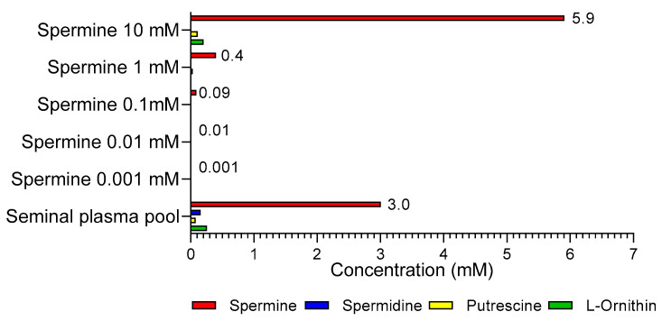
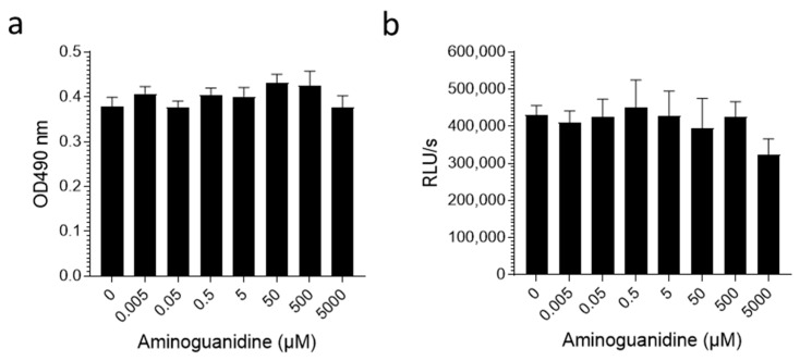
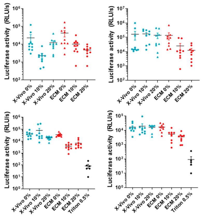
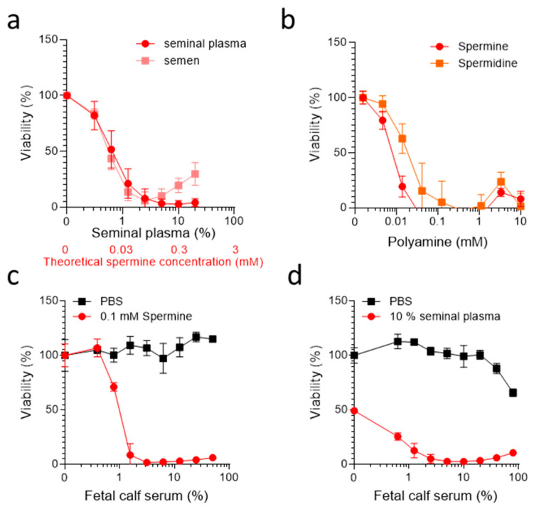
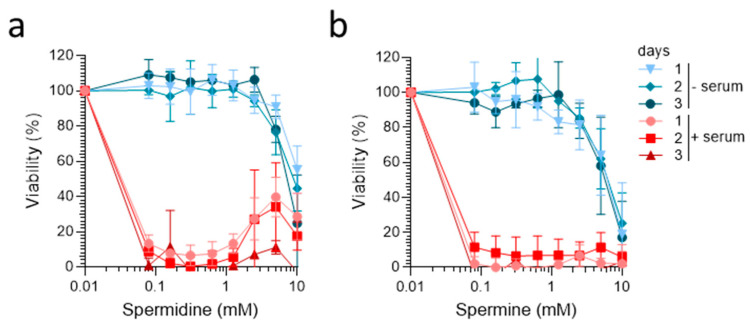
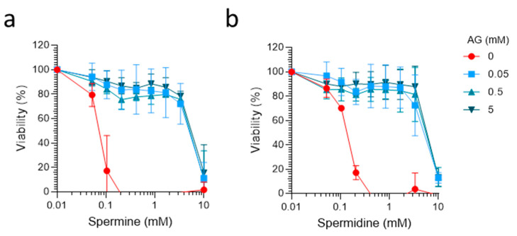
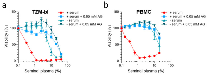
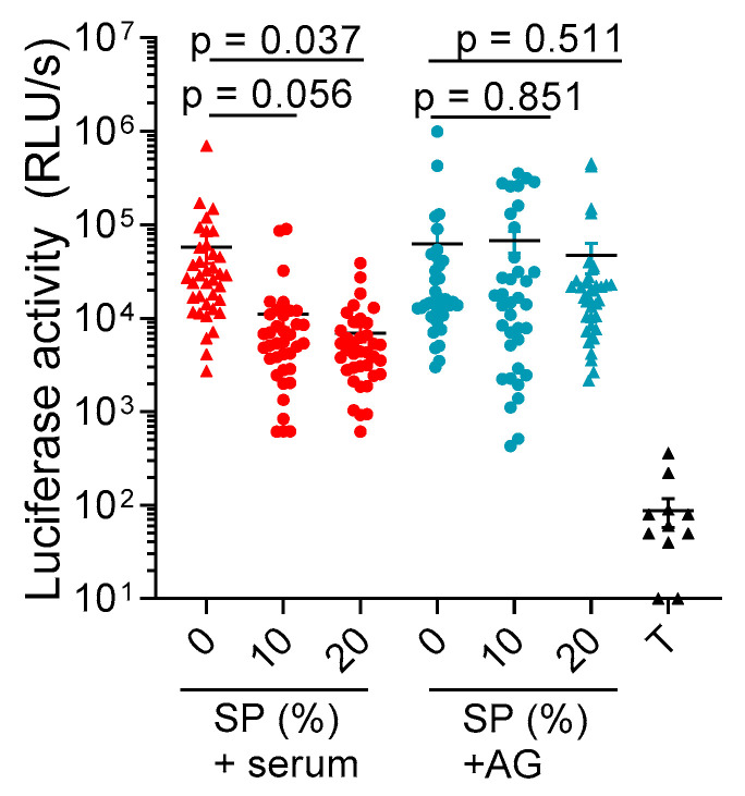
Studies of human semen in cell or tissue culture are hampered by the high cytotoxic activity of this body fluid. The components responsible for the cell damaging activity of semen are amine oxidases, which convert abundant polyamines, such as spermine or spermidine in seminal plasma into toxic intermediates. Amine oxidases are naturally present at low concentrations in seminal plasma and at high concentrations in fetal calf serum, a commonly used cell culture supplement. Here, we show that, in the presence of fetal calf serum, seminal plasma, as well as the polyamines spermine and spermidine, are highly cytotoxic to immortalized cells, primary blood mononuclear cells, and vaginal tissue. Thus, experiments investigating the effect of polyamines and seminal plasma on cellular functions should be performed with great caution, considering the confounding cytotoxic effects. The addition of the amine oxidase inhibitor aminoguanidine to fetal calf serum and/or the utilization of serum-free medium greatly reduced this serum-induced cytotoxicity of polyamines and seminal plasma in cell lines, primary cells, and tissues and, thus, should be implemented in all future studies analyzing the role of polyamines and semen on cellular functions.

Keywords: cytotoxicity; polyamines; seminal fluid; spermine.


Conflict of interest statement

The authors declare no conflict of interest.

Figures

Figure A1
Figure A1
Spermine detection in pooled human seminal plasma. Polyamine concentration was determined in the pooled seminal plasma of 50 individual donors using SPE-LC-MS/MS according to Magnes et al. (2014) [34].
Figure A2
Figure A2
Aminoguanidine is not cytotoxic. The TZM-bl cells (a) or isolated human PBMCs (b) were treated with indicated AG concentrations for 2 days. Viability was determined by MTT assay (a) or CellTiter-Glo™ assay (b). Shown are data derived from triplicate treatment ± SD. Here, RLU/s is defined as relative light units per second.
Figure A3
Figure A3
The AG in a serum-free medium strongly reduces seminal plasma-derived cytotoxic effects in primary vaginal tissue. Here, 2 × 2 × 1 mm3 vaginal tissue blocks derived from four individual donors were incubated with 10% or 20% seminal plasma (or buffer) in the presence of a chemically defined, serum-free medium (X-vivo15) supplemented with 0.05 mM AG or in the presence of ECM growth medium supplemented with 10% FCS. After 3 days, the individual blocks were washed twice with PBS and analyzed using a CellTiter-Glo™ viability assay. Here, 0.5% triton was used as a toxic control. Black lines indicate averages, and RLU/s is defined as relative light units per second.
Figure 1
Figure 1
Semen, seminal plasma, spermine, and spermidine are cytotoxic in the presence of FCS. (a) The TZM-bl cells (2 × 10³) were treated with serially diluted pooled seminal plasma or semen in the presence of 10% FCS. (b) The TZM-bl (2 × 10³) cells were treated with serially diluted synthetic spermine or spermidine in the presence of 10% FCS. (c,d) The TZM-bl cells (2 × 10³) were exposed to 0.1 mM spermine or PBS (c), or 10% pooled SP and PBS (d) in the presence of DMEM supplemented with indicated concentrations of FCS. (ad) The viability was determined 2 days later by MTT assay. Data shown are average % values derived from three individual experiments (a,b) or one representative experiment (c,d) performed in biological triplicates ± SD.
Figure 2
Figure 2
Spermine and spermidine are not cytotoxic under serum-free conditions. The TZM-bl cells (2 × 10³) were incubated with the indicated concentrations of (a) spermidine and (b) spermine in the presence of medium with 10% FCS (+serum) or chemically defined serum-free medium (−serum). Cell viability was determined after incubation for 1, 2, and 3 days using MTT assay. Data shown are average % values derived from three individual experiments performed in biological triplicates ± SD.
Figure 3
Figure 3
The diamine oxidase inhibitor aminoguanidine prevents FCS-mediated cytotoxic effects of spermine and spermidine. Here, FCS (100%) was preincubated with the indicated concentrations of aminoguanidine (AG) for 24 h. Tenfold dilutions of these samples were then used as cell culture supplement for TZM-bl cells, which were incubated with spermine (a) and spermidine (b) for 2 days. Viability was determined by MTT assay. Data shown are average % values derived from 3 individual experiments performed in biological triplicates ± SD.
Figure 4
Figure 4
Aminoguanidine combined with serum-free medium prevents seminal plasma-derived cytotoxic effects. (a,b) The TZM-bl (2 × 10³) cells (a) or PBMCs (1 × 10⁵) (b) were treated with the indicated amounts of SP in the presence of growth medium supplemented with 10% FCS, growth medium supplemented with 10% AG-treated FCS (pretreatment with 0.5 mM AG), a chemically-defined, serum-free medium, or a chemically-defined, serum-free medium supplemented with 0.05 mM AG. Viability was determined after 2 days by MTT assay (a) or 3 days using CellTiter-Glo™ assay (b). Data shown are average % values derived from one representative experiment performed in biological triplicates ± SD.
Figure 5
Figure 5
AG prevents SP-induced cytotoxic effects in vaginal tissue blocks. Here, 40 (2 × 2 × 1 mm3) vaginal tissue blocks derived from 4 individual donors were incubated with buffer, 10%, or 20% SP, in the presence of a chemically-defined, serum-free medium supplemented with 0.05 mM AG or in the presence of normal growth medium supplemented with 10% FCS. After 3 days, the individual blocks were washed twice with PBS and intracellular ATP levels were analyzed using a CellTiter-Glo™ viability assay. Additionally, 0.5% triton-X100 (T) was used as a positive toxicity control. Black lines indicate arithmetic means ± SEM, while RLU/s is defined as relative light units per second. The p-values are obtained by means of linear contrast hypothesis tests following two-way ANOVA.

References

    1. Haugen T.B., Grotmol T. PH of Human Semen. Int. J. Androl. 1998;21:105–108. doi: 10.1046/j.1365-2605.1998.00108.x. - DOI - PubMed
    1. Szczykutowicz J., Kałuza A., Kaźmierowska-Niemczuk M., Ferens-Sieczkowska M. The Potential Role of Seminal Plasma in the Fertilization Outcomes. BioMed Res. Int. 2019;2019:5397804. doi: 10.1155/2019/5397804. - DOI - PMC - PubMed
    1. Valsa J., Skandhan K.P., Sumangala B., Jaya V. Time Bound Changes (in 24 h) in Human Sperm Motility and Level of Calcium and Magnesium in Seminal Plasma. Alex. J. Med. 2016;52:235–241. doi: 10.1016/j.ajme.2015.09.005. - DOI
    1. Gao J., Yuan R., Yang S., Wang Y., Huang Y., Yan L., Jiang H., Qiao J. Age-Related Changes in Human Conventional Semen Parameters and Sperm Chromatin Structure Assay-Defined Sperm DNA/Chromatin Integrity. Reprod. Biomed. Online. 2021;42:973–982. doi: 10.1016/j.rbmo.2021.02.006. - DOI - PubMed
    1. Ricci E., Al-Beitawi S., Cipriani S., Alteri A., Chiaffarino F., Candiani M., Gerli S., Viganó P., Parazzini F. Dietary Habits and Semen Parameters: A Systematic Narrative Review. Andrology. 2018;6:104–116. doi: 10.1111/andr.12452. - DOI - PubMed

LinkOut - more resources