. 2022 Aug 2;23(15):8591.
doi: 10.3390/ijms23158591.

Reg-1α, a New Substrate of Calpain-2 Depending on Its Glycosylation Status

Affiliations

Reg-1α, a New Substrate of Calpain-2 Depending on Its Glycosylation Status

Marie-Christine Lebart et al. Int J Mol Sci. .

Abstract

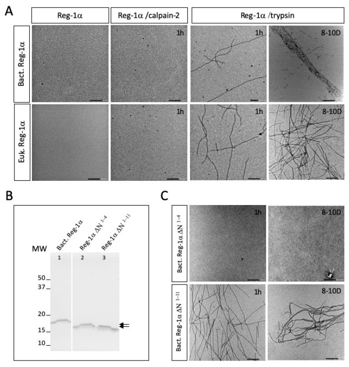
Reg-1α/lithostathine, a protein mainly associated with the digestive system, was previously shown to be overexpressed in the pre-clinical stages of Alzheimer's disease. In vitro, the glycosylated protein was reported to form fibrils at physiological pH following the proteolytic action of trypsin. However, the nature of the protease able to act in the central nervous system is unknown. In the present study, we showed that Reg-1α can be cleaved in vitro by calpain-2, the calcium activated neutral protease, overexpressed in neurodegenerative diseases. Using chemical crosslinking experiments, we found that the two proteins can interact with each other. Identification of the cleavage site using mass spectrometry, between Gln4 and Thr5, was found in agreement with the in silico prediction of the calpain cleavage site, in a position different from the one reported for trypsin, i.e., Arg11-Ile12 peptide bond. We showed that the cleavage was impeded by the presence of the neighboring glycosylation of Thr5. Moreover, in vitro studies using electron microscopy showed that calpain-cleaved protein does not form fibrils as observed after trypsin cleavage. Collectively, our results show that calpain-2 cleaves Reg-1α in vitro, and that this action is not associated with fibril formation.

Keywords: Reg-1α; calpain; cleavage; fibril; trypsin.


Conflict of interest statement

The authors declare no conflict of interest.

Figures

Figure 1
Figure 1
Reg-1α is a new calpain-2 substrate in vitro. (A) NH2 terminal sequence of human Reg-1α, including the signal peptide (blue box) compared to the isoform expressed in bacteria including the His-tag (purple box). The glycosylated Thr is noted in bold, and the position of the trypsin cleavage is indicated by a green arrow. (B) Intact and cleaved molecules with samples corresponding to the test of various ratio (w/w) of enzyme/substrate (1/10, 1/20 and 1/50) during different periods of time (0, 15, 30 and 45 min) were analyzed on 13.5% acrylamide gel and visualized using the Stain-Free Imaging Technology (see Section 4). (C) The uncleaved/cleaved (enzyme/substrate ratio 1/10) products were examined by western blotting using anti-Histidine and anti-Reg-1α antibodies. Note the presence of dimers (*) and cleaved dimers (arrowhead) which resist complete reduction.
Figure 2
Figure 2
Interaction of Reg-1α with calpain-2. The cross-linking reactions (after 0, 15, and 30 min), namely Reg-1α + calpain-2 in its inactive form (Reg-1α /C2I) (red frame) and Reg-1α alone (Reg-1α /Reg-1α) were carried out as described in Section 4. SDS-PAGE was performed on a 4-15% acrylamide gel, and the presence of cross-linked products was evidenced after transfer of the proteins on a PVDF membrane by double immunodetection with anti-Reg-1α (A) and anti-calpain-2 (B) antibodies. The covalent products are indicated by arrowheads. Note the presence of Reg1α monomers (*), dimers (**), trimers (***) initially present and tetramers (****) visible only after the crosslinking reaction of Reg-1α alone.
Figure 3
Figure 3
Prediction of the calpain-2 cleavage site of Reg-1α using GPS-CCD (calpain cleavage detector). (A,B) Result obtained using the GPS-CCD software [32] with the sequence of the recombinant human Reg-1α without (A) or with (B) the NH2-terminal His-tag and initial Met (natural protein/recombinant protein expressed in bacteria) and (C) with the human Reg-1α sequence, including the signal peptide (before processing). The position of the calpain-2 cleavage site predicted with the higher score is highlighted in yellow and the exact position noted in red. Note that the results of the score were obtained with the higher threshold.
Figure 4
Figure 4
Identification of the calpain-2 cleavage site using MALDI-TOF mass spectrometry. (A) MALDI-TOF spectra showing Reg-1α treated with calpain-2 in the linear mode (m/z values from 7000 to 25,000). Inset, profile of the uncleaved protein and (B) the cleaved fragment in the reflectron mode (m/z 600–3000). (C) Enlargement of the spectrum of (B) obtained in the region of m/z 1960–2030. Note the presence of both sodium [M + Na]+ and potassium [M + K]+ adducts. The molecular weights, in Da, are shown above the peaks of each major entity.
Figure 5
Figure 5
O-glycosylation of Reg-1α protects it from the calpain-2-mediated cleavage in vitro. (A) MALDI spectra of the eukaryotic Reg-1α (Euk. Reg-1α) obtained in the linear mode (m/z 16,000–19,000); (B) Both recombinant isoforms obtained from eukaryotic (Euk. Reg-1α) and bacterial (Bact. Reg-1α) sources were tested for calpain-2 (lanes 1–4) or trypsin cleavage (lanes 5–8) (ratio 1:10 and 1:100), respectively; samples were analyzed on 13.5% acrylamide gel and visualized using the Stain-Free Imaging Technology (see Section 4). Note that the protein from eukaryotic source displays a slightly higher molecular weight (see arrows) compared to the protein from bacteria due to the presence of glycosylation; arrowhead indicates the presence of calpain-2 (lanes 2,4). (C,D), MALDI reflectron mode spectra (m/z 300–650) of the glycosylated form of Reg-1α (Euk. Reg-1α) before (C) and after calpain-2 cleavage (Euk. Reg-1α /C2) (D). The position of the calpain inhibitor (E-64) used to stop the reaction is indicated.
Figure 6
Figure 6
Reg-1α fibrils are only formed post cleavage of the undecapeptide. (A) The two recombinant forms (Bact. or Euk. Reg-1α) of the protein were analyzed after calpain-2 or trypsin cleavage at 1h and 8–10 days (8–10D) using electron microscopy. (B) Gel electrophoresis of the COOH-terminal domains of Reg-1α (Reg-1α∆N molecules) after purification using Ni-NTA-agarose beads (see Section 4). Lane 1, intact Reg-1α from bacteria source; lanes 2–3, supernatant obtained post Ni-NTA resin incubation after calpain-2 (Reg-1α∆N1-4) or trypsin (Reg-1αΔN1-11) cleavage. Note the difference in the molecular weight of the two purified fragments (double arrow). (C) Electron micrographs of the purified Reg-1α∆N molecules, post calpain-2 (top panel) and trypsin (bottom panel) cleavage after 1 h and 8–10D. Scale bars: 200 nm.

Similar articles

Cited by

References

    1. Iovanna J.L., Dagorn J.-C. The multifunctional family of secreted proteins containing a C-type lectin-like domain linked to a short N-terminal peptide. Biochim. Biophys. Acta. 2005;1723:8–18. doi: 10.1016/j.bbagen.2005.01.002. - DOI - PubMed
    1. Chen Z., Downing S., Tzanakakis E.S. Four Decades After the Discovery of Regenerating Islet-Derived (Reg) Proteins: Current Understanding and Challenges. Front. Cell Dev. Biol. 2019;7:235. doi: 10.3389/fcell.2019.00235. - DOI - PMC - PubMed
    1. Multigner L., De Caro A., Lombardo D., Campese D., Sarles H. Pancreatic stone protein, a phosphoprotein which inhibits calcium carbonate precipitation from human pancreatic juice. Biochem. Biophys. Res. Commun. 1983;110:69–74. doi: 10.1016/0006-291X(83)91261-5. - DOI - PubMed
    1. Geider S., Baronnet A., Cerini C., Nitsche S., Astier J.P., Michel R., Boistelle R., Berland Y., Dagorn J.C., Verdier J.M. Pancreatic lithostathine as a calcite habit modifier. J. Biol. Chem. 1996;271:26302–26306. doi: 10.1074/jbc.271.42.26302. - DOI - PubMed
    1. Bernard J.P., Adrich Z., Montalto G., De Caro A., De Reggi M., Sarles H., Dagorn J.C. Inhibition of nucleation and crystal growth of calcium carbonate by human lithostathine. Gastroenterology. 1992;103:1277–1284. doi: 10.1016/0016-5085(92)91516-7. - DOI - PubMed

LinkOut - more resources