

Vascularization in Bioartificial Parenchymal Tissue: Bioink and Bioprinting Strategies
- PMID: 35955720
- PMCID: PMC9369172
- DOI: 10.3390/ijms23158589
Vascularization in Bioartificial Parenchymal Tissue: Bioink and Bioprinting Strategies
Abstract
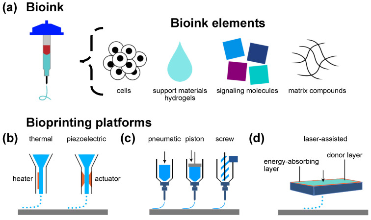
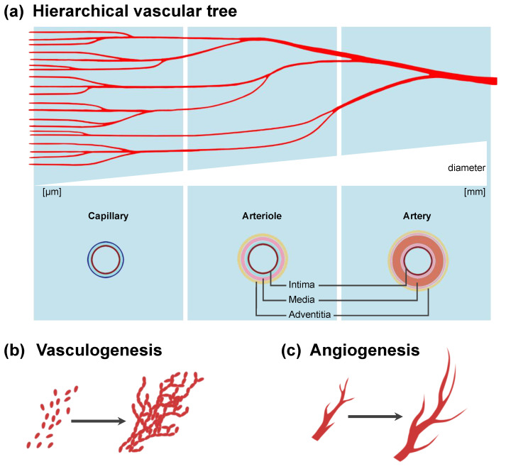
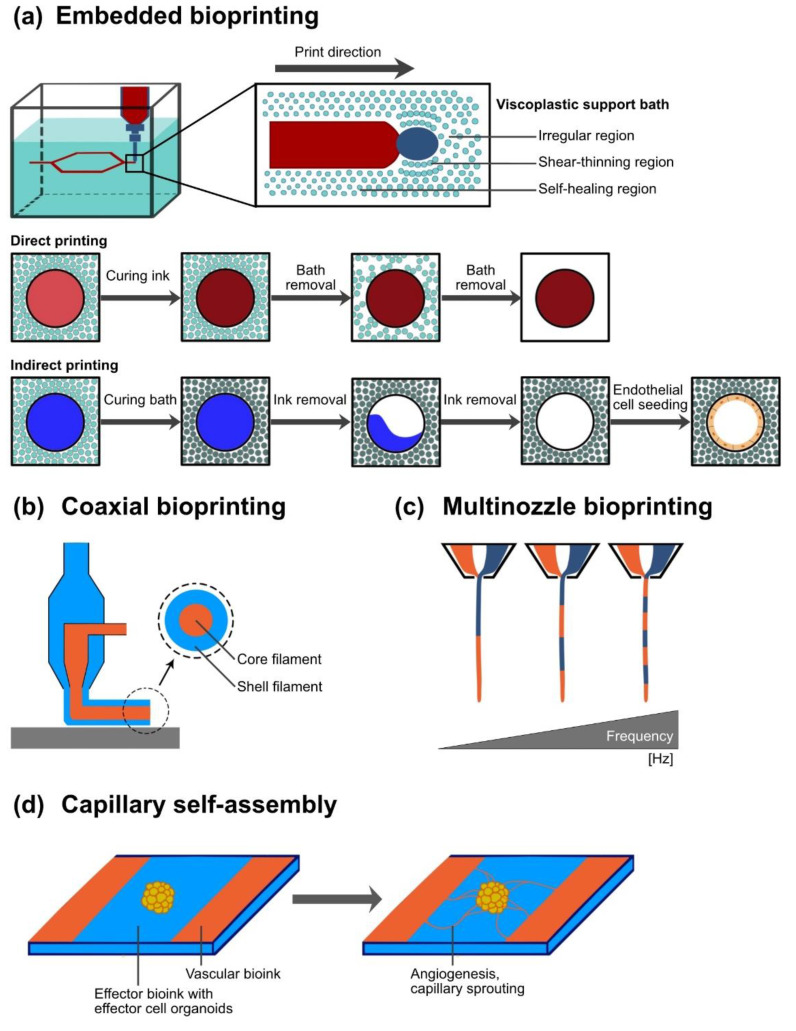
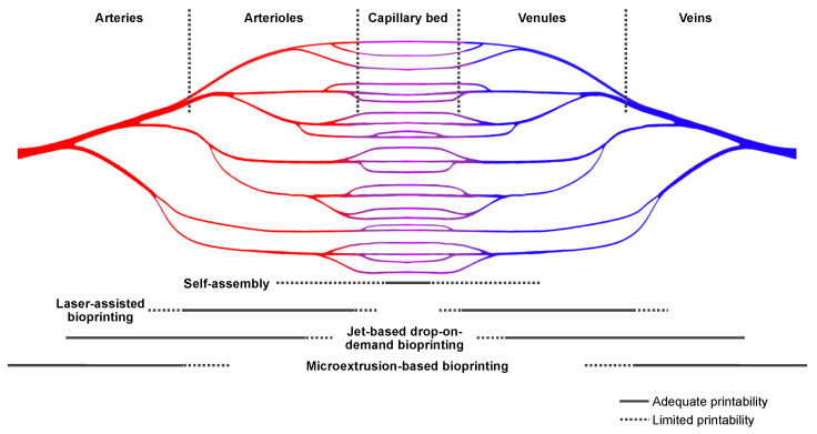
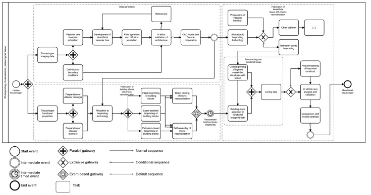
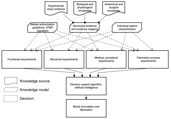
Among advanced therapy medicinal products, tissue-engineered products have the potential to address the current critical shortage of donor organs and provide future alternative options in organ replacement therapy. The clinically available tissue-engineered products comprise bradytrophic tissue such as skin, cornea, and cartilage. A sufficient macro- and microvascular network to support the viability and function of effector cells has been identified as one of the main challenges in developing bioartificial parenchymal tissue. Three-dimensional bioprinting is an emerging technology that might overcome this challenge by precise spatial bioink deposition for the generation of a predefined architecture. Bioinks are printing substrates that may contain cells, matrix compounds, and signaling molecules within support materials such as hydrogels. Bioinks can provide cues to promote vascularization, including proangiogenic signaling molecules and cocultured cells. Both of these strategies are reported to enhance vascularization. We review pre-, intra-, and postprinting strategies such as bioink composition, bioprinting platforms, and material deposition strategies for building vascularized tissue. In addition, bioconvergence approaches such as computer simulation and artificial intelligence can support current experimental designs. Imaging-derived vascular trees can serve as blueprints. While acknowledging that a lack of structured evidence inhibits further meta-analysis, this review discusses an end-to-end process for the fabrication of vascularized, parenchymal tissue.
Keywords: additive manufacturing; bioartificial organs; bioink; biomaterial; bioprinting; regenerative medicine; tissue engineering; vascularization.
Conflict of interest statement
The authors declare no conflict of interest.
Figures
References
-
- Salg G.A., Giese N.A., Schenk M., Huttner F.J., Felix K., Probst P., Diener M.K., Hackert T., Kenngott H.G. The emerging field of pancreatic tissue engineering: A systematic review and evidence map of scaffold materials and scaffolding techniques for insulin-secreting cells. J. Tissue Eng. 2019;10:2041731419884708. doi: 10.1177/2041731419884708. - DOI - PMC - PubMed
-
- Ramezankhani R., Torabi S., Minaei N., Madani H., Rezaeiani S., Hassani S.N., Gee A.P., Dominici M., Silva D.N., Baharvand H., et al. Two Decades of Global Progress in Authorized Advanced Therapy Medicinal Products: An Emerging Revolution in Therapeutic Strategies. Front. Cell Dev. Biol. 2020;8:547653. doi: 10.3389/fcell.2020.547653. - DOI - PMC - PubMed
Publication types
MeSH terms
LinkOut - more resources
Full Text Sources
