. 2022 Aug 2;23(15):8587.
doi: 10.3390/ijms23158587.

Cellular Redox Metabolism Is Modulated by the Distinct Localization of Cyclic Nucleotide Phosphodiesterase 5A Isoforms

Affiliations

Cellular Redox Metabolism Is Modulated by the Distinct Localization of Cyclic Nucleotide Phosphodiesterase 5A Isoforms

Silvia Cardarelli et al. Int J Mol Sci. .

Abstract

3'-5' cyclic nucleotide phosphodiesterases (PDEs) are a family of evolutionarily conserved cAMP and/or cGMP hydrolyzing enzymes, components of transduction pathways regulating crucial aspects of cell life. Among them, cGMP-specific PDE5-being a regulator of vascular smooth muscle contraction-is the molecular target of several drugs used to treat erectile dysfunction and pulmonary hypertension. Production of full-length murine PDE5A isoforms in the milk-yeast Kluyveromyces lactis showed that the quaternary assembly of MmPDE5A1 is a mixture of dimers and tetramers, while MmPDE5A2 and MmPDE5A3 only assembled as dimers. We showed that the N-terminal peptide is responsible for the tetramer assembly of MmPDE5A1, while that of the MmPDE5A2 is responsible for its mitochondrial localization. Overexpression of the three isoforms alters at different levels the cAMP/cGMP equilibrium as well as the NAD(P)+/NAD(P)H balance and induces a metabolic switch from oxidative to fermentative. In particular, the mitochondrial localization of MmPDE5A2 unveiled the existence of a cAMP-cGMP signaling cascade in this organelle, for which we propose a metabolic model that could explain the role of PDE5 in some cardiomyopathies and some of the side effects of its inhibitors.

Keywords: Kluyveromyces lactis; cGMP-specific phosphodiesterase; glycolytic/respiratory flux; rag phenotype; redox balance.


Conflict of interest statement

The authors declare no conflict of interest.

Figures
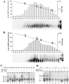
Figure 1
Figure 1
cGMP activity and quaternary assembly of purified recombinant MmPDE5 isoforms. A calibration curve with known MW markers has been superimposed on panels (A,B): apoferritin (Apo, 443 kDa), β-amylase (βAm, 200 kDa), alcohol dehydrogenase (Adh, 150 kDa), bovine serum albumin (BSA, 66 kDa). (A) cGMP-hydrolysing activity and native WB from size exclusion chromatography (SEC) eluted fractions of purified MmPDE5A2. (B) cGMP-hydrolyzing activity and native WB from SEC-eluted fractions of purified MmPDE5A3. Fractions were obtained from 300 μg of affinity purified MmPDE5A2 and MmPDE5A3 applied on a Superose 12 HR 10/30 column and eluted at a flow rate of 0.20 mL/min. This SEC fractionation has been repeated at each purification and one representative example is shown in these figures. (C,D) Native PAGE pattern of purified MmPDE5A1, A2 and A3 stained with Coomassie (~1.0 μg). The proteins were pre-incubated for up to 3 h at 30 °C with dithiotreitol (DTT), cGMP, Sildenafil (Sild) (C), and diamide (D) at the concentration specified in the figures. D1, D2, and D3 indicate the three conformational forms of the dimer and T1, T2, and T3 those of the tetramer. Native page analyses of MmPDE5 isoforms under different conditions/incubations with different reagents have been performed and repeated at least five times. One representative example is shown for both figures (C,D).
Figure 2
Figure 2
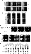
Growth tests of K. lactis wild type strain (CBS2359) and its transformants with MmPDE5A1, MmPDE5A2, MmPDE5A3, and in-gel native staining assays of their protein extracts. (A) Cells were incubated in the presence of oxygen (+O2) or under anaerobic conditions (−O2). (B) Growth test of the above strains plus selected colonies of the PDE5A2-strain in which the p3xFlagPde5A2 has been lost (revertant rev.1, rev.2, rev.3). A2.2 and A2.3 are two other re-transformants of the WT (CBS2359) strain with p3xFlagPde5A2. (C) Growth test of WT, WT + empty vector (WTp), pPde5A2gfp, and p3xFlagPde5A2 transformed strains. All strains were grown in YP + 2% glucose (Glc), plus 5 mM H2O2, (Glc + H2O2) plus 100 μM G418 (Glc + G418), or 5% glucose plus 5 μM antimycin A (Glc + AA), 2% glycerol (Gly). (D) Growth test of ragx1, WT and transformed strains with MmPDE5A1 (A1), MmPDE5A2 (A2), MmPDE5A3 (A3). Strains were grown in YP medium + 4 mM Acetate (Acetate), glucose 2% (Glc) containing 1.2 M NaCl (NaCl) or 30 μM methylene blue (Glc + MB) or 5% glucose plus 5 μM antimycin A (Glc + AA). Cells, grown to early stationary phase, were adjusted to 108 cells mL−1 and 5 μL of serial 10-fold dilutions were spotted onto the indicated medium. Growth was followed for 3–5 days. The initial concentration was 107 cells mL−1. (E) K. lactis in-gel native staining for alcohol dehydrogenase (ADH) and Glc-6-phosphate dehydrogenase (G6PDH). Cells were grown to early stationary phase in YP containing 2% glucose + MB + G418 (Glc + MB + G418), 2%glucose + G418 (Glc + G418) or 2% glucose (Glc). Extracts from these cultures, fractioned in native PAGE, were stained for ADH and G6PDH. Black arrows indicate the migrating positions of KlAdh3 and KlAdh4, white arrow indicates the faster migrating band of G6PDH that corresponds to the less active dimeric form. Native PAGE analyses of ADH and G6PDH activities from K. lactis cells harboring PDE5 isoforms have been repeated at least three times. S.D. among the same bands of each lane was comprised between 4% and 13%. One representative native gel is shown in the figure.
Figure 3
Figure 3
Role of the coenzymes NAD(P)+/NAD(P)H in the onset of fermentation in the K. lactis WT strain, quantification of cyclic nucleotide PDE activity, cAMP content and effects of the ragx1 mutation on the quaternary structure of MmPDE5 isoforms. (A) Schematic summary of the role of NAD+ vs. NADP+ during the glycolytic–fermentative metabolism, their implication in the pentose phosphate pathway (PPP), their oxidation by NDE1 (which has higher affinity for NADH than for NADPH). (B) A model showing the opposite role of the coenzymes on the assembly and activity of G6PDH (C) Effect of cytosolic NAD(P)H on G6DPH isoforms during the respiratory to fermentative transition (from Glc 0.1% to 4%; E stands for Ethanol). Effect of NADPH oxidation by MB during fermentation (YP+ 2% Glc) on G6PDH isoforms. (D) Absorbance spectra and G6PDH content of WT cell extracts supplemented with increasing concentrations of NAD+ (up to 10 mM). (E) Absorbance spectra and G6PDH content of WT cell extracts supplemented with increasing concentrations of NADP+ (up to 10 mM). (F) Extracts from 10 mL cultures of ragx1, WT, MmPDE5A1 (A1), MmPDE5A2 (A2), and MmPDE5A3 (A3) transformed strains grown to early stationary phase were used to determine the cAMP hydrolyzing activity (left panel), cGMP hydrolyzing activity (center panel) and cAMP content (right panel). Reported values are the means from three independent determinations (S.D. was always between 2% and 12%). Student’s t-test was used to determined statistical significance with respect to WT: ** p < 0.01; *** p < 0.001 (G) Quaternary assembly of recombinant MmPDE5A isomers produced in the ragx1 mutant. Native PAGE pattern of purified MmPDE5A1, MmPDE5A2, and MmPDE5A3 stained with Coomassie (~1.0 μg). Legend as in Figure 1C,D. MmPDE5A1 protein purified from the WT strain has been loaded as a control. Native page analyses of MmPDE5 isoforms under different conditions/incubations are the means of at least three independent experiments. One representative gel is shown in the figure.
Figure 4
Figure 4
Fluorescence microscopy images of K. lactis cells overexpressing MmPDE5AGFP isoforms. (A) Intracellular localization of MmPDE5A1, A2, and A3 isoforms revealed in cell transformed with GFP-tagged isoform constructs (green). Cultures were grown to early stationary phase in YP containing 2%Glc + G418; cells stained with DAPI (blue) and MitoTracker (red) were visualized under the confocal microscope. Scale bar: 10 µm. (B) Colocalization analysis on K. lactis cells overexpressing MmPDE5A-GFP isoforms. Cells overexpressing GFP-tagged MmPDE5A1, A2, and A3 isoforms (green) were grown to early stationary phase in 2%Glc + G418 and stained with MitoTracker (red). Images showing a representative single cell, were acquired using a Zeiss LSM 800 System. Colocalization signal (yellow) was quantified using the Pearson correlation coefficient (p). Original magnification 20×; Inset 10×; Scale bar: 5 μm.
Figure 5
Figure 5
Schematic model of the metabolic changes induced in K. lactis by MmPDE5A2. The mitochondrial localization of MmPDE5A2 highlighted the existence of a Cyr1-cAMP/cGMP signaling system in mitochondria dedicated to balancing O2 availability and oxidative phosphorylation with Krebs cycle turnover. cGMP hydrolysis by MmPDE5A2 activates an O2-dependent mechanism, increasing the production of ATP by the respiratory chain while at the same time blocking the fermentation, either directly or through the cAMP-specific endogenous KlPde2. Under physiological conditions, the mitochondrial role of PDE5A2 may be fulfilled by the endogenous KlPde1. The adenylate cyclase Cyr1 is controlled by Ras/Gpa2-Gpr1 and NAD(P)H is re-oxidized by the trans-dehydrogenase Nde1. The carbonic anhydrase Nce103 is located also in the inner mitochondrial space (IMS). G6P, glucose-6 phosphate; F6P, fructose-6 phosphate, F1-6P, fructose1-6 diphosphate; G-3P, glyceraldehyde-3 phosphate; 1-3dPG, 1-3diphosphoglycerate; DHAP, dihydroxyacetone phosphate.

Similar articles

Cited by

References

    1. Beavo J.A., Conti M., Heaslip R.J. Multiple cyclic nucleotide phosphodiesterases. Mol. Pharmacol. 1994;46:399–405. - PubMed
    1. Bender A.T., Beavo J.A. Cyclic nucleotide phosphodiesterases: Molecular regulation to clinical use. Pharmacol. Rev. 2006;3:488–520. doi: 10.1124/pr.58.3.5. - DOI - PubMed
    1. Keravis T., Lugnier C. Cyclic nucleotide phosphodiesterase (PDE) isozymes as targets of the intracellular signalling network: Benefits of PDE inhibitors in various diseases and perspectives for future therapeutic development. Br. J. Pharmacol. 2012;165:1288–1305. doi: 10.1111/j.1476-5381.2011.01729.x. - DOI - PMC - PubMed
    1. Maurice D.H., Ke H., Ahmad F., Wang Y., Chung J., Manganiello V.C. Advances in targeting cyclic nucleotide phosphodiesterases. Nat. Rev. Drug Discov. 2014;13:290–314. doi: 10.1038/nrd4228. - DOI - PMC - PubMed
    1. Omori K., Kotera J. Overview of PDEs and their regulation. Circ. Res. 2007;100:309–327. doi: 10.1161/01.RES.0000256354.95791.f1. - DOI - PubMed

LinkOut - more resources