. 2022 Aug 2;23(15):8578.
doi: 10.3390/ijms23158578.

VIP/VPAC Axis Expression in Immune-Mediated Inflammatory Disorders: Associated miRNA Signatures

Affiliations

VIP/VPAC Axis Expression in Immune-Mediated Inflammatory Disorders: Associated miRNA Signatures

Amalia Lamana et al. Int J Mol Sci. .

Abstract

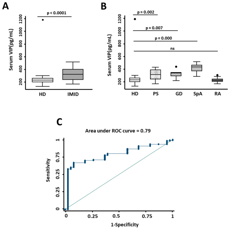
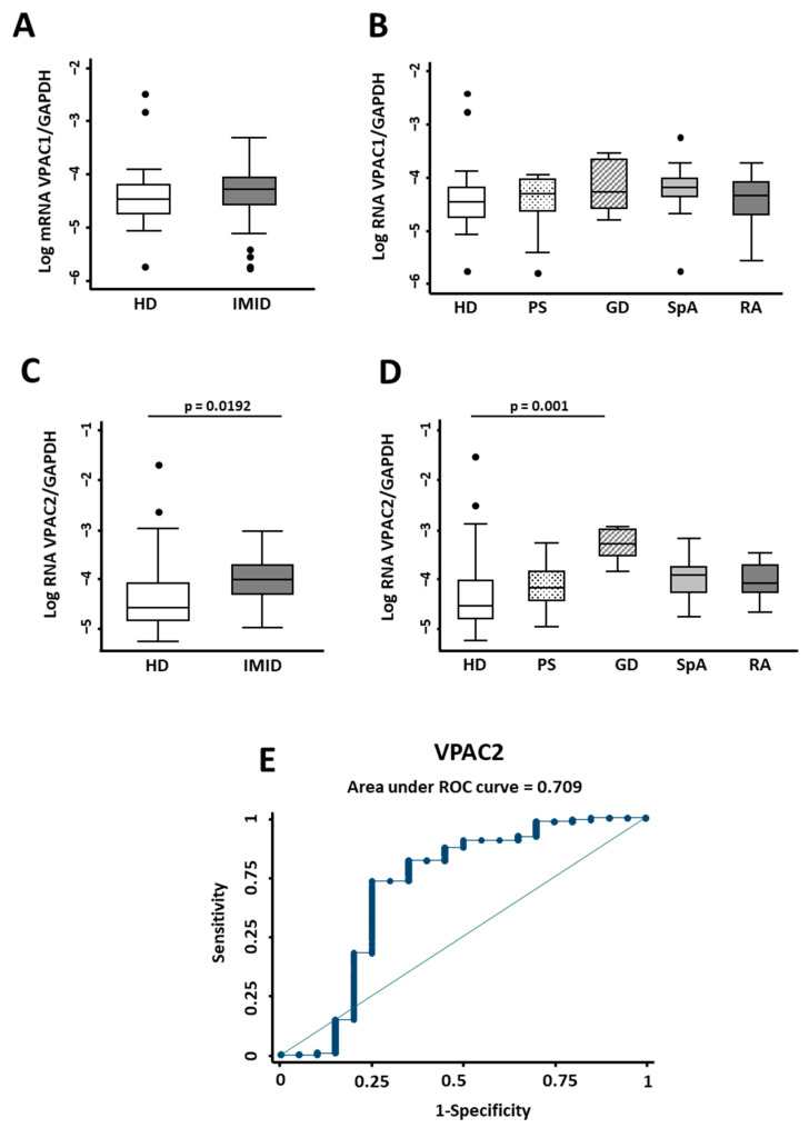
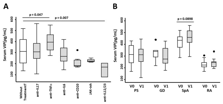
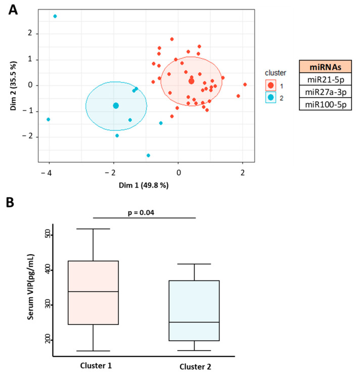
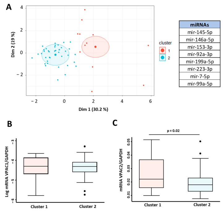
Few studies have considered immune-mediated inflammatory disorders (IMID) together, which is necessary to adequately understand them given they share common mechanisms. Our goal was to investigate the expression of vasoactive intestinal peptide (VIP) and its receptors VPAC1 and VPAC2 in selected IMID, analyze the effect of biological therapies on them, and identify miRNA signatures associated with their expression. Serum VIP levels and mRNA of VPAC and miRNA expression in peripheral blood mononuclear cells were analyzed from 52 patients with psoriasis, rheumatoid arthritis, Graves’ disease, or spondyloarthritis and from 38 healthy subjects. IMID patients showed higher levels of VIP and increased expression of VPAC2 compared to controls (p < 0.0001 and p < 0.0192, respectively). Receiver operating characteristic curve analysis showed that the levels of VIP or VPAC2 expression were adequate discriminators capable of identifying IMID. Treatment of IMID patients with anti-TNFα and anti-IL12/23 significantly affected serum VIP levels. We identified miRNA signatures associated with levels of serum VIP and VPAC2 expression, which correlated with IMID diagnosis of the patients. The results indicate that the expression of VIP/VPAC2 is able of identify IMIDs and open up a line of research based on the association between the VIP/VPAC axis and miRNA signatures in immune-mediated diseases.

Keywords: Graves’ disease; VIP; VPAC receptors; immune-mediated inflammatory disorders; microRNAs; psoriasis; rheumatoid arthritis; spondyloarthritis.


Conflict of interest statement

The authors declare no conflict of interest.

Figures

Figure 1
Figure 1
Expression of serum VIP levels in inflammatory/autoimmune pathologies. (A) Determination of serum VIP levels (pg/mL) by ELISA of 38 healthy donors (HD) and 52 patients with immune-mediated inflammatory diseases (IMID). Statistical significance was determined using the variable of serum VIP levels normalized by inverse square root and Student’s t-test to obtain p-value < 0.0001. (B) Serum VIP levels (pg/mL) of 38 healthy donors (HD) and 15 patients with psoriasis (PS), 8 with Graves’ disease (GD), 15 with spondyloarthritis (SpA), and 14 with rheumatoid arthritis (RA) are shown. Statistical significance was calculated using the variable of serum VIP levels normalized by inverse square root and applying ANOVA and Bonferroni correction for multiple comparisons to obtain p-values as indicated. In all panels, data are presented as the interquartile range (p75: upper edge of the box, p25: bottom edge of the box, and p50: midline) and p90 and p10 (lines below and above the box) of the serum VIP levels. Dots represent outliers. Significance threshold was set at p < 0.05. (C) The graph shows the receiver operating characteristic (ROC) curve analysis to assess the ability of serum VIP levels to discriminate between patients with IMID and healthy donors. Area under ROC curve is indicated in the graph.
Figure 2
Figure 2
Expression of the VIP/receptor axis in inflammatory/autoimmune pathologies. (A) The relative expression of VPAC1/GAPDH mRNA normalized by log transformation of 38 healthy donors (HD) and 52 patients with immune-mediated inflammatory diseases (IMID) is shown. Statistical significance was determined with Student’s t-test. No significant differences were observed. (B) Log of relative expression of VPAC1/GAPDH mRNA of 38 healthy donors (HD) and 15 patients with psoriasis (PS), 8 with Graves’ disease (GD), 15 with spondyloarthritis (SpA), and 14 with rheumatoid arthritis (RA) is shown. (C) Log of relative expression of VPAC2/GAPDH mRNA of 38 healthy donors (HD) and 52 patients with immune-mediated inflammatory diseases (IMID) is shown. Statistical significance was calculated using relative expression of VPAC2/GAPDH mRNA normalized by inverse square root using Student’s t-test. The p-value is shown in the figure. (D) Log of relative expression of VPAC2/GAPDH mRNA of 38 healthy donors (HD) and 15 patients with psoriasis (PS), 8 with Graves’ disease (GD), 15 with spondyloarthritis (SPA), and 14 with rheumatoid arthritis (RA) is shown. Statistical significance was calculated using inverse square root of relative expression of VPAC2/GAPDH mRNA by applying the ANOVA test and Bonferroni correction for multiple comparisons. Significant differences between groups are shown in the figure. In all panels, data are presented as the interquartile range (p75: upper edge of the box, p25: bottom edge of the box, and p50: midline) and p90 and p10 (lines below and above the box) of the serum VIP levels. Dots represent outliers. Significance threshold was set at p < 0.05. (E) The graph shows the ROC curve analysis to assess the ability of relative expression of VPAC2/GAPDH to discriminate between patients with IMID and healthy donors. Area under ROC curve is indicated in the graph.
Figure 3
Figure 3
Effect of biological therapies on the expression of serum VIP levels in patients with immune-mediated inflammatory diseases (IMIDs). (A) Serum VIP levels (pg/mL) of 38 healthy donors without treatment and 9 IMID patients with anti-IL17 treatment, 15 with anti-TNFα treatment, 10 with anti-IL6 treatment, 8 with anti-CD20 treatment, 4 with JAK inhibitors, and 2 with anti-IL12/23 treatment are shown. Statistical significance was calculated using the variable of serum VIP levels normalized by inverse square root and applying ANOVA and Bonferroni correction for multiple comparisons to obtain the p-values as indicated. In all panels, data are presented as the interquartile range (p75: upper edge of the box, p25: bottom edge of the box, and p50: midline) and p90 and p10 (lines below and above the box) of the serum VIP levels. Dots represent outliers. Significance threshold was set at p < 0.05. (B) Serum VIP levels (pg/mL) by ELISA of 15 patients with psoriasis (PS), 8 with Graves’ disease (GD), 15 with spondyloarthritis (SpA), and 14 with rheumatoid arthritis (RA) before (V0) and after 2–4 months (V1) of treatment with biological therapies are shown. To analyze the differences in serum VIP levels before and after treatment, paired sign test was used. Significant differences between groups are shown in the figure.
Figure 4
Figure 4
Association of miRNA profile in patients with immune-mediated inflammatory diseases (IMID) with serum levels of VIP. (A) Graphical representation of cluster analysis based on miR215p, miR27a-5p, and miR100-5p expression in 45 IMID patients. K-means clustering based on Euclidean distance and principal component analysis (PCA) was applied. (B) Serum VIP levels (pg/mL) of IMID patients according to their association with miRNA expression cluster. Statistical significance was calculated using the variable of serum VIP levels normalized by inverse square root and applying Student’s t-test. Data are presented as the interquartile range (p75: upper edge of the box, p25: bottom edge of the box, and p50: midline) and p90 and p10 (lines below and above the box) of the serum VIP levels. Significance threshold was set at p < 0.05.
Figure 5
Figure 5
miRNA signature associated with VPAC receptors gene expression. (A) Cluster analysis in 52 IMID patients based on the expression of miRNAs miR145-5p, miR146A-5p, miR153-3p, miRNA92a-3p, miR199a-5p, miR223-3p, miR7-5p, and miR99a-5p. K-means clustering based on Euclidean distance and PCA was used for graphical representation. Log of relative expression (2−ΔCt) of VPAC1/GAPDH (B) and VPAC2/GAPDH mRNA (C) according to their association with miRNA expression cluster. Statistical significance was calculated using Student’s t-test. For both datasets, data are presented as the interquartile range (p75: upper edge of the box, p25: bottom edge of the box, and p50: midline) and p90 and p10 (lines below and above the box) of the relative gene expression. Dots represent outliers. Significance threshold was set at p < 0.05.

Similar articles

Cited by

  • Circulating miRNAs in Graves' ophthalmopathy: a systematic review.
    Syrakouli E, Geropoulos G, Laskou S, Anestiadou E, Syrakouli A, Koimtzis G, Geropoulos V, Giannis D, Sapalidis K, Psarras K. Syrakouli E, et al. Endocrine. 2025 Jun 28. doi: 10.1007/s12020-025-04286-9. Online ahead of print. Endocrine. 2025. PMID: 40580399
  • Effects of Neuropeptides on Dendritic Cells in the Pathogenesis of Psoriasis.
    Zhang J, Zhao S, Xing X, Shang L, Cao J, He Y. Zhang J, et al. J Inflamm Res. 2023 Jan 6;16:35-43. doi: 10.2147/JIR.S397079. eCollection 2023. J Inflamm Res. 2023. PMID: 36636251 Free PMC article. Review.
  • Genetic variants regulating the immune response improve the prediction of COVID-19 severity provided by clinical variables.
    Delgado-Wicke P, Fernández de Córdoba-Oñate S, Roy-Vallejo E, Alegría-Carrasco E, Rodríguez-Serrano DA, Lamana A, Montes N, Nicolao-Gómez A, Carracedo-Rodríguez R, Marcos-Jiménez A, Díaz-Fernández P, Galván-Román JM, Rabes-Rodríguez L, Sanz-Alba M, Álvarez-Rodríguez J, Villa-Martí A, Rodríguez-Franco C, Villapalos-García G, Zubiaur P, Abad-Santos F, de Los Santos I, Gomariz RP, García-Vicuña R, Muñoz-Calleja C, González-Álvaro I, Fernández-Ruiz E; PREDINMUN-COVID Group. Delgado-Wicke P, et al. Sci Rep. 2024 Sep 5;14(1):20728. doi: 10.1038/s41598-024-71476-2. Sci Rep. 2024. PMID: 39237611 Free PMC article.

References

    1. El-Gabalawy H., Guenther L.C., Bernstein C.N. Epidemiology of immune-mediated inflammatory diseases: Incidence, prevalence, natural history, and comorbidities. J. Rheumatol. Suppl. 2010;85:2–10. doi: 10.3899/jrheum.091461. - DOI - PubMed
    1. Russell A.S., Gulliver W.P., Irvine E.J., Albani S., Dutz J.P. Quality of life in patients with immune-mediated inflammatory diseases. J. Rheumatol. Suppl. 2011;88:7–19. doi: 10.3899/jrheum.110899. - DOI - PubMed
    1. McInnes I.B., Gravallese E.M. Immune-mediated inflammatory disease therapeutics: Past, present and future. Nat. Rev. Immunol. 2021;21:680–686. doi: 10.1038/s41577-021-00603-1. - DOI - PMC - PubMed
    1. Theofilopoulos A.N., Kono D.H., Baccala R. The multiple pathways to autoimmunity. Nat. Immunol. 2017;18:716–724. doi: 10.1038/ni.3731. - DOI - PMC - PubMed
    1. David T., Ling S.F., Barton A. Genetics of immune-mediated inflammatory diseases. Clin. Exp. Immunol. 2018;193:3–12. doi: 10.1111/cei.13101. - DOI - PMC - PubMed

MeSH terms

Substances