

Fibroblast Growth Factors and Cellular Communication Network Factors: Intimate Interplay by the Founding Members in Cartilage
- PMID: 35955724
- PMCID: PMC9369280
- DOI: 10.3390/ijms23158592
Fibroblast Growth Factors and Cellular Communication Network Factors: Intimate Interplay by the Founding Members in Cartilage
Abstract
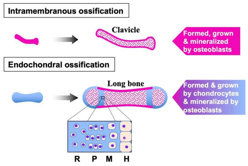
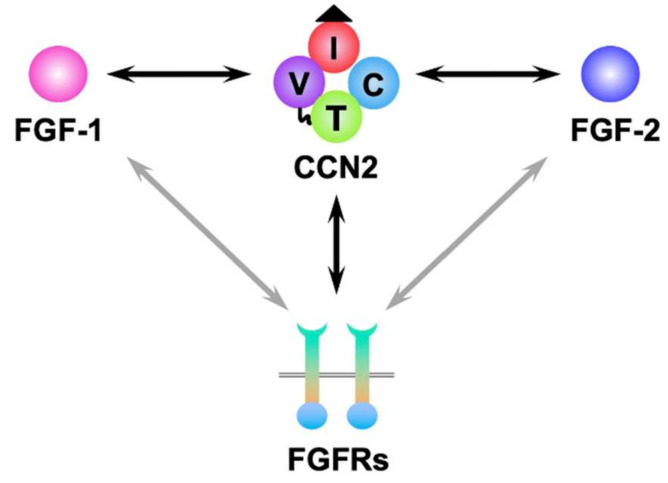
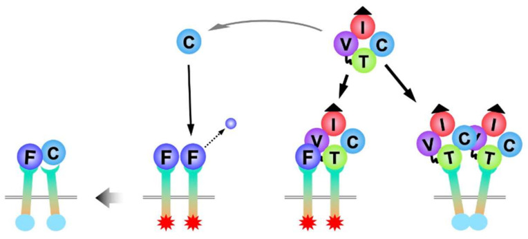
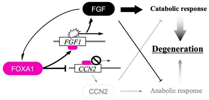
Fibroblast growth factors (FGFs) constitute a large family of signaling molecules that act in an autocrine/paracrine, endocrine, or intracrine manner, whereas the cellular communication network factors (CCN) family is composed of six members that manipulate extracellular signaling networks. FGFs and CCNs are structurally and functionally distinct, except for the common characteristics as matricellular proteins. Both play significant roles in the development of a variety of tissues and organs, including the skeletal system. In vertebrates, most of the skeletal parts are formed and grow through a process designated endochondral ossification, in which chondrocytes play the central role. The growth plate cartilage is the place where endochondral ossification occurs, and articular cartilage is left to support the locomotive function of joints. Several FGFs, including FGF-2, one of the founding members of this family, and all of the CCNs represented by CCN2, which is required for proper skeletal development, can be found therein. Research over a decade has revealed direct binding of CCN2 to FGFs and FGF receptors (FGFRs), which occasionally affect the biological outcome via FGF signaling. Moreover, a recent study uncovered an integrated regulation of FGF and CCN genes by FGF signaling. In this review, after a brief introduction of these two families, molecular and genetic interactions between CCN and FGF family members in cartilage, and their biological effects, are summarized. The molecular interplay represents the mutual involvement of the other in their molecular functions, leading to collaboration between CCN2 and FGFs during skeletal development.
Keywords: CCN2; cartilage; cellular communication network factor; fibroblast growth factor; skeletal development.
Conflict of interest statement
The authors declare no conflict of interest.
Figures
Similar articles
-
Recent research on the growth plate: Advances in fibroblast growth factor signaling in growth plate development and disorders.J Mol Endocrinol. 2014 Aug;53(1):T11-34. doi: 10.1530/JME-14-0012. J Mol Endocrinol. 2014. PMID: 25114206 Review.
-
Fibroblast growth factors: from molecular evolution to roles in development, metabolism and disease.J Biochem. 2011 Feb;149(2):121-30. doi: 10.1093/jb/mvq121. Epub 2010 Oct 12. J Biochem. 2011. PMID: 20940169 Free PMC article. Review.
-
Fibroblast growth factor expression in the postnatal growth plate.Bone. 2007 Mar;40(3):577-86. doi: 10.1016/j.bone.2006.10.013. Epub 2006 Dec 13. Bone. 2007. PMID: 17169623
-
Fibroblast growth factors in skeletal development.Curr Top Dev Biol. 2019;133:195-234. doi: 10.1016/bs.ctdb.2018.11.020. Epub 2019 Jan 3. Curr Top Dev Biol. 2019. PMID: 30902253 Review.
-
Role of FGFs/FGFRs in skeletal development and bone regeneration.J Cell Physiol. 2012 Dec;227(12):3731-43. doi: 10.1002/jcp.24083. J Cell Physiol. 2012. PMID: 22378383 Review.
Cited by
-
Fibroblast growth factor 5 protects against spinal cord injury through activating AMPK pathway.J Cell Mol Med. 2023 Dec;27(23):3706-3716. doi: 10.1111/jcmm.17934. Epub 2023 Nov 10. J Cell Mol Med. 2023. PMID: 37950418 Free PMC article.
-
Role of Fibroblast Growth Factors in Neurological Disorders: Insight into Therapeutic Approaches and Molecular Mechanisms.Mol Neurobiol. 2025 Apr 26. doi: 10.1007/s12035-025-04962-x. Online ahead of print. Mol Neurobiol. 2025. PMID: 40281300 Review.
-
Role of signaling pathways in age-related orthopedic diseases: focus on the fibroblast growth factor family.Mil Med Res. 2024 Jun 21;11(1):40. doi: 10.1186/s40779-024-00544-5. Mil Med Res. 2024. PMID: 38902808 Free PMC article. Review.
-
Do not overwork: cellular communication network factor 3 for life in cartilage.J Cell Commun Signal. 2023 Jun;17(2):353-359. doi: 10.1007/s12079-023-00723-4. Epub 2023 Feb 6. J Cell Commun Signal. 2023. PMID: 36745317 Free PMC article. Review.
-
Bone and Cartilage Biology.Int J Mol Sci. 2023 Mar 9;24(6):5264. doi: 10.3390/ijms24065264. Int J Mol Sci. 2023. PMID: 36982339 Free PMC article.
References
-
- Ornitz D.M., Marie P.J. Fibroblast growth factors in skeletal development. Curr. Top. Dev. Biol. 2019;133:195–234. - PubMed
Publication types
MeSH terms
Substances
Grants and funding
LinkOut - more resources
Full Text Sources
Miscellaneous
