Review
. 2022 Aug 2;23(15):8599.
doi: 10.3390/ijms23158599.

Beyond Cancer: Regulation and Function of PD-L1 in Health and Immune-Related Diseases

Affiliations
Review

Beyond Cancer: Regulation and Function of PD-L1 in Health and Immune-Related Diseases

Amke C Beenen et al. Int J Mol Sci. .

Abstract

Programmed Cell Death 1 Ligand 1 (PD-L1, CD274, B7-H1) is a transmembrane protein which is strongly involved in immune modulation, serving as checkpoint regulator. Interaction with its receptor, Programmed Cell Death Protein 1 (PD-1), induces an immune-suppressive signal, which modulates the activity of T cells and other effector cells. This mediates peripheral tolerance and contributes to tumor immune escape. PD-L1 became famous due to its deployment in cancer therapy, where blockage of PD-L1 with the help of therapeutic antagonistic antibodies achieved impressive clinical responses by reactivating effector cell functions against tumor cells. Therefore, in the past, the focus has been placed on PD-L1 expression and its function in various malignant cells, whereas its role in healthy tissue and diseases apart from cancer remained largely neglected. In this review, we summarize the function of PD-L1 in non-cancerous cells, outlining its discovery and origin, as well as its involvement in different cellular and immune-related processes. We provide an overview of transcriptional and translational regulation, and expression patterns of PD-L1 in different cells and organs, and illuminate the involvement of PD-L1 in different autoimmune diseases as well as in the context of transplantation and pregnancy.

Keywords: PD-1–PD-L1 axis; PD-L1; immune checkpoint; non-cancerous tissues.


Conflict of interest statement

The authors declare no conflict of interest.

Figures

Figure 1
Figure 1
Inactivation of T-cell function through PD-L1 mediated blockage: In parallel to the activation of the TCR, PD-L1 engages PD-1, which delivers an inhibitory signal into the T cell. PD-L1 can also bind CD80 in cis, forming a heterodimer and dissolving the CD80 homodimer. This prevents the CD80–CTLA-4 interaction, while preserving CD28 binding capabilities. PD-1–PD-L1 interaction activates PD-1, leading to the dephosphorylation of CD28 and preventing T-cell co-stimulation through CD28. Black arrows depict an interaction between proteins, whereas the red hammers and arrows indicate an inhibitory and an activating effect, respectively.
Figure 2
Figure 2
Modulation of PD-L1 Expression through Interferons and Cytokines: PD-L1 expression is initiated via different pathways, with IFN signaling being of major importance. Phosphorylation of STAT factors and subsequent signal transduction into the nucleus trigger expression of IRF1, which in turn activates the PD-L1 promotor. NFκB, activated via TLR4 signaling or IL-18, can also activate the promotor directly. TNF acts synergistically with IFN via inducing IFNγ receptor expression. PD-L1 expression is attenuated via different inhibitors able to interfere at various signaling steps. The importance of the different modulators differs depending on the cell type; please refer to the text for more detailed explanations. TNF-R: TNF-receptor; IFN-R: IFN-receptor; IL2-RG: interleukin-receptor with common gamma chain.
Figure 3
Figure 3
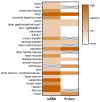
Organ PD-L1 mRNA and protein expression: RNA and protein expression are derived from the Human Protein Atlas (www.proteinatlas.org, accessed on 1 July 2022) [69,70]. The RNA expression summary is based on normalized expression (nTPM) values. Crossed-out gray cells indicate no available data. 1 Some authors identified a different organ expression, please refer to the main text.

Similar articles

Cited by

References

    1. Peterson E.J., Maltzman J.S. 12—T-Cell Activation and Tolerance. In: Rich R.R., Fleisher T.A., Shearer W.T., Schroeder H.W., Frew A.J., Weyand C.M., editors. Clinical Immunology. 5th ed. Elsevier; London, UK: 2019. pp. 183–196.e181. - DOI
    1. Corse E., Gottschalk R.A., Allison J.P. Strength of TCR–Peptide/MHC Interactions and In Vivo T Cell Responses. J. Immunol. 2011;186:5039–5045. doi: 10.4049/jimmunol.1003650. - DOI - PubMed
    1. van der Merwe P.A., Dushek O. Mechanisms for T cell receptor triggering. Nat. Rev. Immunol. 2011;11:47–55. doi: 10.1038/nri2887. - DOI - PubMed
    1. Lenschow D.J., Walunas T.L., Bluestone J.A. CD28/B7 System of T Cell Costimulation. Annu. Rev. Immunol. 1996;14:233–258. doi: 10.1146/annurev.immunol.14.1.233. - DOI - PubMed
    1. Wei S.C., Duffy C.R., Allison J.P. Fundamental Mechanisms of Immune Checkpoint Blockade Therapy. Cancer Discov. 2018;8:1069–1086. doi: 10.1158/2159-8290.CD-18-0367. - DOI - PubMed