. 2022 Aug 2;23(15):8602.
doi: 10.3390/ijms23158602.

p-Coumaric acid, Kaempferol, Astragalin and Tiliroside Influence the Expression of Glycoforms in AGS Gastric Cancer Cells

Affiliations

p-Coumaric acid, Kaempferol, Astragalin and Tiliroside Influence the Expression of Glycoforms in AGS Gastric Cancer Cells

Iwona Radziejewska et al. Int J Mol Sci. .

Abstract

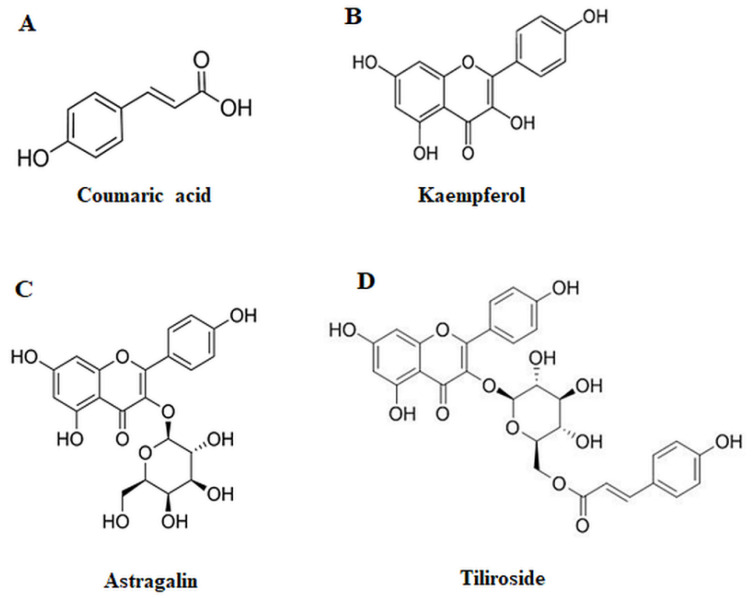
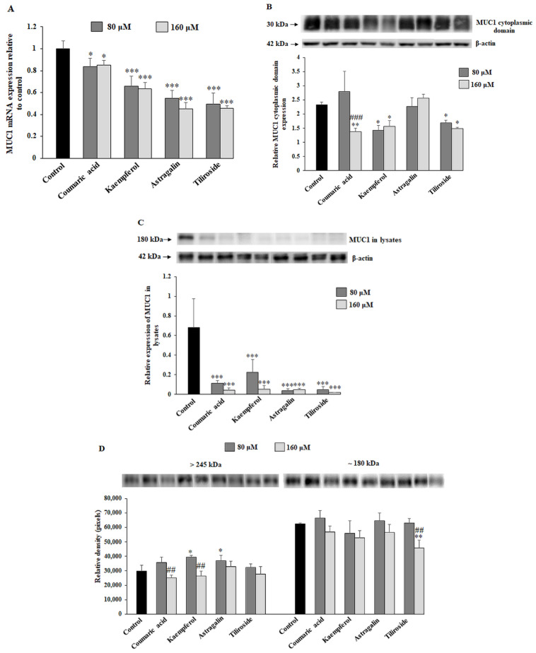
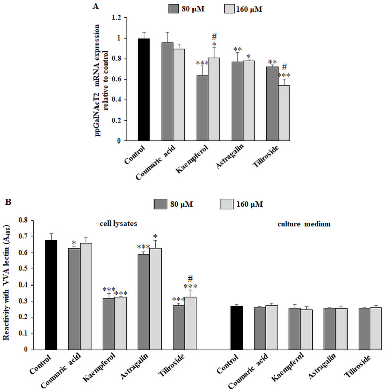
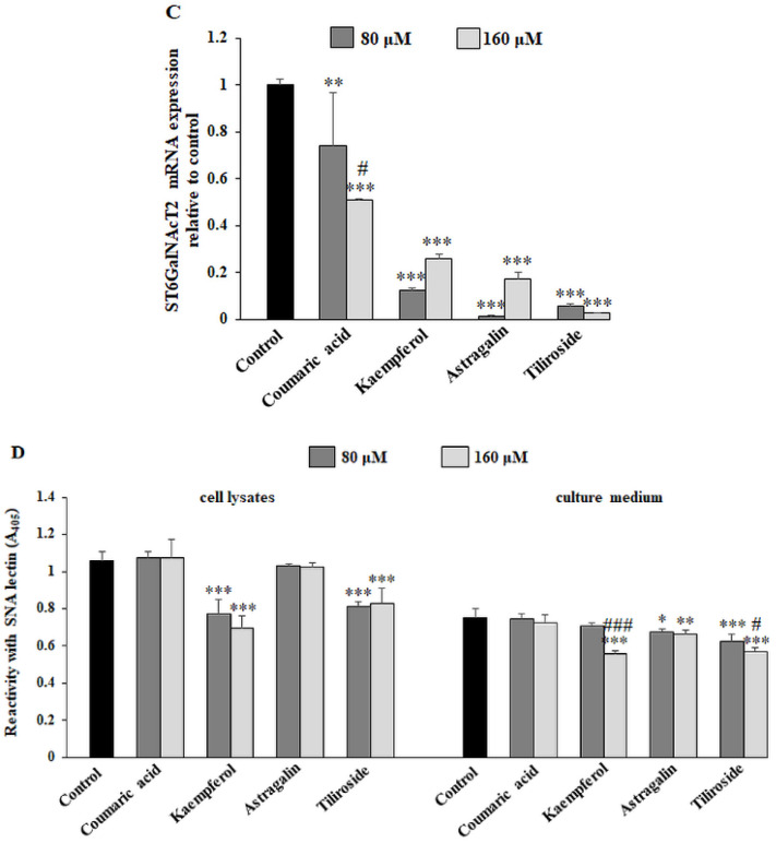
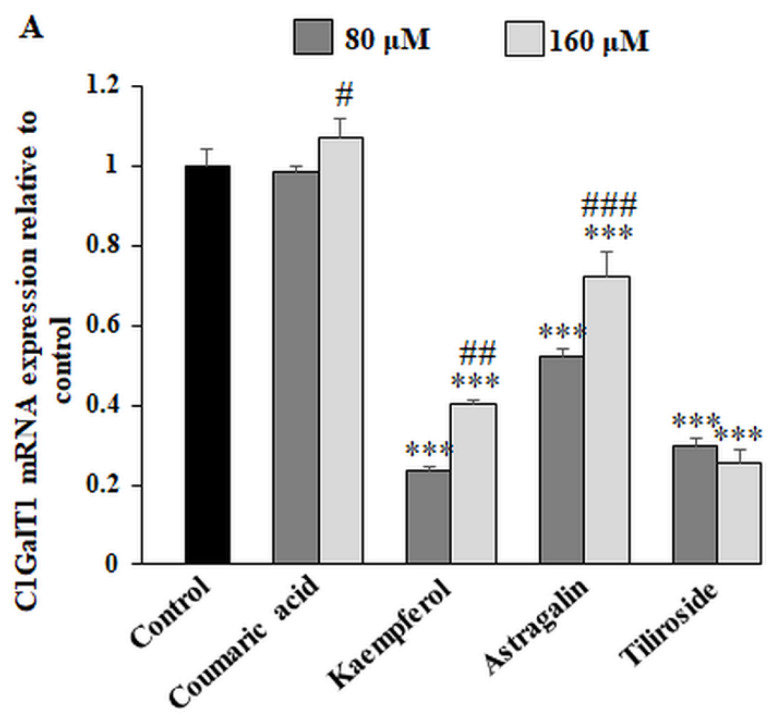
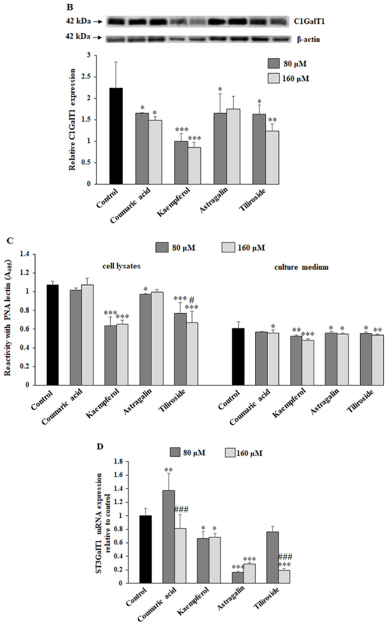
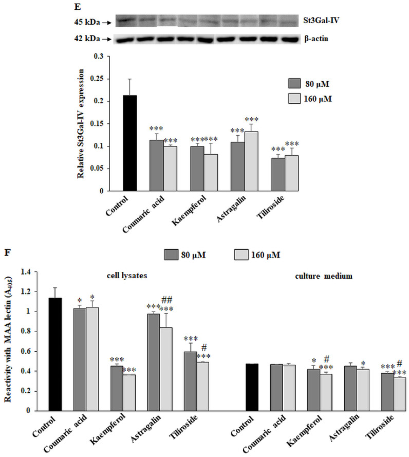
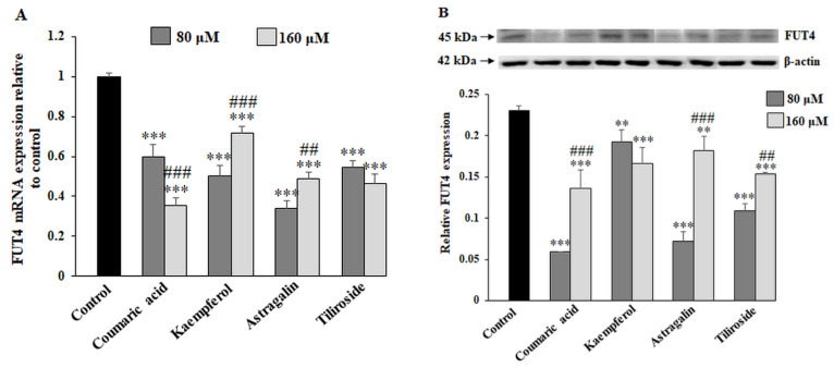
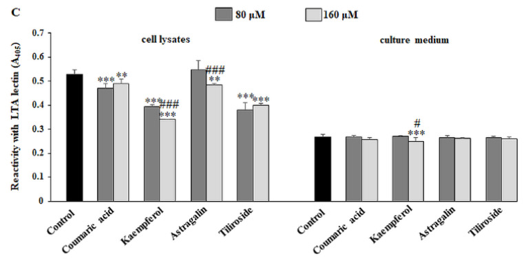
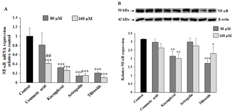
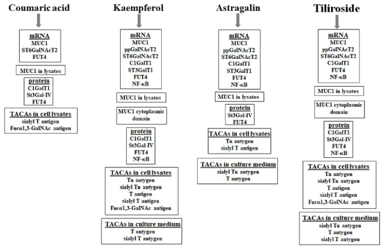
Abnormal glycosylation of cancer cells is considered a key factor of carcinogenesis related to growth, proliferation, migration and invasion of tumor cells. Many plant-based polyphenolic compounds reveal potential anti-cancer properties effecting cellular signaling systems. Herein, we assessed the effects of phenolic acid, p-coumaric acid and flavonoids such as kaempferol, astragalin or tiliroside on expression of selected cancer-related glycoforms and enzymes involved in their formation in AGS gastric cancer cells. The cells were treated with 80 and 160 µM of the compounds. RT-PCR, Western blotting and ELISA tests were performed to determine the influence of polyphenolics on analyzed factors. All the examined compounds inhibited the expression of MUC1, ST6GalNAcT2 and FUT4 mRNAs. C1GalT1, St3Gal-IV and FUT4 proteins as well as MUC1 domain, Tn and sialyl T antigen detected in cell lysates were also lowered. Both concentrations of kaempferol, astragalin and tiliroside also suppressed ppGalNAcT2 and C1GalT1 mRNAs. MUC1 cytoplasmic domain, sialyl Tn, T antigens in cell lysates and sialyl T in culture medium were inhibited only by kaempferol and tiliroside. Nuclear factor NF-κB mRNA expression decreased after treatment with both concentrations of kaempferol, astragalin and tiliroside. NF-κB protein expression was inhibited by kaempferol and tiliroside. The results indicate the rationality of application of examined polyphenolics as potential preventive agents against gastric cancer development.

Keywords: MUC1; gastric cancer; glycosylation; polyphenolics.


Conflict of interest statement

The authors declare no conflict of interest. The funders had no role in the design of the study; in the collection, analyses or interpretation of data; in the writing of the manuscript; or in the decision to publish the results.

Figures

Figure 1
Figure 1
Structures of selected compounds—p-coumaric acid (A), kaempferol (B), astragalin (C) and tiliroside (D).
Figure 2
Figure 2
Viability of AGS gastric cancer cells treated for 24 h with 20–160 μM concentrations of coumaric acid, kaempferol, astragalin and tiliroside. Mean values ± SD are the mean of triplicate culture.
Figure 3
Figure 3
The effect of coumaric acid, kaempferol, astragalin and tiliroside on MUC1 mRNA, MUC1 cytoplasmic domain, MUC1 expression in cell lysates and culture medium. The AGS cancer cells were incubated for 24 h with 80 and 160 μM concentrations of the polyphenolics. mRNA was determined by RT-PCR (A). The results are shown as a relative fold change in mRNA expression of gene in comparison to the gene in control, where expression was set at 1. ± SD are the mean of triplicate cultures. * p ˂ 0.05, *** p ˂ 0.001. MUC1 cytoplasmic domain (B), MUC1 in cell lysates (C) and in culture medium (D) were assessed by Western blot analysis. β-actin served as a protein loading control. The intensities of the bands were quantified by densitometric analysis. Data represent the mean ± SD of triplicate culture. * p ˂ 0.05, ** p ˂ 0.01, *** p ˂ 0.001 compared to untreated control; ## p ˂ 0.01, ### p < 0.001 compared to lower concentration (80 μM) of specific polyphenolic compound.
Figure 4
Figure 4
The effect of coumaric acid, kaempferol, astragalin and tiliroside on ppGalNAcT2 mRNA, Tn antigen in cell lysates and culture medium, ST6GalNAcT2 mRNA, and sialyl Tn antigen expression in cell lysates and culture medium. The AGS cancer cells were incubated for 24 h with 80 and 160 μM concentrations of the polyphenolics. mRNA was determined by RT-PCR ((A) for ppGalNAcT2 and (C) for ST6GalNAcT2). The results are shown as a relative fold change in mRNA expression of genes in comparison to the genes in control where expression was set at 1. ± SD are the mean of triplicate cultures. * p ˂ 0.05, ** p ˂ 0.01, *** p ˂ 0.001 compared to untreated control; # p < 0.05 compared to lower concentration (80 μM) of specific polyphenolic compound. Tn and sialyl Tn antigens’ relative expressions in cell lysates and culture medium were analyzed by ELISA tests with biotinylated lectins (VVA recognizing Tn structure (B) and SNA recognizing sialyl Tn antigen (D)). The results are expressed as absorbance at 405 nm after reactivity with proper lectin. Values ± SD are the mean from three independent assays. * p ˂ 0.05, ** p ˂ 0.01, *** p ˂ 0.001 compared to untreated control; # p < 0.05, ### p < 0.001 compared to lower concentration (80 μM) of specific polyphenolic compound.
Figure 4
Figure 4
The effect of coumaric acid, kaempferol, astragalin and tiliroside on ppGalNAcT2 mRNA, Tn antigen in cell lysates and culture medium, ST6GalNAcT2 mRNA, and sialyl Tn antigen expression in cell lysates and culture medium. The AGS cancer cells were incubated for 24 h with 80 and 160 μM concentrations of the polyphenolics. mRNA was determined by RT-PCR ((A) for ppGalNAcT2 and (C) for ST6GalNAcT2). The results are shown as a relative fold change in mRNA expression of genes in comparison to the genes in control where expression was set at 1. ± SD are the mean of triplicate cultures. * p ˂ 0.05, ** p ˂ 0.01, *** p ˂ 0.001 compared to untreated control; # p < 0.05 compared to lower concentration (80 μM) of specific polyphenolic compound. Tn and sialyl Tn antigens’ relative expressions in cell lysates and culture medium were analyzed by ELISA tests with biotinylated lectins (VVA recognizing Tn structure (B) and SNA recognizing sialyl Tn antigen (D)). The results are expressed as absorbance at 405 nm after reactivity with proper lectin. Values ± SD are the mean from three independent assays. * p ˂ 0.05, ** p ˂ 0.01, *** p ˂ 0.001 compared to untreated control; # p < 0.05, ### p < 0.001 compared to lower concentration (80 μM) of specific polyphenolic compound.
Figure 5
Figure 5
The effect of coumaric acid, kaempferol, astragalin and tiliroside on C1GalT1 mRNA, C1GalT1 protein, T antigen in cell lysates and culture medium, ST3GalT1 mRNA, St3Gal-IV protein and sialyl T antigen in cell lysates and culture medium. The AGS cancer cells were incubated for 24 h with 80 and 160 μM concentrations of the polyphenolics. mRNA was determined by RT-PCR ((A) for C1GalT1 and (D) for ST3GalT1). The results are shown as a relative fold change in mRNA expression of genes in comparison to the genes in control, where expression was set at 1. ±SD are the mean of triplicate cultures. * p ˂ 0.05, ** p ˂ 0.01, *** p ˂ 0.001 compared to untreated control; # p < 0.05, ## p ˂ 0.01, ### p < 0.001 compared to lower concentration (80 μM) of specific polyphenolic compound. C1GalT1 (B) and St3Gal-IV (E) protein expressions were assessed by Western blot analysis. β-actin served as a protein loading control. The intensities of the bands were quantified by densitometric analysis. Data represent the mean ± SD of triplicate culture. * p ˂ 0.05, ** p ˂ 0.01, *** p ˂ 0.001 compared to untreated control. T and sialyl T antigens’ relative expressions in cell lysates and culture medium were analyzed by ELISA tests with biotinylated lectins (PNA recognizing T structure (C) and MAA recognizing sialyl T antigen (F)). The results are expressed as absorbance at 405 nm after reactivity with proper lectin. Values ± SD are the mean from three independent assays. * p ˂ 0.05, ** p ˂ 0.01, *** p ˂ 0.001 compared to untreated control; # p < 0.05, ## p < 0.01 compared to lower concentration (80 μM) of specific polyphenolic.
Figure 5
Figure 5
The effect of coumaric acid, kaempferol, astragalin and tiliroside on C1GalT1 mRNA, C1GalT1 protein, T antigen in cell lysates and culture medium, ST3GalT1 mRNA, St3Gal-IV protein and sialyl T antigen in cell lysates and culture medium. The AGS cancer cells were incubated for 24 h with 80 and 160 μM concentrations of the polyphenolics. mRNA was determined by RT-PCR ((A) for C1GalT1 and (D) for ST3GalT1). The results are shown as a relative fold change in mRNA expression of genes in comparison to the genes in control, where expression was set at 1. ±SD are the mean of triplicate cultures. * p ˂ 0.05, ** p ˂ 0.01, *** p ˂ 0.001 compared to untreated control; # p < 0.05, ## p ˂ 0.01, ### p < 0.001 compared to lower concentration (80 μM) of specific polyphenolic compound. C1GalT1 (B) and St3Gal-IV (E) protein expressions were assessed by Western blot analysis. β-actin served as a protein loading control. The intensities of the bands were quantified by densitometric analysis. Data represent the mean ± SD of triplicate culture. * p ˂ 0.05, ** p ˂ 0.01, *** p ˂ 0.001 compared to untreated control. T and sialyl T antigens’ relative expressions in cell lysates and culture medium were analyzed by ELISA tests with biotinylated lectins (PNA recognizing T structure (C) and MAA recognizing sialyl T antigen (F)). The results are expressed as absorbance at 405 nm after reactivity with proper lectin. Values ± SD are the mean from three independent assays. * p ˂ 0.05, ** p ˂ 0.01, *** p ˂ 0.001 compared to untreated control; # p < 0.05, ## p < 0.01 compared to lower concentration (80 μM) of specific polyphenolic.
Figure 6
Figure 6
The effect of coumaric acid, kaempferol, astragalin and tiliroside on FUT4 mRNA, FUT4 protein and Fucα1,3-GalNAc antigen. The AGS cancer cells were incubated for 24 h with 80 and 160 μM concentrations of the polyphenolics. mRNA was determined by RT-PCR (A). The results are shown as a relative fold change in mRNA expression of genes in comparison to the genes in control, where expression was set at 1. ± SD are the mean of triplicate cultures. *** p ˂ 0.001 compared to untreated control; ## p ˂ 0.01, ### p < 0.001 compared to lower concentration (80 μM) of specific polyphenolic compound. FUT4 protein expression was assessed by Western blot analysis (B). β-actin served as a protein loading control. The intensities of the bands were quantified by densitometric analysis. Data represent the mean ± SD of triplicate culture. ** p ˂ 0.01, *** p ˂ 0.001 compared to untreated control; # p < 0.05, ### p < 0.001 compared to lower concentration (80 μM) of specific polyphenolic compound. Fucα1,3-GalNAc antigen relative expression in cell lysates and culture medium was analyzed by ELISA tests with biotinylated lectins (LTA recognizing fucosylated antigen) (C). The results are expressed as absorbance at 405 nm after reactivity with the lectin. Values ± SD are the mean from three independent assays. ** p ˂ 0.01, *** p ˂ 0.001 compared to untreated control; # p < 0.05, ### p < 0.001 compared to lower concentration (80 μM) of specific polyphenolic compound.
Figure 6
Figure 6
The effect of coumaric acid, kaempferol, astragalin and tiliroside on FUT4 mRNA, FUT4 protein and Fucα1,3-GalNAc antigen. The AGS cancer cells were incubated for 24 h with 80 and 160 μM concentrations of the polyphenolics. mRNA was determined by RT-PCR (A). The results are shown as a relative fold change in mRNA expression of genes in comparison to the genes in control, where expression was set at 1. ± SD are the mean of triplicate cultures. *** p ˂ 0.001 compared to untreated control; ## p ˂ 0.01, ### p < 0.001 compared to lower concentration (80 μM) of specific polyphenolic compound. FUT4 protein expression was assessed by Western blot analysis (B). β-actin served as a protein loading control. The intensities of the bands were quantified by densitometric analysis. Data represent the mean ± SD of triplicate culture. ** p ˂ 0.01, *** p ˂ 0.001 compared to untreated control; # p < 0.05, ### p < 0.001 compared to lower concentration (80 μM) of specific polyphenolic compound. Fucα1,3-GalNAc antigen relative expression in cell lysates and culture medium was analyzed by ELISA tests with biotinylated lectins (LTA recognizing fucosylated antigen) (C). The results are expressed as absorbance at 405 nm after reactivity with the lectin. Values ± SD are the mean from three independent assays. ** p ˂ 0.01, *** p ˂ 0.001 compared to untreated control; # p < 0.05, ### p < 0.001 compared to lower concentration (80 μM) of specific polyphenolic compound.
Figure 7
Figure 7
The effect of coumaric acid, kaempferol, astragalin and tiliroside on NF-κB mRNA and protein. The AGS cancer cells were incubated for 24 h with 80 and 160 μM concentrations of the polyphenolic compounds. mRNA was determined by RT-PCR (A). The results are shown as a relative fold change in mRNA expression of genes in comparison to the genes in control where expression was set at 1. ± SD are the mean of triplicate cultures. *** p ˂ 0.001 compared to untreated control; ## p ˂ 0.01 compared to lower concentration (80 μM) of specific polyphenolic. NF-κB protein expression was assessed by Western blot analysis (B). β-actin served as a protein loading control. The intensities of the bands were quantified by densitometric analysis. Data represent the mean ± SD of triplicate culture. * p ˂ 0.05, ** p ˂ 0.01, *** p ˂ 0.001 compared to untreated control.
Figure 8
Figure 8
Summarized inhibitory effect of coumaric acid, kaempferol, astragalin and tiliroside on specific glycoforms and NF-κB in AGS gastric cancer cells.

Similar articles

Cited by

References

    1. Johnson F.M., Beckman M. Updates on management of gastric cancer. Curr. Oncol. Rep. 2019;21:67. doi: 10.1007/s11912-019-0820-4. - DOI - PubMed
    1. Correra P. Gastric cancer: Overview. Gastroenterol. Clin. N. Am. 2013;42:211–217. doi: 10.1016/j.gtc.2013.01.002. - DOI - PMC - PubMed
    1. Maiuolo J., Musolino V., Gliozzi M., Carresi C., Oppedisano F., Nucera S., Scarano F., Scicchitano M., Guarnieri L., Bosco F., et al. The employment of genera Vaccinium, Citrus, Olea, and Cynara polyphenols for the reduction of selected anti-cancer drug side effects. Nutrients. 2022;14:1574. doi: 10.3390/nu14081574. - DOI - PMC - PubMed
    1. Niedzwiecki A., Roomi M.W., Kalinovsky T., Rath M. Anticancer efficacy of polyphenols and their combinations. Nutrients. 2016;8:552. doi: 10.3390/nu8090552. - DOI - PMC - PubMed
    1. Pinho S.S., Carvalho S., Marcos-Pinto R., Magalhaes A., Oliveira C., Gu J., Dinis-Ribeiro M., Carneiro F., Seruca R., Reis C.A. Gastric cancer: Adding glycosylation to the equation. Trends Mol. Med. 2013;19:664–676. doi: 10.1016/j.molmed.2013.07.003. - DOI - PubMed