

Unique Features of River Lamprey (Lampetra fluviatilis) Myogenesis
- PMID: 35955736
- PMCID: PMC9368804
- DOI: 10.3390/ijms23158595
Unique Features of River Lamprey (Lampetra fluviatilis) Myogenesis
Abstract
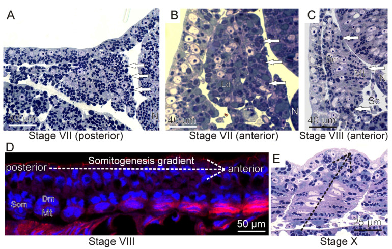
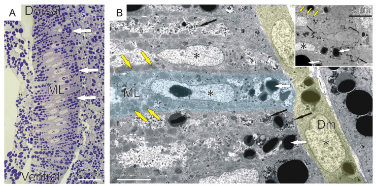
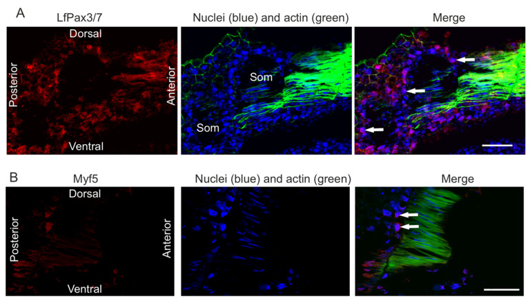
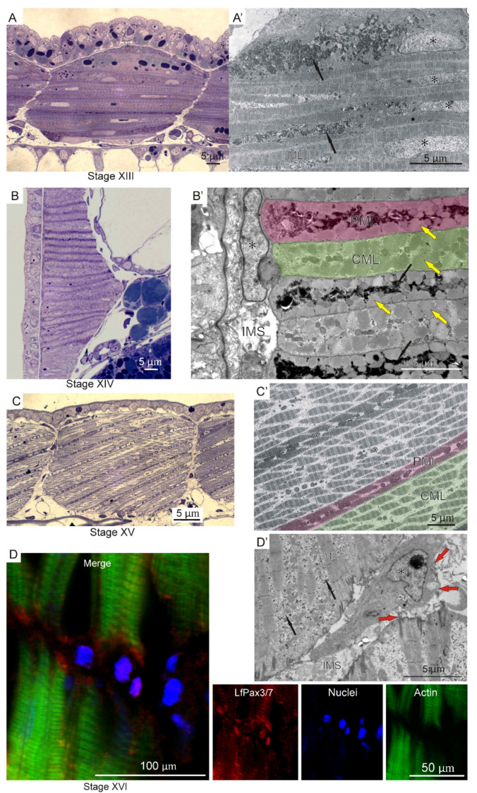
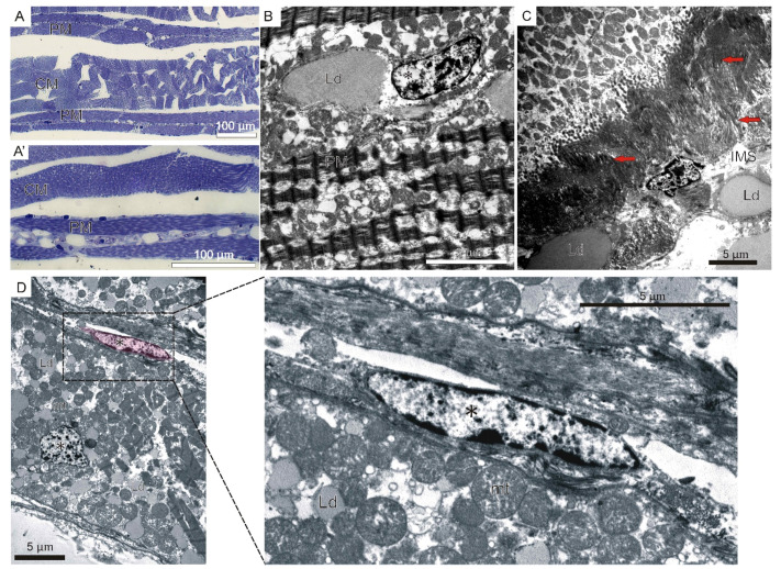
The river lamprey (L. fluviatilis) is a representative of the ancestral jawless vertebrate group. We performed a histological analysis of trunk muscle fiber differentiation during embryonal, larval, and adult musculature development in this previously unstudied species. Investigation using light, transmission electron (TEM), and confocal microscopy revealed that embryonal and larval musculature differs from adult muscle mass. Here, we present the morphological analysis of L. fluviatilis myogenesis, from unsegmented mesoderm through somite formation, and their differentiation into multinucleated muscle lamellae. Our analysis also revealed the presence of myogenic factors LfPax3/7 and Myf5 in the dermomyotome. In the next stages of development, two types of muscle lamellae can be distinguished: central surrounded by parietal. This pattern is maintained until adulthood, when parietal muscle fibers surround the central muscles on both sides. The two types show different morphological characteristics. Although lampreys are phylogenetically distant from jawed vertebrates, somite morphology, especially dermomyotome function, shows similarity. Here we demonstrate that somitogenesis is a conservative process among all vertebrates. We conclude that river lamprey myogenesis shares features with both ancestral and higher vertebrates.
Keywords: Lampetra fluviatilis; MfPax3/7; Mrf5; embryogenesis; larval musculature; myogenesis; myogenic factors; river lamprey; somitogenesis.
Conflict of interest statement
The authors declare no conflict of interest.
Figures


Similar articles
-
Expression and interaction of muscle-related genes in the lamprey imply the evolutionary scenario for vertebrate skeletal muscle, in association with the acquisition of the neck and fins.Dev Biol. 2011 Feb 1;350(1):217-27. doi: 10.1016/j.ydbio.2010.10.029. Epub 2010 Oct 28. Dev Biol. 2011. PMID: 21035440
-
Evolutionary perspectives from development of mesodermal components in the lamprey.Dev Dyn. 2007 Sep;236(9):2410-20. doi: 10.1002/dvdy.21177. Dev Dyn. 2007. PMID: 17477393 Review.
-
Phylogeography of the European brook lamprey (Lampetra planeri) and the European river lamprey (Lampetra fluviatilis) species pair based on mitochondrial data.J Fish Biol. 2020 Apr;96(4):905-912. doi: 10.1111/jfb.14279. Epub 2020 Feb 26. J Fish Biol. 2020. PMID: 32039478
-
Downstream migration of early larvae of the European river lamprey Lampetra fluviatilis.Dokl Biol Sci. 2014 Nov;459:344-7. doi: 10.1134/S0012496614060039. Epub 2015 Jan 6. Dokl Biol Sci. 2014. PMID: 25560212 No abstract available.
-
Lamprey vision: Photoreceptors and organization of the retina.Semin Cell Dev Biol. 2020 Oct;106:5-11. doi: 10.1016/j.semcdb.2019.10.008. Epub 2019 Nov 9. Semin Cell Dev Biol. 2020. PMID: 31711759 Review.
Cited by
-
Histomorphological and Dynamical Changes in Female River Lampreys during Maturation under Controlled Conditions as a Part of Lamprey Restoration Programs.Animals (Basel). 2024 Aug 29;14(17):2516. doi: 10.3390/ani14172516. Animals (Basel). 2024. PMID: 39272301 Free PMC article.
-
Shared features of blastula and neural crest stem cells evolved at the base of vertebrates.Nat Ecol Evol. 2024 Sep;8(9):1680-1692. doi: 10.1038/s41559-024-02476-8. Epub 2024 Jul 26. Nat Ecol Evol. 2024. PMID: 39060477 Free PMC article.
References
-
- Kujawa R. River Lamprey [Pol. Minóg Rzeczny] UWM; Olsztyn, Poland: 2017.
-
- Borowiec B.G., Docker M.F., Johnson N.S., Moser M.L., Zielinski B., Wilkie M.P. Exploiting the Physiology of Lampreys to Refine Methods of Control and Conservation. J. Great Lakes Res. 2021;47:S723–S741. doi: 10.1016/j.jglr.2021.10.015. - DOI
MeSH terms
LinkOut - more resources
Full Text Sources
