. 2022 Aug 3;23(15):8607.
doi: 10.3390/ijms23158607.

MATE1 Deficiency Exacerbates Dofetilide-Induced Proarrhythmia

Affiliations

MATE1 Deficiency Exacerbates Dofetilide-Induced Proarrhythmia

Muhammad Erfan Uddin et al. Int J Mol Sci. .

Abstract

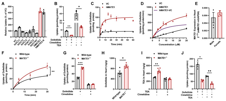
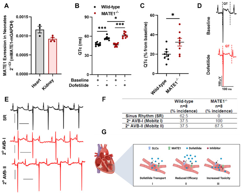
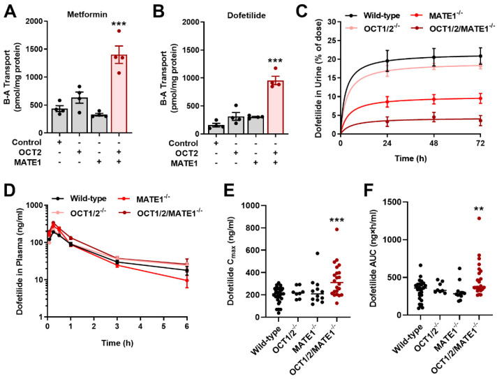
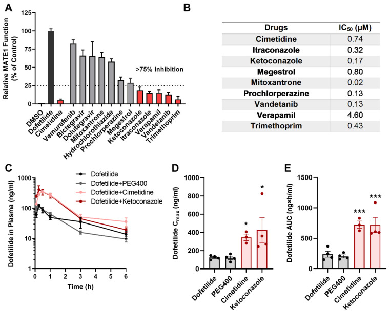
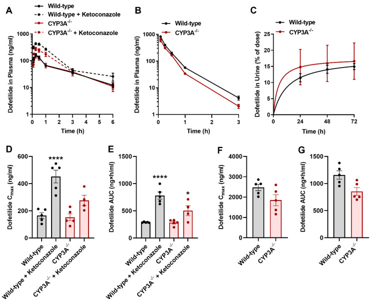
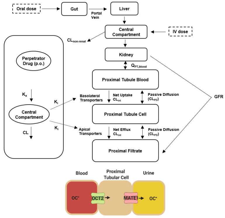
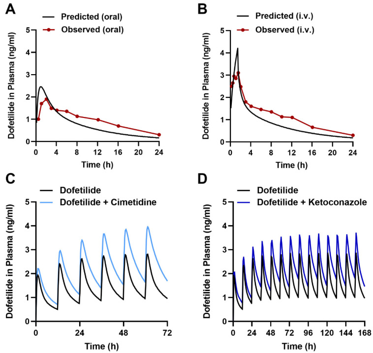
Dofetilide is a rapid delayed rectifier potassium current inhibitor widely used to prevent the recurrence of atrial fibrillation and flutter. The clinical use of this drug is associated with increases in QTc interval, which predispose patients to ventricular cardiac arrhythmias. The mechanisms involved in the disposition of dofetilide, including its movement in and out of cardiomyocytes, remain unknown. Using a xenobiotic transporter screen, we identified MATE1 (SLC47A1) as a transporter of dofetilide and found that genetic knockout or pharmacological inhibition of MATE1 in mice was associated with enhanced retention of dofetilide in cardiomyocytes and increased QTc prolongation. The urinary excretion of dofetilide was also dependent on the MATE1 genotype, and we found that this transport mechanism provides a mechanistic basis for previously recorded drug-drug interactions of dofetilide with various contraindicated drugs, including bictegravir, cimetidine, ketoconazole, and verapamil. The translational significance of these observations was examined with a physiologically-based pharmacokinetic model that adequately predicted the drug-drug interaction liabilities in humans. These findings support the thesis that MATE1 serves a conserved cardioprotective role by restricting excessive cellular accumulation and warrant caution against the concurrent administration of potent MATE1 inhibitors and cardiotoxic substrates with a narrow therapeutic window.

Keywords: PBPK modeling; arrhythmia; dofetilide; organic cation transporters.


Conflict of interest statement

The authors declare that they have no competing interests.

Figures

Figure 1
Figure 1
Inhibition of MATE1 enhances cardiac accumulation of dofetilide. (A) Transport of [3H] dofetilide (1 µM, 15 min) in cells overexpressing the human transporters OCT1, OCT2, OCT3, OAT1, OAT3, OCTN1, MATE1, or MATE2-K (2 min). Relative uptake is expressed as percentage change compared with empty vector controls (n = 3). (B) Relative uptake of [3H] dofetilide and [14C] TEA in HEK293 cells overexpressing human MATE1 in the presence and absence of cimetidine (25 µM). (C) Time dependent uptake (2–60 min) of [3H] dofetilide (1 µM) in HEK293 cells stably transfected with vector control (VC) or MATE1. (D) Transport kinetics of [3H] dofetilide in cells overexpressing human MATE1. The Michaelis-Menten constant (Km) and the maximal uptake rate (Vmax) values for the kinetics of dofetilide (1–25 µM) was determined after an incubation time of 2 min. Km and Vmax values for transport activity are 6.72 ± 1.71 µM, and 544.40 ± 55.70 pmol/min/mg, respectively. (E) Expression of the MATE1 gene in hearts isolated from untreated wild-type male and female mice (n = 4 per group). (F) Time dependent uptake of [3H] dofetilide (2 µM) in ex vivo cardiomyocytes isolated from wild-type or MATE1-deficient female mice (n = 4–6 per group). (G) Ex vivo concentrations of [3H] dofetilide (2 µM) in cardiomyocytes isolated from wild-type or MATE1-deficient female mice (n = 3 per group) for 30 min in the presence or absence of cimetidine (25 µM) pretreatment. (H) Concentration of dofetilide in whole heart tissue from wild-type or MATE1-deficient male mice 15 min after a single i.v. injection of dofetilide via the caudal vein at a dose of 2.5 mg/kg (n = 4 per group). (I) Concentration of TEA in whole heart tissue from wild-type and MATE1-deficient male mice (n = 4–5 per group) with or without treatment of with cimetidine (100 mg/kg) 30 min before an i.v. administration of [14C] TEA (0.2 mg/kg). Heart samples were collected 15 min after TEA administration. (J) Uptake of 2 µM [3H] dofetilide and [14C] TEA in AC16 human cardiomyocytes (n = 3) for 20 min in the presence or absence of cimetidine (25 µM) pretreatment (15 min). All experimental values are presented as mean ± SEM. Statistical analysis was performed using an unpaired two-sided Student’s t-test with Welch’s correction: * p < 0.05, ** p < 0.01, *** p < 0.001.
Figure 2
Figure 2
MATE1 deficiency exacerbates dofetilide-induced proarrhythmia. (A) Expression of the MATE1 gene in heart (n = 3) and kidneys (n = 4) isolated from untreated neonatal wild-type mice (day 1). (B) QTc interval and (C) percent QTc interval changes in neonatal wild-type or MATE1-deficient mice (day one) 15 min after a single i.p. injection of dofetilide at a dose of 0.5 mg/kg (n = 8 per group). All experimental values are presented as mean ± SEM. Statistical analysis was performed using an unpaired two-sided Student’s t test with Welch’s correction: * p < 0.05, *** p < 0.001, compared to baseline values. (D) In vivo surface ECG illustrating changes in QTc after dofetilide treatment in neonatal wild-type mice. (E) Dofetilide-induced second-degree AV blocks in neonatal MATE1-deficient mice. (F) Incidence of second-degree AV blocks (Mobitz I and Mobitz II) in neonatal wild-type and MATE1-deficient mice (n = 8 per group) after treatment with dofetilide. (G) Schematic diagram illustrating the proposed MATE1-dependent regulation of dofetilide transport (i) as a modulator of accumulation in cardiac myocytes leading to reduced (ii) or increased (iii) intracellular concentrations and reduced or increased electrophysiologic activity.
Figure 3
Figure 3
Inhibition of MATE1 reduces renal elimination of dofetilide. Characterization of the basolateral to apical (B-A) transport of [14C] metformin (A) [3H] dofetilide (B) in single transfected MDCK-VC, MDCK-OCT2, MDCK-MATE1, and double-transfected MDCK-OCT2-MATE1 cell lines. Transcellular transport was quantified by measuring the amount of metformin or dofetilide added basolaterally to the monolayers and appearing in the apical compartment after a 60-min incubation. Statistical analysis was performed using an unpaired two-sided Student’s t-test with Welch’s correction: *** p <  0.001 vs. MDCK-VC. (C) Urinary excretion of dofetilide in female wild-type, OCT1/2-deficient, MATE1-deficient, and OCT1/2/MATE1-deficient mice (n = 5) following a single i.v. dose of dofetilide (2.5 mg/kg). (D) Plasma concentration-time profile of dofetilide in female wild-type, OCT1/2-deficient, MATE1-deficient, and OCT1/2/MATE1-deficient mice (n = 5) receiving a single oral dose of dofetilide (5 mg/kg). (E) Peak concentration (Cmax) of dofetilide and (F) area under the curve (AUC) of dofetilide after a single oral dose in female mice of varying transporter genotypes. Statistical analysis was performed using one-way ANOVA with Dunnett’s post hoc test: ** p < 0.01, *** p < 0.001, compared with wild-type mice. All data represent the mean ± SEM.
Figure 4
Figure 4
Drugs contraindicated for combined use with dofetilide inhibit MATE1 function. (A) FDA-listed contraindicated drugs of dofetilide were assessed at a concentration of 25 µM in HEK293 cells overexpressing human MATE1. [3H] Dofetilide (1 µM) and cimetidine were used as positive control substrate or inhibitor, respectively. Data are represented as the percentage residual MATE1 activity as compared with the vehicle control (DMSO) group (n = 3 per group). (B) IC50 values of different contraindicated drugs. (C) Plasma concentration-time curves profile of dofetilide in male wild-type mice receiving vehicle (PEG400), cimetidine (100 mg/kg), or ketoconazole (50 mg/kg) 30 min before dofetilide (n = 5 per group). (D,E) Pharmacokinetic parameters of dofetilide in male wild-type mice in the presence or absence of pretreatment with vehicle or contraindicated drugs. All data represent the mean ± SEM. Statistical analysis was performed using one-way ANOVA with Dunnett’s post hoc test: * p < 0.05, *** p < 0.001, compared with dofetilide alone control group.
Figure 5
Figure 5
CYP3A inhibition does not influence the pharmacokinetics of dofetilide. (A) Plasma concentration-time profile of dofetilide (5 mg/kg, p.o.) in male wild-type or CYP3A-deficient mice pretreated with ketoconazole (100 mg/kg) 30 min before dofetilide (n = 5 per group). (B) Plasma concentration-time curves profile of dofetilide receiving an i.v. dose of 2.5 mg/kg in male wild-type or CYP3A-deficient mice (n = 5). (C) Urinary excretion of dofetilide in male wild-type and CYP3A-deficient mice (n = 5) following a single dose of dofetilide (2.5 mg/kg, i.v.). Pharmacokinetic parameters of dofetilide in male wild-type and CYP3A-deficient mice receiving an oral (5 mg/kg) (D,E), and i.v. (2.5 mg/kg) dose (F,G). Statistical analysis was performed using one-way ANOVA with Dunnett’s post hoc test: * p < 0.05, **** p < 0.0001 compared with wild-type mice receiving vehicle alone. All data represent the mean ± SEM.
Figure 6
Figure 6
Structure of the physiologically based pharmacokinetic (PBPK) model for dofetilide. Abbreviations: CL, clearance; GFR, glomerular filtration rate; OC+, organic cation; Ka, absorption rate constant; Ki, inhibition constant; Q, blood flow.
Figure 7
Figure 7
PBPK modeling predicts transporter-mediated interactions with dofetilide in humans. Observed and predicted plasma concentration-time profile after a single dose of 0.5 mg dofetilide oral administration (A), and 90-min i.v. infusion (B) at a single dose of 0.5 mg in adult healthy volunteers. Predicted plasma concentration-time profiles receiving multiple oral doses of dofetilide (0.5 mg, BID) with and without cimetidine (400 mg, BID) (C) and ketoconazole (400 mg, QD) (D) in healthy volunteers.

References

    1. Naccarelli G.V., Varker H., Lin J., Schulman K.L. Increasing Prevalence of Atrial Fibrillation and Flutter in the United States. Am. J. Cardiol. 2009;104:1534–1539. doi: 10.1016/j.amjcard.2009.07.022. - DOI - PubMed
    1. Morillo C.A., Banerjee A., Perel P., Wood D., Jouven X. Atrial Fibrillation: The Current Epidemic. J. Geriatr. Cardiol. 2017;14:195–203. doi: 10.11909/j.issn.1671-5411.2017.03.011. - DOI - PMC - PubMed
    1. Chugh S.S., Havmoeller R., Narayanan K., Singh D., Rienstra M., Benjamin E.J., Gillum R.F., Kim Y.H., McAnulty J.H., Zheng Z.J., et al. Worldwide Epidemiology of Atrial Fibrillation: A Global Burden of Disease 2010 Study. Circulation. 2014;129:837–847. doi: 10.1161/CIRCULATIONAHA.113.005119. - DOI - PMC - PubMed
    1. Krijthe B.P., Kunst A., Benjamin E.J., Lip G.Y., Franco O.H., Hofman A., Witteman J.C., Stricker B.H., Heeringa J. Projections on the Number of Individuals with Atrial Fibrillation in the European Union, from 2000 to 2060. Eur. Heart J. 2013;34:2746–2751. doi: 10.1093/eurheartj/eht280. - DOI - PMC - PubMed
    1. Alonso A., Krijthe B.P., Aspelund T., Stepas K.A., Pencina M.J., Moser C.B., Sinner M.F., Sotoodehnia N., Fontes J.D., Janssens A.C., et al. Simple Risk Model Predicts Incidence of Atrial Fibrillation in a Racially and Geographically Diverse Population: The CHARGE-AF Consortium. J. Am. Heart Assoc. 2013;2:e000102. doi: 10.1161/JAHA.112.000102. - DOI - PMC - PubMed

LinkOut - more resources