

Molecular Mechanisms of Cytotoxicity of NCX4040, the Non-Steroidal Anti-Inflammatory NO-Donor, in Human Ovarian Cancer Cells
- PMID: 35955744
- PMCID: PMC9369271
- DOI: 10.3390/ijms23158611
Molecular Mechanisms of Cytotoxicity of NCX4040, the Non-Steroidal Anti-Inflammatory NO-Donor, in Human Ovarian Cancer Cells
Abstract
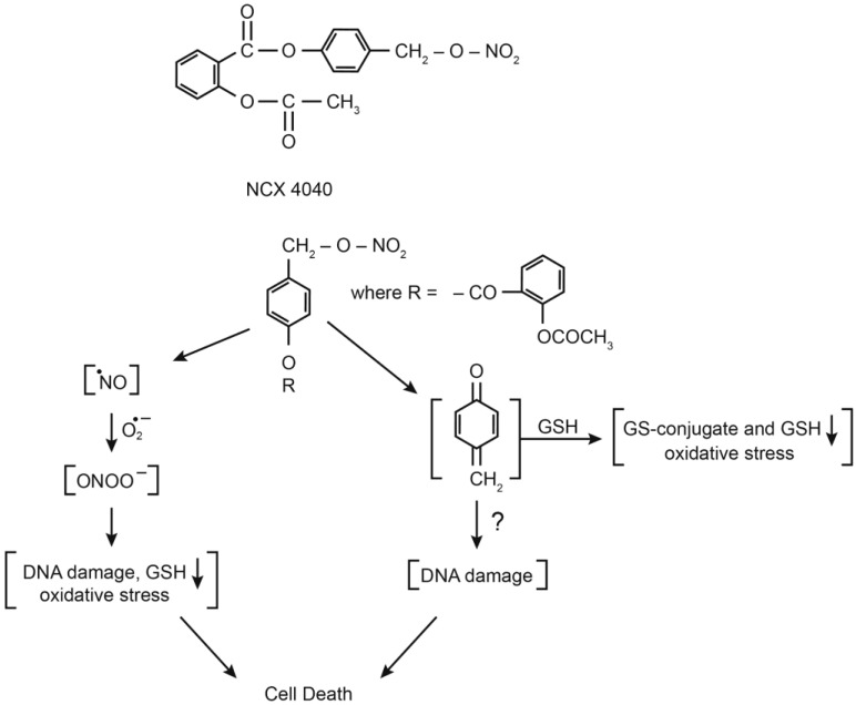
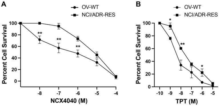
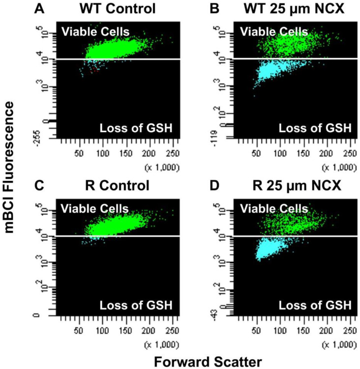
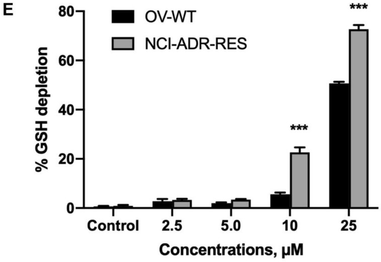
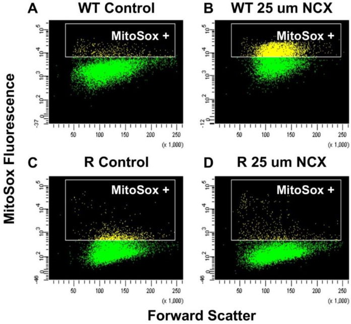
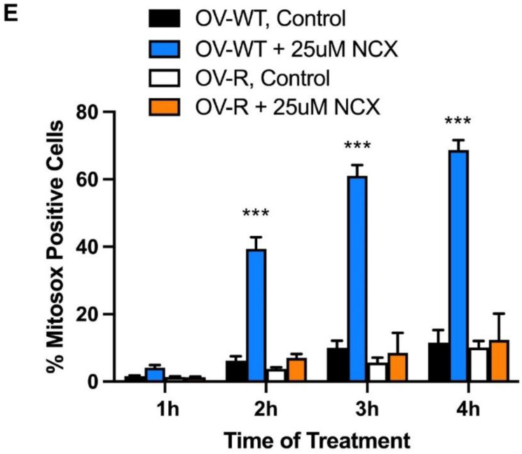
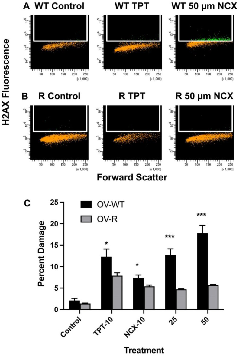
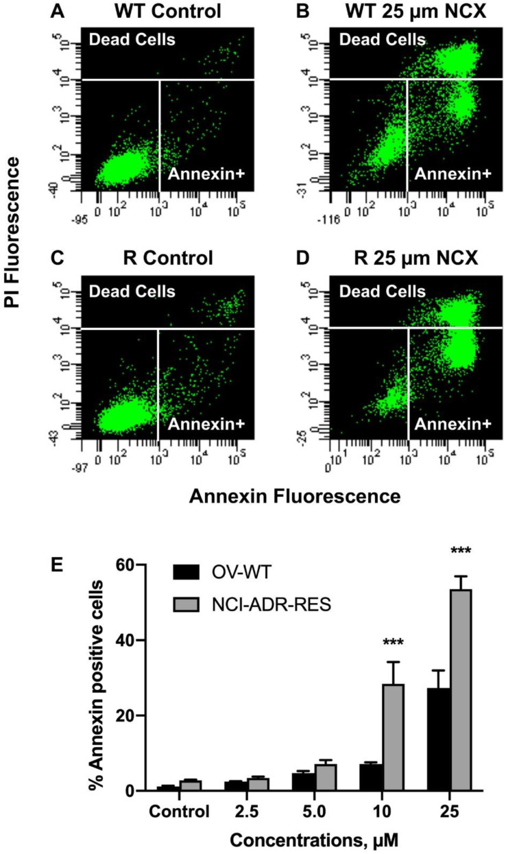
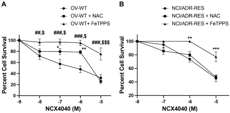
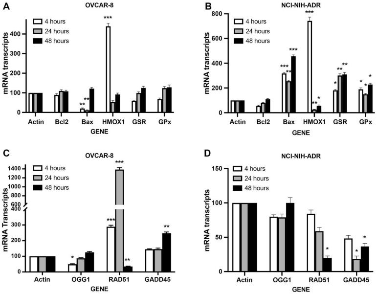
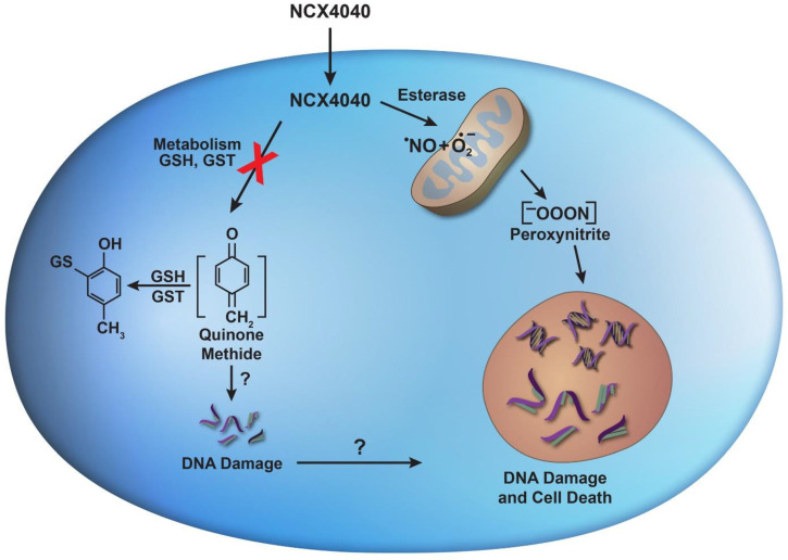
NCX4040, the non-steroidal anti-inflammatory-NO donor, is cytotoxic to several human tumors, including ovarian tumor cells. We have found that NCX4040 is also cytotoxic against both OVCAR-8 and its adriamycin resistant (NCI/ADR-RES) tumor cell lines. Here, we have examined mechanism(s) for the cytotoxicity of NCX4040 in OVCAR-8 and NCI/ADR-RES cell lines. We found that NCX4040 induced significant apoptosis in both cell lines. Furthermore, NCX4040 treatment caused significant depletion of cellular glutathione, causing oxidative stress due to the formation of reactive oxygen/nitrogen species (ROS/RNS). Significantly more ROS/RNS were detected in OVCAR-8 cells than in NCI/ADR-RES cells which may have resulted from increased activities of SOD, glutathione peroxidase and transferases expressed in NCI/ADR-RES cells. NCX4040 treatment resulted in the formation of double-strand DNA breaks in both cells; however, more of these DNA breaks were detected in OVCAR-8 cells. RT-PCR studies indicated that NCX4040-induced DNA damage was not repaired as efficiently in NCI/ADR-RES cells as in OVCAR-8 cells which may lead to a differential cell death. Pretreatment of OVCAR-8 cells with N-acetylcysteine (NAC) significantly decreased cytotoxicity of NCX4040 in OVCAR-8 cells; however, NAC had no effects on NCX4040 cytotoxicity in NCI/ADR-RES cells. In contrast, FeTPPS, a peroxynitrite scavenger, completely blocked NCX4040-induced cell death in both cells, suggesting that NCX4040-induced cell death could be mediated by peroxynitrite formed from NCX4040 following cellular metabolism.
Keywords: DNA damage; NCX4040; nitric oxide; peroxynitrite; reactive oxygen species.
Conflict of interest statement
The authors declare no actual or potential conflict of interest in the work reported in this manuscript.
Figures
Similar articles
-
Mechanisms of Cell Death Induced by Erastin in Human Ovarian Tumor Cells.Int J Mol Sci. 2024 Aug 8;25(16):8666. doi: 10.3390/ijms25168666. Int J Mol Sci. 2024. PMID: 39201357 Free PMC article.
-
Ferroptosis-Mediated Cell Death Induced by NCX4040, The Non-Steroidal Nitric Oxide Donor, in Human Colorectal Cancer Cells: Implications in Therapy.Cells. 2023 Jun 14;12(12):1626. doi: 10.3390/cells12121626. Cells. 2023. PMID: 37371096 Free PMC article.
-
Gene Expression Profiling Elucidates Cellular Responses to NCX4040 in Human Ovarian Tumor Cells: Implications in the Mechanisms of Action of NCX4040.Cancers (Basel). 2022 Dec 31;15(1):285. doi: 10.3390/cancers15010285. Cancers (Basel). 2022. PMID: 36612280 Free PMC article.
-
Nitro aspirin (NCX4040) induces apoptosis in PC3 metastatic prostate cancer cells via hydrogen peroxide (H2O2)-mediated oxidative stress.Free Radic Biol Med. 2019 Nov 1;143:494-509. doi: 10.1016/j.freeradbiomed.2019.08.025. Epub 2019 Aug 22. Free Radic Biol Med. 2019. PMID: 31446057 Free PMC article.
-
A case study in misidentification of cancer cell lines: MCF-7/AdrR cells (re-designated NCI/ADR-RES) are derived from OVCAR-8 human ovarian carcinoma cells.Cancer Lett. 2007 Jan 8;245(1-2):350-2. doi: 10.1016/j.canlet.2006.01.013. Epub 2006 Feb 28. Cancer Lett. 2007. PMID: 16504380
Cited by
-
Mechanisms of Cell Death Induced by Erastin in Human Ovarian Tumor Cells.Int J Mol Sci. 2024 Aug 8;25(16):8666. doi: 10.3390/ijms25168666. Int J Mol Sci. 2024. PMID: 39201357 Free PMC article.
-
Ferroptosis-Mediated Cell Death Induced by NCX4040, The Non-Steroidal Nitric Oxide Donor, in Human Colorectal Cancer Cells: Implications in Therapy.Cells. 2023 Jun 14;12(12):1626. doi: 10.3390/cells12121626. Cells. 2023. PMID: 37371096 Free PMC article.
-
Gene Expression Profiling Elucidates Cellular Responses to NCX4040 in Human Ovarian Tumor Cells: Implications in the Mechanisms of Action of NCX4040.Cancers (Basel). 2022 Dec 31;15(1):285. doi: 10.3390/cancers15010285. Cancers (Basel). 2022. PMID: 36612280 Free PMC article.
-
Cancer Metabolism: The Role of ROS in DNA Damage and Induction of Apoptosis in Cancer Cells.Metabolites. 2023 Jun 27;13(7):796. doi: 10.3390/metabo13070796. Metabolites. 2023. PMID: 37512503 Free PMC article. Review.
-
Ferroptosis in Toxicology: Present and Future.Int J Mol Sci. 2025 Jul 11;26(14):6658. doi: 10.3390/ijms26146658. Int J Mol Sci. 2025. PMID: 40724908 Free PMC article. Review.
References
-
- Murad F. Nitric oxide signaling: Would you believe that a simple free radical could be a second messenger, autacoid, paracrine substance, neurotransmitter, and hormone? Recent Prog. Horm. Res. 1998;53 - PubMed
MeSH terms
Substances
LinkOut - more resources
Full Text Sources
Medical
Research Materials
