Review
. 2022 Aug 3;23(15):8613.
doi: 10.3390/ijms23158613.

The Biochemical Mechanism of Fork Regression in Prokaryotes and Eukaryotes-A Single Molecule Comparison

Affiliations
Review

The Biochemical Mechanism of Fork Regression in Prokaryotes and Eukaryotes-A Single Molecule Comparison

Piero R Bianco. Int J Mol Sci. .

Abstract

The rescue of stalled DNA replication forks is essential for cell viability. Impeded but still intact forks can be rescued by atypical DNA helicases in a reaction known as fork regression. This reaction has been studied at the single-molecule level using the Escherichia coli DNA helicase RecG and, separately, using the eukaryotic SMARCAL1 enzyme. Both nanomachines possess the necessary activities to regress forks: they simultaneously couple DNA unwinding to duplex rewinding and the displacement of bound proteins. Furthermore, they can regress a fork into a Holliday junction structure, the central intermediate of many fork regression models. However, there are key differences between these two enzymes. RecG is monomeric and unidirectional, catalyzing an efficient and processive fork regression reaction and, in the process, generating a significant amount of force that is used to displace the tightly-bound E. coli SSB protein. In contrast, the inefficient SMARCAL1 is not unidirectional, displays limited processivity, and likely uses fork rewinding to facilitate RPA displacement. Like many other eukaryotic enzymes, SMARCAL1 may require additional factors and/or post-translational modifications to enhance its catalytic activity, whereas RecG can drive fork regression on its own.

Keywords: DNA repair; DNA replication; HARP; RPA; RecG; SMARCAL1; SSB protein; fork regression; fork reversal; stalled replication fork.


Conflict of interest statement

The author declares no conflict of interest.

Figures

Figure 1
Figure 1
The key elements of the fork regression reaction. (AC) An impeded fork is shown in (A) with the impediment, leading to cleavage of the nascent leading (B) or lagging strands (C). The net result is that the architecture of the fork is lost. (D) The terminology of the fork regression reaction. A DNA replication fork is shown impeded by an obstacle. To facilitate repair, the position of the fork is moved in the direction opposite to that of the fork’s movement, that is, in a net backward direction. This is defined as fork regression. The product of regression is known as the “chicken foot intermediate” which is essentially a Holliday junction. The opposite of fork regression is fork reversal, which takes place once the fork impediment has been removed. Reversal results in the restoration of the nascent fork structure, which ultimately results in the resumption of DNA replication. Black strands, parental DNA; blue and orange dashed strands indicate nascent leading strands and lagging strands, respectively; green strands represent nascent annealed parental strands. (B) The DNA enzymatic requirements of fork regression. For an enzyme to catalyze an efficient reaction, it must be an atypical DNA helicase that unwinds the nascent heteroduplex arms of the fork and couples this unwinding to duplex reannealing that occurs both in the wake of the enzyme, as well as ahead of the advancing molecular machine. (C) The force requirements of fork regression. The atypical DNA helicase must couple the energy associated with ATP binding, hydrolysis, and product release to the generation of mechanical force required to both regress the fork and displace proteins bound to single- (cyan sphere) or double-stranded (purple hexagon) regions of the fork. (E) To catalyze regression, the enzyme must couple DNA unwinding (red arrows) to duplex rewinding (green arrows). (F) In addition to being able to function as an atypical DNA helicase, the enzyme must also generate sufficient force to displace single- and/or double-strand DNA binding proteins during the fork regression reaction.
Figure 2
Figure 2
RecG binds to forks to catalyze regression. A ribbon diagram of a homology model of E. coli RecG bound to a fork substrate. The relevant regions are colored for clarity and the DNA strands are colored as in Figure 1. For comparison, a schematic of the fork is shown above, with DNA strand coloring the same as that in the structure. The model was built using Swiss-Model and PDB file 1GM5 as a template [60,72].
Figure 3
Figure 3
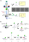
The magnetic tweezer, single-molecule assay that is used to dissect fork transactions. (A) Schematic of the assay developed by the Croquette group to monitor changes in the DNA substrate with single-base-pair resolution (left panel) [41,91]. In this approach, a single DNA molecule containing a 1200 bp hairpin is attached to a super-paramagnetic bead at one end and to a coverslip surface at the opposite end. The position of the bead attached to the DNA is monitored in real-time by imaging it using a CCD camera. These images change (right middle panel) as the DNA molecule is affected by enzyme action and the images are correlated with changes in the Z-height when compared to the reference bead (R), which is fixed to the coverslip surface. During the assay, the position of the beads is carefully controlled by the application of a magnetic field (the magnetic tweezers), with the amount of force applied being proportional to the strength of the field. If an enzyme binds to the fork and catalyzes duplex unwinding, this is visualized as an increase in Z-height as a function of time and is interpreted as fork reversal (top right panel). In contrast, if an enzyme binds to the fork and catalyzes DNA annealing, this is observed as a decrease in Z-height as a function of time and this is interpreted as fork regression (bottom right panel). (B,C) In situ construction of DNA substrates relevant to fork transactions. (B) The construction of forks with gaps in the leading or lagging strands is facilitated by the application of force, which results in complete unzippering of the hairpin. This is followed by the introduction of oligonucleotides that anneal, in separate reactions, to the opposing fork arms. When the force is decreased, forks with gaps in either the nascent leading or lagging strand arms are revealed. (C) The assembly of a fork with 3 duplex arms. Here, a primer (red) is annealed to the ssDNA arm close to the coverslip surface (step 1). Then, as in panel (B), the hairpin is completely unzippered by the magnetic tweezers. This is followed by the introduction of a second primer (green), which anneals to a position several hundred base pairs distal to the first (step 2). When the force is decreased, the fork structure reforms with primers annealed to opposing arms (step 3). When DNA polymerase and dNTPs are introduced, the primers are extended, resulting in the formation of a fork with three 600 bp duplex arms (step 4).
Figure 4
Figure 4
Kinetics and directionality of fork arm reannealing. (A,B) Reactions catalyzed by E. coli RecG [41]. (C,D) The reaction catalyzed by SMARCAL1 [46]. Representative time courses for the annealing reaction for each enzyme are shown in panels (A,C). The DNA substrate is the hairpin from Figure 3A. (B) RecG catalyzes fork regression only. Analysis of annealing reactions by RecG. Panels (C,D) are reprinted from Cell Reports, 3 (6), R. Betous, F. B. Couch, A. C. Mason, B. F. Eichman, M. Manosas and D. Cortez, Substrate-Selective Repair and Restart of Replication Forks by DNA Translocases, pages 1958–1969, Copyright (2013), with permission from Elsevier.
Figure 5
Figure 5
Single-strand DNA binding proteins affect fork regression enzymes differently. (AC) RecG; (DF) SMARCAL1. (A) RecG drives annealing against forces as high as 35 pN. (B) RecG couples SSB displacement to DNA annealing (rewinding). (C) The linker region of SSB is critical for efficient displacement by RecG. The mutants used are SSBΔC8 (last 8 residues deleted), SSB155 (22 residues deleted from the linker), and SSB125 (entire linker deleted). (D) RPA enhances the minimal processivity of SMARCAL1. (E,F) RPA enhances the processivity of SMARCAL1 and does not impact the annealing rate. SMARCAL1-Δ34 is a mutant that lacks the RPA binding domain. Panels (DF) are adapted from Cell Reports, 3 (6), R. Betous, F. B. Couch, A. C. Mason, B. F. Eichman, M. Manosas and D. Cortez, Substrate-Selective Repair and Restart of Replication Forks by DNA Translocases, pages 1958–1969, Copyright (2013), with permission from Elsevier.
Figure 6
Figure 6
Extrusion of a Holliday junction, a key intermediate in fork regression. (A,B) reactions catalyzed by E. coli RecG [41] and SMARCAL1 [46], respectively. The DNA substrate for each enzyme was constructed as in Figure 3C. Panel (B) is reprinted from Cell Reports, 3 (6), R. Betous, F. B. Couch, A. C. Mason, B. F. Eichman, M. Manosas and D. Cortez, Substrate-Selective Repair and Restart of Replication Forks by DNA Translocases, pages 1958–1969, Copyright (2013), with permission from Elsevier.

Similar articles

Cited by

References

    1. Fijalkowska I.J., Schaaper R.M., Jonczyk P. DNA replication fidelity in Escherichia coli: A multi-DNA polymerase affair. FEMS Microbiol. Rev. 2012;36:1105–1121. doi: 10.1111/j.1574-6976.2012.00338.x. - DOI - PMC - PubMed
    1. Merchut-Maya J.M., Bartek J., Maya-Mendoza A. Regulation of replication fork speed: Mechanisms and impact on genomic stability. DNA Repair. 2019;81:102654. doi: 10.1016/j.dnarep.2019.102654. - DOI - PubMed
    1. Skarstad K., Katayama T. Regulating DNA replication in bacteria. Cold Spring Harb Perspect. Biol. 2013;5:a012922. doi: 10.1101/cshperspect.a012922. - DOI - PMC - PubMed
    1. Lewis J.S., Jergic S., Dixon N.E. The E. coli DNA Replication Fork. Enzymes. 2016;39:31–88. - PubMed
    1. Dewar J.M., Walter J.C. Mechanisms of DNA replication termination. Nat. Rev. Mol. Cell Biol. 2017;18:507–516. doi: 10.1038/nrm.2017.42. - DOI - PMC - PubMed

MeSH terms

LinkOut - more resources