. 2022 Aug 3;23(15):8646.
doi: 10.3390/ijms23158646.

Transcriptional and Post-Translational Regulation of Junctional Adhesion Molecule-B (JAM-B) in Leukocytes under Inflammatory Stimuli

Affiliations

Transcriptional and Post-Translational Regulation of Junctional Adhesion Molecule-B (JAM-B) in Leukocytes under Inflammatory Stimuli

Priscilla E Day-Walsh et al. Int J Mol Sci. .

Abstract

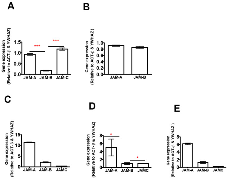
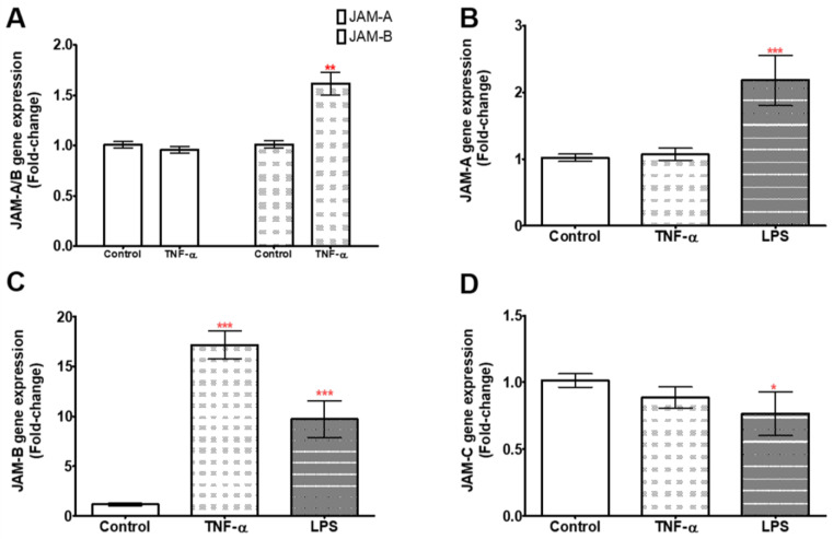
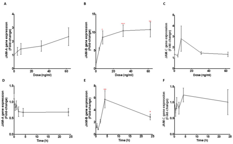
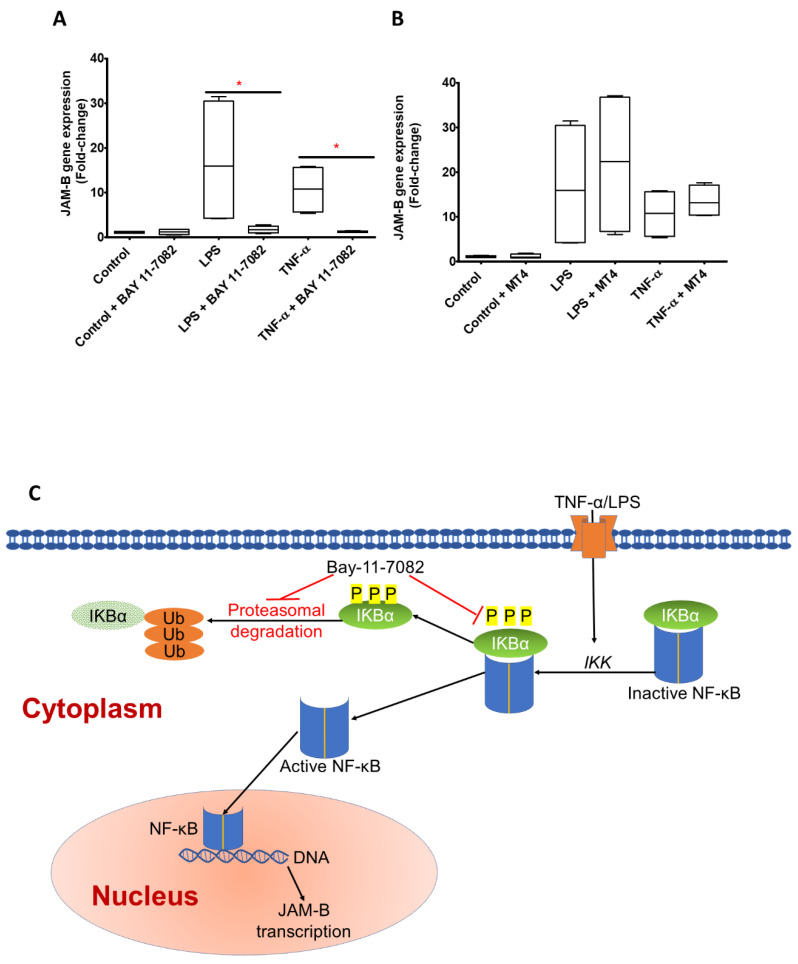
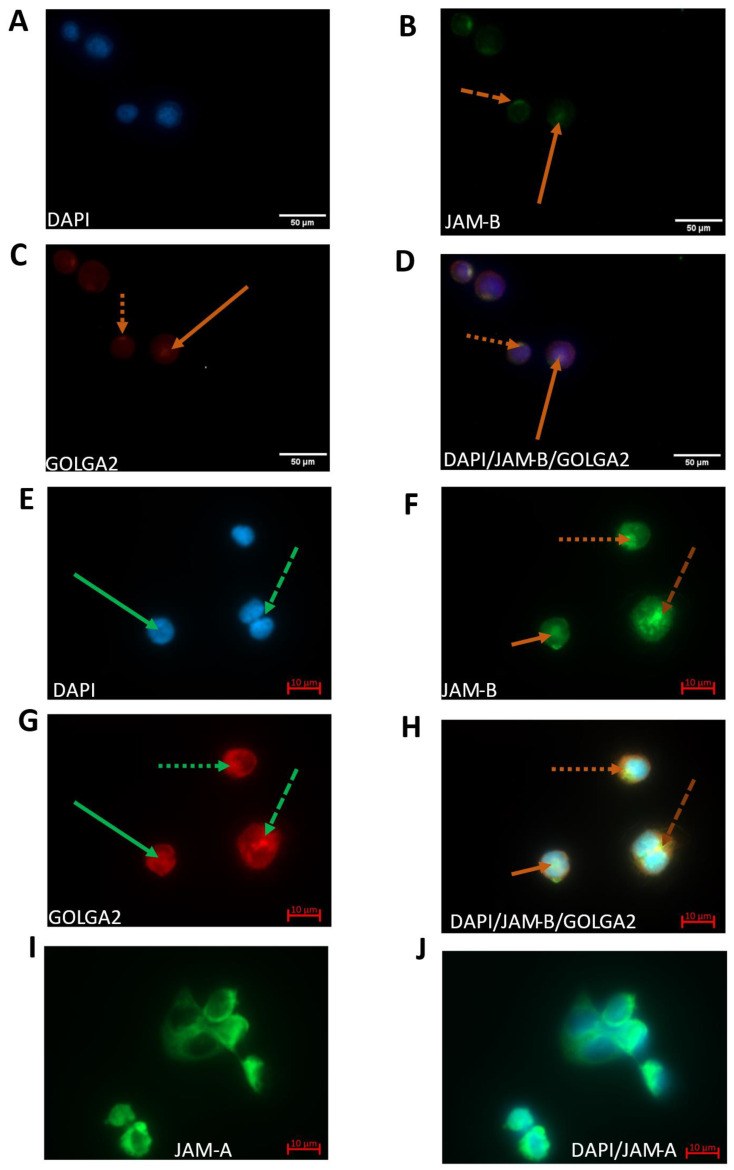
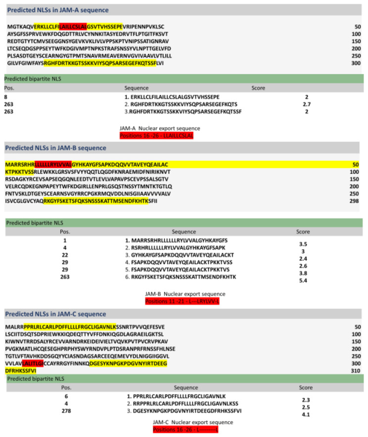
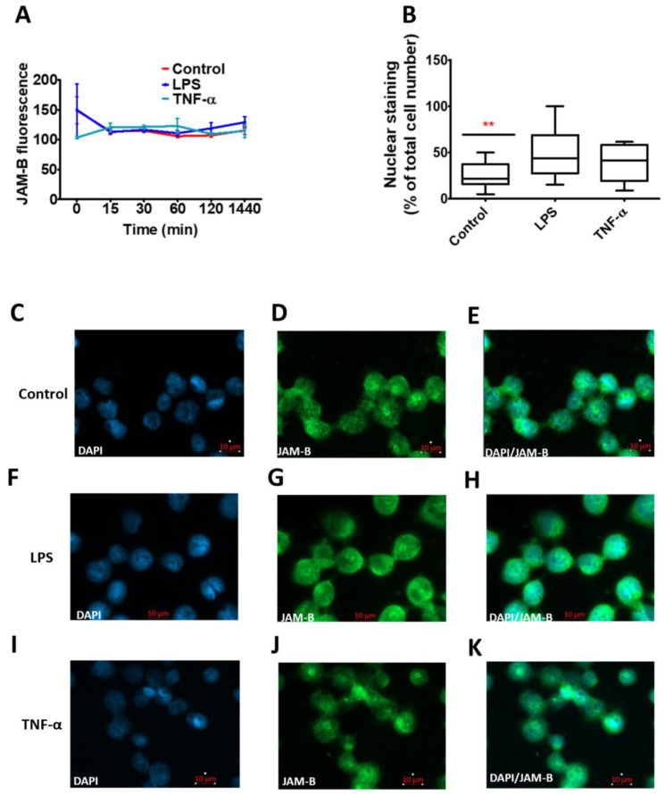
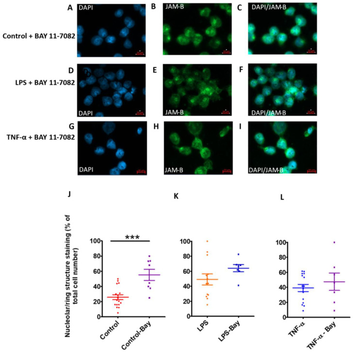
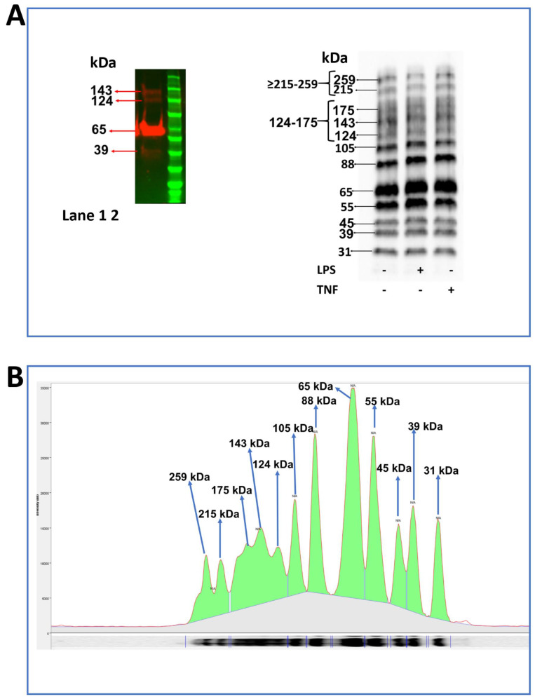
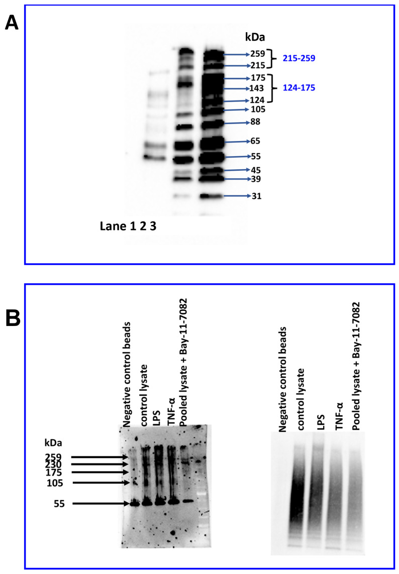
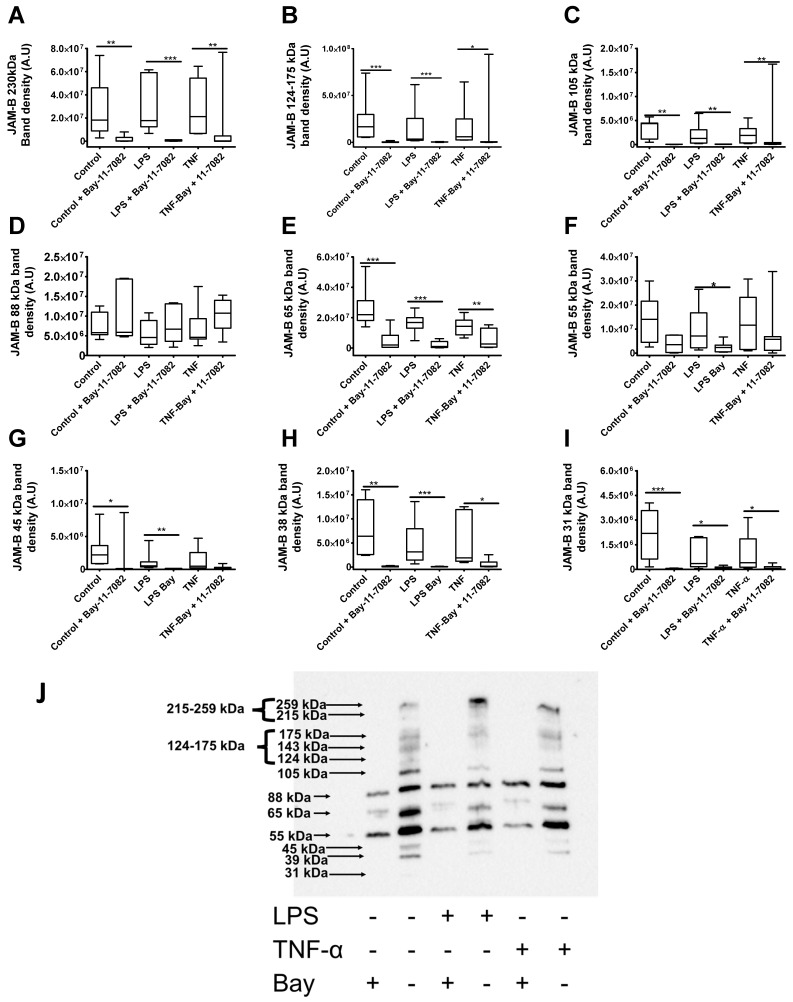
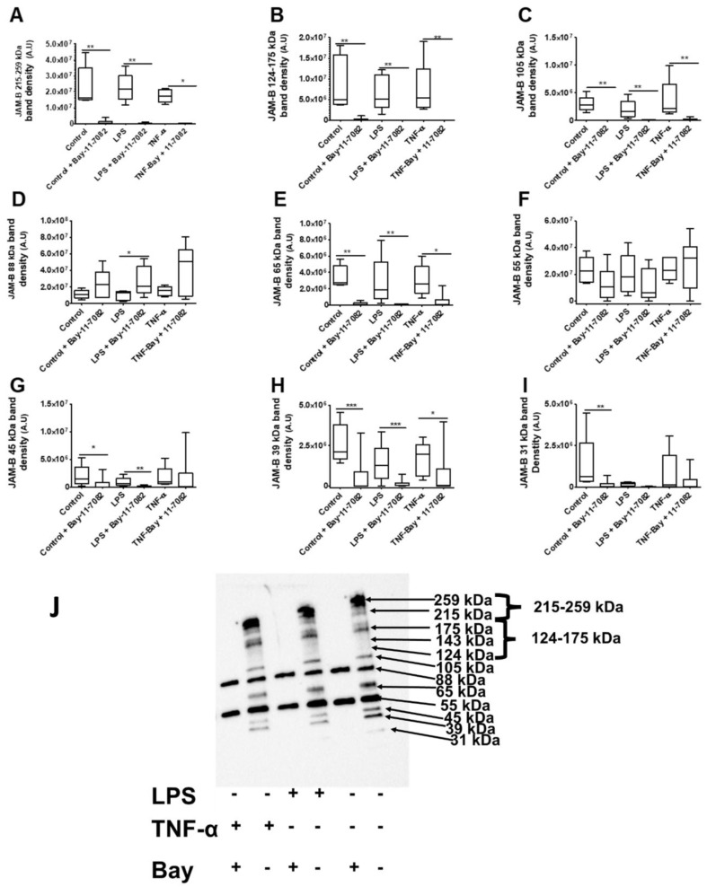
Junctional adhesion molecules (JAMs; comprising JAM-A, -B and -C) act as receptors for viruses, mediate cell permeability, facilitate leukocyte migration during sterile and non-sterile inflammation and are important for the maintenance of epithelial barrier integrity. As such, they are implicated in the development of both communicable and non-communicable chronic diseases. Here, we investigated the expression and regulation of JAM-B in leukocytes under pathogen- and host-derived inflammatory stimuli using immunoassays, qPCR and pharmacological inhibitors of inflammatory signalling pathways. We show that JAM-B is expressed at both the mRNA and protein level in leukocytes. JAM-B protein is localised to the cytoplasm, Golgi apparatus and in the nucleus around ring-shaped structures. We also provide evidence that JAM-B nuclear localisation occurs via the classical importin-α/β pathway, which is likely mediated through JAM-B protein nuclear localisation signals (NLS) and export signals (NES). In addition, we provide evidence that under both pathogen- and host-derived inflammatory stimuli, JAM-B transcription is regulated via the NF-κB-dependent pathways, whereas at the post-translational level JAM-B is regulated by ubiquitin-proteosome pathways. Anaphase-promoting ubiquitin ligase complex (APC/C) and herpes simplex virus-associated ubiquitin-specific protease (HAUSP/USP) were identified as candidates for JAM-B ubiquitination and de-ubiquitination, respectively. The expression and regulation of JAM-B in leukocytes reported here is a novel observation and contrasts with previous reports. The data reported here suggest that JAM-B expression in leukocytes is under the control of common inflammatory pathways.

Keywords: barrier function; cell adhesion; cell migration; cell permeability; host; inflammation; pathogen; tight junctions.


Conflict of interest statement

None of the authors have declared a conflict of interest.

Figures

Figure 1
Figure 1
Relative gene expression of JAMs in (A) THP-1 monocytes, (B) PMA-differentiated THP-1 macrophages, (C) primary human monocytes, (D) differentiated primary macrophages and (E) peripheral blood mononuclear cells (PBMC). * indicates p ≤ 0.05 *** indicates p ≤ 0.0001 in comparison to each other. n = 3 for cell lines and n = 1 donor with 3 replicates for primary cells as such statistics not carried out for primary cells. Data are mean ± SEM.
Figure 2
Figure 2
Fold-changes in JAM gene expression in response to TNF-α and LPS treatments for 24 h relative to the geometric mean of YWHAZ and ACT-β expression. All data are the relative expression of the JAMs treated with TNF-α or LPS compared to the respective non-treated control. (A) JAM-A and JAM-B expression in THP-1-differentiated macrophages. (BD) Relative expression of JAM-A, JAM-B and JAM-C, respectively, in THP-1 monocytes. *** indicates p ≤ 0.0001, ** indicates p ≤ 0.01, * indicates p ≤ 0.05. n = 9 for TNF-α treatments and n = 6 for LPS treatments. Data are mean ± SEM.
Figure 3
Figure 3
Gene expression responses of the JAMs to varying LPS doses relative to the geometric mean of YWHAZ and ACT-β expression (AC) and to 10 ng/mL LPS over a 24 h time course with sampling at 0, 0.5, 1, 2 and 4 h and then at 24 h in THP-1 monocytes (DF). (A,D) JAM-A gene expression, (B,E) JAM-B gene expression and (C,F) JAM-C gene expression. The comparisons in (A) to (C) are between the control without LPS and the different LPS doses while those in (D) to (C) are between expression at time 0 and at various times. *** indicates p ≤ 0.0001, ** indicates p ≤ 0.001, * indicates p ≤ 0.05.
Figure 4
Figure 4
Regulation of JAM-B gene expression relative to the geometric mean of YWHAZ and ACT-β expression. (A) In THP1-1 monocytes, treatment with 10 µM of the NF-κB inhibitor Bay-11-7082 for 24 h did not further reduce JAM-B gene expression in the control samples but reduced JAM-B gene expression in the LPS (10 ng/mL) and TNF-α (10 ng/mL) treated samples (p = 0.029, n = 4). (B) Addition of the p38α/β MAPK inhibitor (MT4) had no effect on JAM-B gene expression in control, LPS or TNF-α incubations. (C) Mechanisms through which the Bay-11-7082 inhibitor prevents TNF-α or LPS induced JAM-B gene expression mediated by NF-κβ. Bay-11-7082 inhibits the phosphorylation of IKβ that is bound to inactive NF-κβ and the formation of poly-ubiquitination chains that aid the degradation of IKβ, i.e., the effect of Bay-11-7082 is to prevent the phosphorylation and ubiquitin-dependent proteasomal degradation of IKβ which remains bound to NF-κB and effectively blocks the NF-κβ nuclear localisation sequence so that NF-κβ does not translocate to the nucleus and become active to induce JAM-B transcription. The observation that treatment with Bay-11-7082 completely abolishes the TNFα- and LPS-induced increases JAM-B gene expression strongly suggests that the TNFα- and LPS-induced increases in JAM-B expression are a consequence of increased phosphorylation and proteasomal degradation of IKβ. * indicates p ≤ 0.05.
Figure 5
Figure 5
Immunofluorescence of DAPI, JAM-B and G130 using a mouse anti-JAM-B monoclonal antibody (SC-293496) and an anti-GM130 polyclonal antibody (PA1-077) in THP-1 monocytes (AD) and macrophages (EJ). (A) DAPI nuclear staining in THP-1 monocytes; (B) JAM-B staining in THP-1 monocytes; (C) G130 Golgi staining in THP-1 monocytes (stains peripheral membrane component of the cis-Golgi stack marker GOLGA2); (D) composite image showing co-localisation of JAM-B and G130; (E) DAPI staining in THP-1-differentiated macrophages; (F) JAM-B staining in THP-1-differentiated macrophages showing polarised staining (dotted line), in splitting nuclei (dashed line), inside the nucleus (solid line); (G) cis-Golgi stack marker GOLGA2; (H) composite image showing DAPI, JAM-B and GOLGA2 staining. At least three technical replicates were carried out. JAM-B staining contrasts with that of JAM-A (I) (JAM-A staining) and (J) (composite JAM-A and DAPI staining).
Figure 6
Figure 6
Nuclear localisation and export signals for the junctional adhesion molecules.
Figure 7
Figure 7
The effects of 10 ng/mL LPS and TNF-α after 24 h incubation on THP-1 macrophage cellular and sub-cellular JAM-B protein expression. (A) Time course assessment of total cellular JAM-B protein expression using immunofluorescence; (B) percentage of JAM-B expression in the punctate loci of the nucleus; (CE) DAPI, JAM-B and composite image in control-treated samples; (FH) DAPI, JAM-B and composite image in LPS-treated samples; (IK) DAPI, JAM-B and composite image in TNF-α-treated samples. ** indicates p ≤ 0.01 in comparison to controls.
Figure 8
Figure 8
The effects of the NF-κB inhibitor Bay-11-7082 (10 µM) after 24 h treatment on JAM-B subcellular localisation in THP-1-differentiated macrophages. (AC) DAPI, JAM-B and composite image in control-treated samples; (DF) DAPI, JAM-B and composite image in LPS-treated samples; (GI) DAPI, JAM-B and composite image in TNF-α-treated samples. (J) JAM-B punctate nuclear localisation quantification showing significant increase in JAM-B expression in the nucleus in the control plus Bay-11-7082-treated samples. (K) JAM-B punctate nuclear localisation quantification showing significant increase in JAM-B expression in the nucleus in the LPS plus Bay-11-7082-treated samples. (L) JAM-B punctate nuclear localisation quantification showing no significant increase in JAM-B expression in the nucleus in the TNF-α plus Bay-11-7082-treated samples. *** indicates p ≤ 0.001.
Figure 9
Figure 9
Western blot probing for JAM-B protein in samples of recombinant JAM-B protein and THP-1 monocyte whole cell lysates. All samples were separated by SDS-PAGE under reducing conditions using DTT. (A) Recombinant protein (left panel, lane 1) run together with protein molecular weight marker (left panel, lane 2) and cell lysates from unstimulated (right panel) and LPS- (right panel) or TNFα- (right panel) stimulated THP-1-cultured cells (right panel); (B) example chromatogram demonstrating band molecular weights and grouping to aid quantification. Representative images of more than three technical replicates.
Figure 10
Figure 10
Immunoblots of JAM-B in lysates of THP-1 monocyte whole cells, nuclear and cytosolic sub-fractions of THP-1 cells with and without treatment with 10 µM Bay-11-7082 inhibitor. (A) Immunoblot of nuclear (lane 1), cytoplasmic (lane 2) and whole cell lysates (lane 3) probed with anti-JAM-B antibodies. (B) Immunoblots of ubiquitin-enriched samples of whole cell lysates prepared by immunoprecipitation probed with anti-JAM-B antibodies (left panel) and anti-ubiquitin antibody (right panel). Negative control beads denote samples obtained following the immunoprecipitation procedure with negative control beads; control lysate was ubiquitin-captured samples from unstimulated/untreated cells, or cells treated with either 10 ng/mL LPS or 10 ng/mL TNF-α are ubiquitin-captured lysates from cells treated with inflammatory stimuli (LPS or TNFα) and in samples pooled from Bay-11-7082 (10 µM)-treated control and LPS- and TNF-α-treated samples.
Figure 11
Figure 11
Effects of the Bay-11-7082 inhibitor on the density of JAM-B protein species in THP-1 monocyte whole cell lysates after incubation with and without inflammatory stimuli (relative to total protein on stain-free gel). (A) ≥230 kDa bands, (B) 124–175 kDa bands, (C) 105 kDa band, (D) 88 kDa band (E) 65 kDa band, (F) 55 kDa band (G) 45 kDa bands, (H) 38 kDa band, (I) 31 kDa band and (J) blot image showing protein band densities in the Bay-11-7082 inhibitor-treated THP-1 whole cell lysates treated with and without inflammatory stimuli. A.U, arbitrary units; * indicates p ≤ 0.05, ** indicates p ≤ 0.01, *** indicates p ≤ 0.001 after Mann–Whitney U test (n = 7). All bands were normalised to the total protein on the stain-free blots.
Figure 12
Figure 12
Effects of the Bay-11-7082 inhibitor on the abundance of JAM-B protein species in THP-1 monocyte cytosolic cell lysates after incubation with and without inflammatory stimuli (relative to total protein on stain-free gel): (A) 215–259 kDa bands, (B) 124–175 kDa bands, (C) 105 kDa band, (D) 88 kDa band, (E) 65 kDa band, (F) 55 kDa band, (G) 45 kDa band, (H) 39 kDa band, (I) 31 kDa band and (J) blot images showing protein band densities in the Bay-11-7082 inhibitor-treated THP-1 cytosolic cell lysates in the presence and absence of inflammatory stimuli. A.U, arbitrary units; * indicates p ≤ 0.05, ** indicates p ≤ 0.01, *** indicates p ≤ 0.001 after Mann–Whitney U test (n = 3).

Similar articles

References

    1. Sugano Y., Takeuchi M., Hirata A., Matsushita H., Kitamura T., Tanaka M., Miyajima A. Junctional adhesion molecule-A, JAM-A, is a novel cell-surface marker for long-term repopulating hematopoietic stem cells. Blood. 2008;111:1167–1172. doi: 10.1182/blood-2007-03-081554. - DOI - PubMed
    1. Li X., Stankovic M., Lee B.P.-L., Aurrand-Lions M., Hahn C.N., Lu Y., Imhof B.A., Vadas M.A., Gamble J.R. JAM-C induces endothelial cell permeability through its association and regulation of β3 integrins. Arterioscler. Thromb. Vasc. Biol. 2009;29:1200–1206. doi: 10.1161/ATVBAHA.109.189217. - DOI - PubMed
    1. Vonlaufen A., Aurrand-Lions M., Pastor C., Lamagna C., Hadengue A., Imhof B., Frossard J.-L. The role of junctional adhesion molecule C (JAM-C) in acute pancreatitis. J. Pathol. 2006;209:540–548. doi: 10.1002/path.2007. - DOI - PubMed
    1. Barton E.S., Forrest J.C., Connolly J.L., Chappell J.D., Liu Y., Schnell F.J., Nusrat A., Parkos C.A., Dermody T.S. Junction adhesion molecule is a receptor for reovirus. Cell. 2001;104:441–451. doi: 10.1016/S0092-8674(01)00231-8. - DOI - PubMed
    1. Williams L., Martin-Padura I., Dejana E., Hogg N., Simmons D. Identification and characterisation of human Junctional Adhesion Molecule (JAM) Mol. Immunol. 1999;36:1175–1188. doi: 10.1016/S0161-5890(99)00122-4. - DOI - PubMed