. 2022 Aug 4;23(15):8680.
doi: 10.3390/ijms23158680.

Structural Evolution of Delta (B.1.617.2) and Omicron (BA.1) Spike Glycoproteins

Affiliations

Structural Evolution of Delta (B.1.617.2) and Omicron (BA.1) Spike Glycoproteins

Ingrid Guarnetti Prandi et al. Int J Mol Sci. .

Abstract

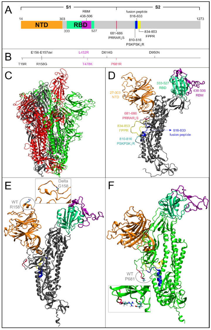
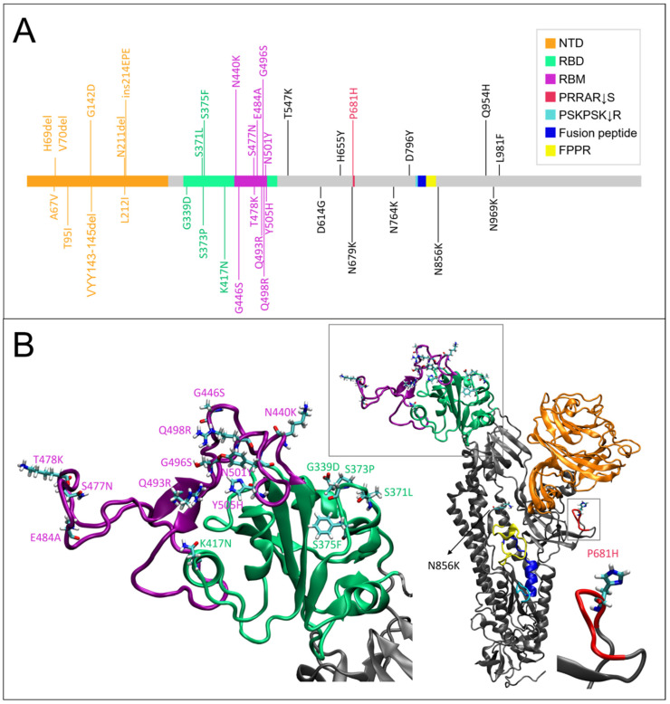
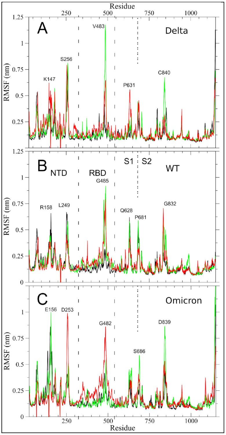
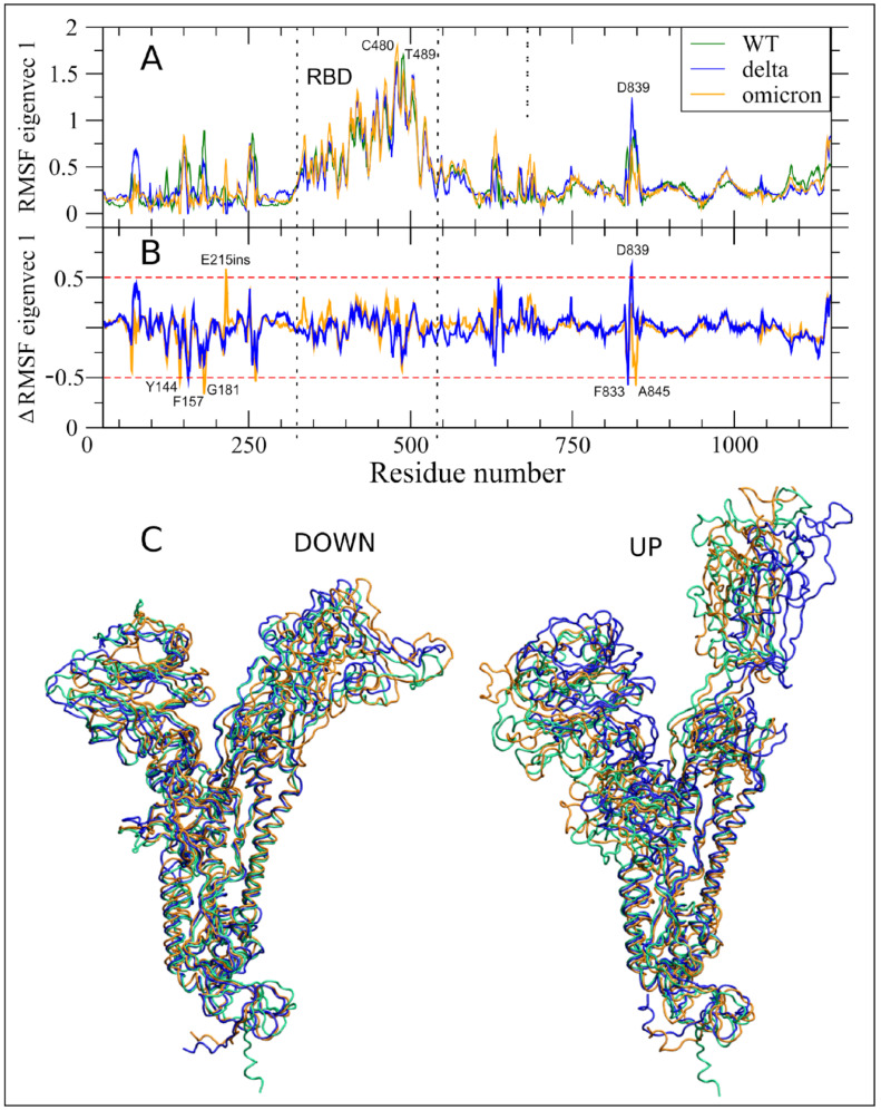
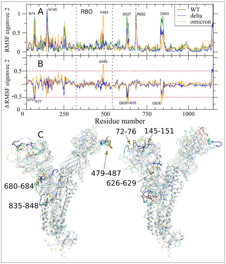
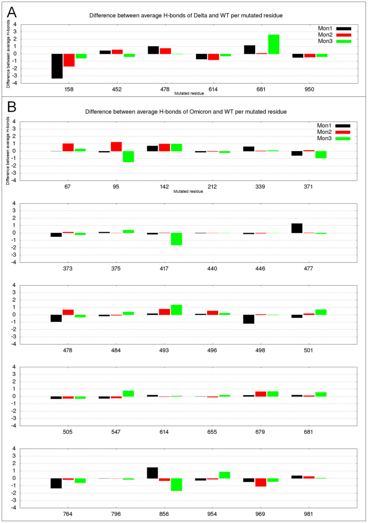
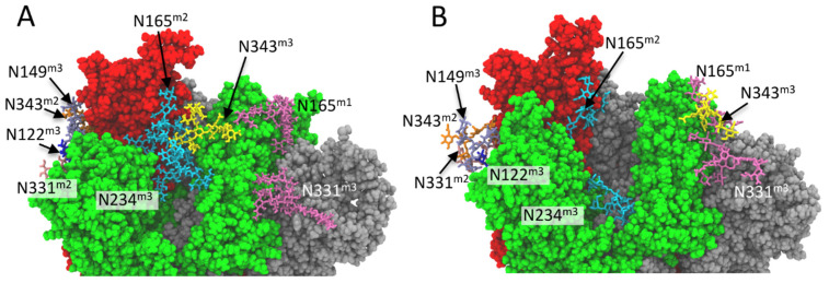
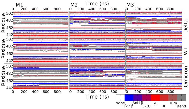
The vast amount of epidemiologic and genomic data that were gathered as a global response to the COVID-19 pandemic that was caused by SARS-CoV-2 offer a unique opportunity to shed light on the structural evolution of coronaviruses and in particular on the spike (S) glycoprotein, which mediates virus entry into the host cell by binding to the human ACE2 receptor. Herein, we carry out an investigation into the dynamic properties of the S glycoprotein, focusing on the much more transmissible Delta and Omicron variants. Notwithstanding the great number of mutations that have accumulated, particularly in the Omicron S glycoprotein, our data clearly showed the conservation of some structural and dynamic elements, such as the global motion of the receptor binding domain (RBD). However, our studies also revealed structural and dynamic alterations that were concentrated in the aa 627-635 region, on a small region of the receptor binding motif (aa 483-485), and the so-called "fusion-peptide proximal region". In particular, these last two S regions are known to be involved in the human receptor ACE2 recognition and membrane fusion. Our structural evidence, therefore, is likely involved in the observed different transmissibility of these S mutants. Finally, we highlighted the role of glycans in the increased RBD flexibility of the monomer in the up conformation of Omicron.

Keywords: COVID-19; SARS-CoV-2; delta; molecular dynamics; omicron; spike; variants.


Conflict of interest statement

The authors declare no conflict of interest.

Figures

Figure 1
Figure 1
Domain organization and structural differences between the WT and Delta variant. (A) Graphical scheme of the spike domains from aa 14 to 1273: N-terminal domain (NTD) is represented in orange; the receptor-binding domain or RBD (aa 333–527) is highlighted in green; the receptor-binding motif, or RBM (aa 438–506), which is within RBD region, is represented in purple; the cleavage sequence at the S1/S2 boundary is colored in red; and the second cleavage site, the fusion peptide and the fusion peptide proximal region (FPPR) are represented in cyan, blue, and yellow, respectively. (B) Representation of Delta mutations, the numbering is aligned according to panel A. (C) Cartoon model visualization of the WT representative frame (629.8 ns of MD simulation) selected by cluster analyses. The monomers are colored in black, red (in up conformation), and green, respectively. (D) WT monomer 1 is represented with the same colors as the schematic domains in panel A. (E) Cartoon representation of WT monomer 1. The ellipse shows the conformation of the WT residue R158 and the rectangle shows the representative frame (655.4 ns) detail of the same region for the Delta variant with G158 mutation. (F) Cartoon representation of WT monomer 3. The ellipse and the rectangle show the conformations of the residue 681 for the WT and the Delta variant, respectively.
Figure 2
Figure 2
Domain organization and mutations of Omicron variant. (A) Graphical scheme of the domains in a spike monomer and Omicron mutations from aa 14 to 1273. (B) Cartoon representation of the representative frame (797 ns) of Omicron monomer (mon1) selected by cluster analyses. The WT numbering was maintained. The boxes detail Omicron mutations, respectively, in the RBD domain (in green) and in the cleavage sequence at the S1/S2 boundary (in red).
Figure 3
Figure 3
SARS-CoV-2 Delta, WT, and Omicron per-residue RMSF are shown in panels (AC), respectively. Spike monomers 1, 2, and 3 are colored in black, red, and green, respectively. Monomer 2 differs from the others by assuming the up conformation (see M&M). The dashed lines indicate the NTD and RBD regions. The dotted line at residue 681 highlights the S1/S2 boundary. The residue numbers of fluctuation peaks are reported as in WT equivalent, not considering the deletion and insertion of mutants.
Figure 4
Figure 4
Essential Dynamics V1 analyses. (A) RMSF of the S protein filtered trajectory along V1 for WT, Delta, and Omicron are reported in dark green, blue, and orange colors, respectively. Residues corresponding to fluctuation peaks along V1 are indicated. (B) Difference between filtered RMSF along V1 between Delta and Omicron as compared to WT are reported in blue and orange colors, respectively. Residues out of the ±0.5 nm threshold (dashed red lines) are indicated. (C) Extreme projections of the monomers in down and up conformations along V1 for WT, Delta and Omicron MD trajectories are in green, blue, and orange colors, respectively.
Figure 5
Figure 5
Long-range correlated motions along Essential Dynamics V2. (A) RMSF of the S protein filtered trajectory along V2 for WT, Delta, and Omicron variants are reported in dark green, blue, and orange colors, respectively. The residues corresponding to fluctuation peaks along V2 are indicated. (B) The difference between the filtered RMSF along V2 between Delta and Omicron as compared to the WT are reported in blue and orange colors, respectively. Residues above the threshold of |±0.5 nm| (dashed red lines) are indicated. (C) Extreme projections of the S protein MD trajectory along V2 for WT, Delta, and Omicron variants are in transparent mode and green, blue, and orange colors, respectively. The six regions showing the long-range correlated movements along this ED eigenvectors are in opaque mode and their residue range is reported.
Figure 6
Figure 6
Altered hydrogen bond number in mutated residues. S monomers 1–3 are colored in black, red, and green colors, respectively. (A) Differences between the average H-bond numbers in Delta and WT; (B) Differences between the average H-bond number in Omicron and WT.
Figure 7
Figure 7
Interactions of N-glycans with RBD domain in up conformation (monomer 2, red color). Monomer colors as in Figure 1C. Enlargement of the region of glycan-glycan and glycan-protein interaction close to RBD of monomer 2 in “up” conformation of the (A) Delta(representative frame at 783.8 ns of MD simulation)and (B) Omicron variant (representative frame at 893.3 ns of MD simulation), respectively. The amino acids are represented in the CPK model and the glycans (without hydrogen atoms) are represented in licorice. For each residue, the monomer to which they belong is reported as superscript.
Figure 8
Figure 8
Secondary structure content as a function of the simulation time in M1, M2, and M3 of Delta, WT, and Omicron in the RBM region.

Similar articles

Cited by

References

    1. Ksiazek T.G., Erdman D., Goldsmith C.S., Zaki S.R., Peret T., Emery S., Tong S., Urbani C., Comer J.A., Lim W., et al. A Novel Coronavirus Associated with Severe Acute Respiratory Syndrome. N. Engl. J. Med. 2003;348:1953–1966. doi: 10.1056/NEJMoa030781. - DOI - PubMed
    1. Zhu N., Zhang D., Wang W., Li X., Yang B., Song J., Zhao X., Huang B., Shi W., Lu R., et al. A Novel Coronavirus from Patients with Pneumonia in China, 2019. N. Engl. J. Med. 2020;382:727–733. doi: 10.1056/NEJMoa2001017. - DOI - PMC - PubMed
    1. Guan W.-J., Ni Z.-Y., Hu Y., Liang W.-H., Ou C.-Q., He J.-X., Liu L., Shan H., Lei C.-L., Hui D.S.C., et al. Clinical Characteristics of Coronavirus Disease 2019 in China. N. Engl. J. Med. 2020;382:1708–1720. doi: 10.1056/NEJMoa2002032. - DOI - PMC - PubMed
    1. World Health Organization. [(accessed on 26 May 2022)]. Available online: https://www.who.int/en/activities/tracking-SARS-CoV-2-variants.
    1. Gorbalenya A.E., Baker S.C., Baric R.S., de Groot R.J., Drosten C., Gulyaeva A.A., Haagmans B.L., Lauber C., Leontovich A.M., Neuman B.W., et al. The Species Severe Acute Respiratory Syndrome-Related Coronavirus: Classifying 2019-NCoV and Naming It SARS-CoV-2. Nat. Microbiol. 2020;5:536–544. doi: 10.1038/s41564-020-0695-z. - DOI - PMC - PubMed

Substances

Supplementary concepts