. 2022 Aug 4;23(15):8688.
doi: 10.3390/ijms23158688.

Roles of Focal Adhesion Kinase PTK2 and Integrin αIIbβ3 Signaling in Collagen- and GPVI-Dependent Thrombus Formation under Shear

Affiliations

Roles of Focal Adhesion Kinase PTK2 and Integrin αIIbβ3 Signaling in Collagen- and GPVI-Dependent Thrombus Formation under Shear

Jingnan Huang et al. Int J Mol Sci. .

Abstract

Glycoprotein (GP)VI and integrin αIIbβ3 are key signaling receptors in collagen-dependent platelet aggregation and in arterial thrombus formation under shear. The multiple downstream signaling pathways are still poorly understood. Here, we focused on disclosing the integrin-dependent roles of focal adhesion kinase (protein tyrosine kinase 2, PTK2), the shear-dependent collagen receptor GPR56 (ADGRG1 gene), and calcium and integrin-binding protein 1 (CIB1). We designed and synthetized peptides that interfered with integrin αIIb binding (pCIB and pCIBm) or mimicked the activation of GPR56 (pGRP). The results show that the combination of pGRP with PTK2 inhibition or of pGRP with pCIB > pCIBm in additive ways suppressed collagen- and GPVI-dependent platelet activation, thrombus buildup, and contraction. Microscopic thrombus formation was assessed by eight parameters (with script descriptions enclosed). The suppressive rather than activating effects of pGRP were confined to blood flow at a high shear rate. Blockage of PTK2 or interference of CIB1 no more than slightly affected thrombus formation at a low shear rate. Peptides did not influence GPVI-induced aggregation and Ca2+ signaling in the absence of shear. Together, these data reveal a shear-dependent signaling axis of PTK2, integrin αIIbβ3, and CIB1 in collagen- and GPVI-dependent thrombus formation, which is modulated by GPR56 and exclusively at high shear. This work thereby supports the role of PTK2 in integrin αIIbβ3 activation and signaling.

Keywords: GPR56; focal adhesion kinase; integrins; platelets; thrombus formation.


Conflict of interest statement

The authors declare no conflict of interest. The funders had no role in the design of the study; in the collection, analyses, or interpretation of data; in the writing of the manuscript, or in the decision to publish the results.

Figures

Figure 1
Figure 1
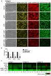
Effect of GPR56 interference on collagen-induced thrombus formation at a high shear rate. Whole blood (700 μL) was pre-incubated with vehicle medium or pGRP peptide (50 μg/mL) for 10 min. After recalcification, blood samples were perfused over microspots of collagen I, III, and IV for 3.5 min at wall-shear rate of 1600 s−1. Brightfield and fluorescence images were taken per microspot at end stage. (A) Shown are representative microscopic images for collagen I of (i) vehicle control runs or (ii) pGRP runs. Scale bar = 10 μm. Complementary images for thrombi on collagen III and collagen IV are shown in Figure S1. (B) Cumulative plots per condition of scaled (0–10) image parameters: P1, platelet adhesion; P2, platelet aggregate coverage; P3–5, thrombus morphology, multilayer and contraction scores; platelet activation markers: P6, PS exposure; P7, P-selectin expression; P8, fibrinogen binding (see Table 1). Shown are means of duplicate runs for three donors. Mean values ± SD (n = 3); n.s., not significant, ** p < 0.005, *** p < 0.001 vs. vehicle (paired Student’s t-test).
Figure 2
Figure 2
Effects of GPR56 and PTK2 interference on collagen-induced platelet aggregation. Washed platelets (250 × 109/L) were incubated with vehicle (control), pGRP peptide (50 μg/mL), and indicated PTK2 inhibitor (2.5–10 μM) for 10 min. Platelet aggregation was monitored by light transmission in response to collagen I (1 μg/mL). (A) Representative traces of collagen-induced aggregation. (B) Dose-dependent effect of PTK2 inhibitors on maximal aggregation. Mean values ± SD (n = 3 donors); n.s., not significant, * p < 0.05, ** p < 0.005, **** p < 0.0001 vs. vehicle (paired Student’s t-test).
Figure 3
Figure 3
Effects of GPR56-binding peptide and PTK2 inhibition on collagen-induced thrombus formation. Whole blood samples were pre-incubated with vehicle medium (control) or indicated PTK2 inhibitor (PF573228 or FAK-IN14 at 2.5–10 μM) with or without pGRP peptide (50 μg/mL) for 10 min. After recalcification, the blood was perfused over collagen I, III, and IV for 3.5 min at a standard shear rate of 1000 s−1. End-stage brightfield and fluorescence images were analyzed for thrombus parameters P1–8. Enlarged images (lower-left corner) are indicated to visualize the formed platelet aggregates. (A) Representative images for collagen I of (i) vehicle control, (ii) PF573228 (10 μM), (iii) FAK-IN14 (10 μM), (iv) pGRP + PF573228 runs, and (v) pGRP + FAK-IN14. Scale bar = 10 μm. Representative images for collagen III and collagen IV are shown in Figure S2. (B) Subtraction heatmap representing control-subtracted scaled (0–10) parameter values for collagen I, III, and IV microspots. The color code represents a decrease (green) or increase (red) in comparison to control runs. Means of duplicate runs for three donors were compared per blood sample. For statistics, see Figure S3.
Figure 4
Figure 4
Shear rate dependency of GPR56 and PTK2 interference. Blood samples pre-incubated with vehicle (control) or PF573228 (5 μM) with/ without pGRP (50 μg/mL) for 10 min and then perfused over collagen microspots for 3.5 or 6 min at a wall-shear rate of 1000 s−1 or 150 s−1. Parameter analysis of recorded images was as for Figure 1. Shown is a subtraction heatmap representing control-subtracted scaled (0–10) parameter values for collagen I, III, and IV microspots. The color code represents a decrease (green) or increase (red) in comparison to vehicle control runs.
Figure 5
Figure 5
Molecular dynamics simulation of the pCIB–CIB1 complex formation. (A) Reported structure of CIB1 in complex with the pCIB peptide, mimicking part of the intracellular αIIb chain. (B) Calculated structure of the pCIB–CIB1 complex obtained by molecular dynamics simulation. (C) Structure of the modified pCIBm-CIB1 complex by molecular dynamics simulation. Color code: hydrogen bonds shown as yellow dashed lines; amino acid residues of the wildtype (B) and mutated (C) peptides are indicated in cyan and magenta, respectively; CIB1 residues are pictured in green; also indicated per peptide is the calculated binding free energy (BFE).
Figure 6
Figure 6
Combined GPR56 and CIB1 peptides affecting collagen-induced thrombus formation. Blood samples were pre-incubated with vehicle medium (control) or indicated peptides pGRP, pCIB, and pCIBm (50 μg/mL each) for 10 min. Thrombus formation on collagen I, III, and IV was monitored. (A) Representative images for collagen I of (i) vehicle control, (ii) pCIBm, (iii) pCIB, and (iv) pGRP + pCIBm, or (v) pGRP + pCIB. Scale bar = 10 μm. (B) Percentual effects of peptides on combined parameters of platelet deposition (P1–2), thrombus characteristics (P3–5), and platelet activation (P6–8) versus the vehicle control condition. Additional images and raw data for collagen III and collagen IV are given in Figures S5 and S6. (C) Subtraction heatmap representing control-subtracted scaled (0–10) parameter values for collagen I, III, and IV microspots. The color code represents a decrease (green) or increase (red) in comparison to controls. Mean values ± SD (n = 3 donors). * p < <0.05, *** p < 0.001, **** p < 0.0001 vs. vehicle (paired Student’s t-test).
Figure 7
Figure 7
Shear rate dependency of GPR56 and CIB1 peptides. Blood samples pre-incubated with vehicle (control) or indicated peptides (50 μg/mL) for 10 min and then perfused over collagen microspots for 3.5 min at 1000 s−1 or for 6 min at 150 s−1. Shown is the subtraction heatmap representing control-subtracted scaled (0–10) image parameter values for collagen I, III, and IV microspots. The color code represents a decrease (green) or increase (red) in comparison to vehicle control runs.
Figure 8
Figure 8
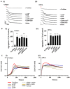
No effect of combined peptides pGRP and pCIB on collagen-induced platelet aggregation or Ca2+ fluxes. (A,B) Platelet preparations (250 × 108/L) were pre-incubated with vehicle control, tirofiban (1 μg/mL) or indicated peptides (50 μg/mL) for 10 min. Platelet aggregation was monitored by light transmission aggregometry in response to 1 μg/mL collagen I or (i) or 1 μg/mL CRP-XL (ii). (A) representative aggregation traces (B) and normalized transmission changes. (C) Fura-2-loaded platelets were pre-incubated with vehicle control, tirofiban (1 μg/mL), or indicated peptides (50 μg/mL) for 10 min, before addition to 96-well plates. After supplementation of 1 mM CaCl2, loaded platelets were automatically stimulated with 10 μg/mL collagen I (i) or 10 μg/mL CRP-XL (ii). Dual wavelength 340/380 nm fluorescence changes per well were recorded in a FlexStation 3. Shown are representative [Ca2+]i traces per agonist. Mean values ± SD (n = 3 donors); n.s., not significant, * p < 0.05, ** p < 0.005, *** p < 0.001, vs. vehicle (paired Student’s t-test).
Figure 9
Figure 9
Scheme of the proposed combined action of GPR56, GPVI, and aIIbb3 signaling in shear-dependent thrombus formation on collagen, consisting of platelet aggregation, secretion, and shape change. (i) GPR56 signaling: small GTP-binding protein RhoA, activated by p115 RhoGEF (guanine nucleotide exchange factor). (ii) GPVI signaling via FcR γ-chain co-receptor: SFK (Src-family kinases) and the tyrosine kinases Syk, Tec, and Btk; phosphatidylinositol 3-kinase (PI3K); leading to activation of PLCγ2, which generates the secondary messengers DAG (diacylglycerol) and IP3 (inositol trisphosphate). (iii) αIIbβ3 outside-in signaling: SFK- and CIB1-mediated activation, the latter triggering PTK2 (focal adhesion kinase FAK); small GTP-binding proteins Rap1b and RhoA transmit parts of the signal. For further explanation, see text.

Similar articles

Cited by

References

    1. Versteeg H.H., Heemskerk J.W., Levi M., Reitsma P.S. New fundamentals in hemostasis. Physiol. Rev. 2013;93:327–358. doi: 10.1152/physrev.00016.2011. - DOI - PubMed
    1. Nagy M., Perrella G., Dalby A., Becerra M., Garcia Quintanilla L., Pike J., Morgan N., Gardiner E., Heemskerk J.W., Azocar L., et al. Flow studies on human GPVI-deficient blood under coagulating and non-coagulating conditions. Blood Adv. 2020;4:2953–2961. doi: 10.1182/bloodadvances.2020001761. - DOI - PMC - PubMed
    1. Huang J., Zhang P., Solari F.A., Sickmann A., Garcia A., Jurk K., Heemskerk J.W. Molecular proteomics and signalling of human platelets in health and disease. Int. J. Mol. Sci. 2021;22:9860. doi: 10.3390/ijms22189860. - DOI - PMC - PubMed
    1. Harbi M.H., Smith C.W., Nicolson P.L., Watson S.P., Thomas M.R. Novel antiplatelet strategies targeting GPVI, CLEC-2 and tyrosine kinases. Platelets. 2021;32:29–41. doi: 10.1080/09537104.2020.1849600. - DOI - PubMed
    1. Sokol S., Skerenova M., Biringer K., Simurda T., Kubisz P., Stasko J. Glycoprotein VI gene variants affect pregnancy loss in patients with platelet hyperaggregability. Clin. Appl. Thromb. Hemost. 2018;24:202S–208S. doi: 10.1177/1076029618802358. - DOI - PMC - PubMed

MeSH terms