. 2022 Aug 5;23(15):8702.
doi: 10.3390/ijms23158702.

c-Kit Induces Migration of Triple-Negative Breast Cancer Cells and Is a Promising Target for Tyrosine Kinase Inhibitor Treatment

Affiliations

c-Kit Induces Migration of Triple-Negative Breast Cancer Cells and Is a Promising Target for Tyrosine Kinase Inhibitor Treatment

José A López-Mejía et al. Int J Mol Sci. .

Abstract

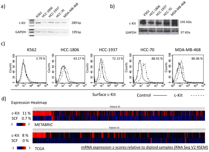
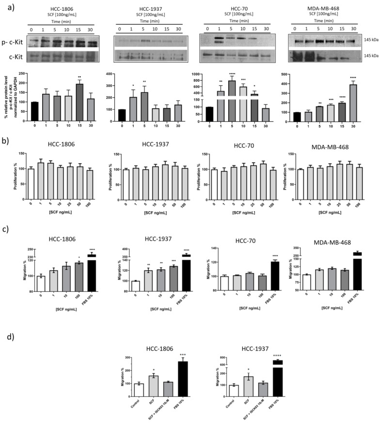
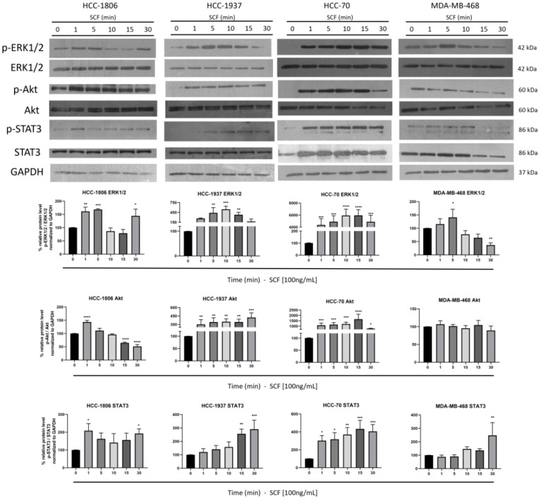
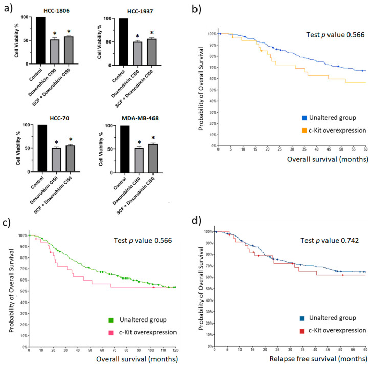
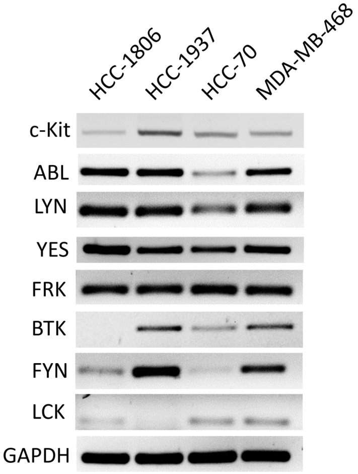
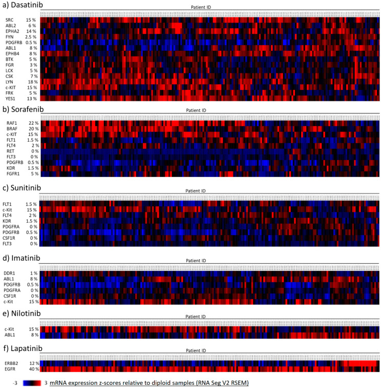
Triple-negative breast cancer (TNBC) is associated with a poor prognosis and the absence of targeted therapy. c-Kit, a receptor tyrosine kinase (RTK), is considered a molecular target for anticancer drugs. Tyrosine kinase inhibitors (TKIs) recognizing c-Kit are used for the treatment of c-Kit-expressing tumors. However, the expression, function, and therapeutic potential of c-Kit have been little explored in TNBC. Here, we studied the expression and effects of c-Kit in TNBC through in vitro and in silico analysis, and evaluated the response to TKIs targeting c-Kit. Analysis of TNBC cells showed the expression of functional c-Kit at the cell membrane. The stimulation of c-Kit with its ligand induced the activation of STAT3, Akt, and ERK1/2, increasing cell migration, but had no effect on cell proliferation or response to Doxorubicin. Analysis of public datasets showed that the expression of c-Kit in tumors was not associated with patient survival. Finally, TNBC cells were susceptible to TKIs, in particular the effect of Nilotinib was stronger than Doxorubicin in all cell lines. In conclusion, TNBC cells express functional c-Kit, which is a targetable molecule, and show a strong response to Nilotinib that may be considered a candidate drug for the treatment of TNBC.

Keywords: c-Kit; nilotinib; receptor tyrosine kinase; stem cell factor; triple-negative breast cancer; tyrosine kinase inhibitor.


Conflict of interest statement

The authors declare no conflict of interest.

Figures

Figure 1
Figure 1
Expression of c-Kit in TNBC cell lines and tumors. The expression of c-Kit was evaluated in HCC-1806, HCC-1937, HCC-70, and MDA-MB-468 TNBC-derived cell lines by (a) RT-PCR, and (b) Western blot; (c) the presence of c-Kit at the cell membrane was evaluated by flow cytometry, the proportion of cells showing surface c-Kit is indicated. K562 cells were included as a c-Kit-expression positive control, and the expression of GAPDH was used as internal control for RT-PCR and Western blot assays. (d) The expression of c-Kit and its ligand SCF was investigated in tumor samples using transcriptomic data from the METABRIC (N = 299 samples), and TCGA (N = 168 samples) datasets. Gene overexpression was considered for samples showing a z-score relative to diploid samples > 2 (EXP > 2). Gene underexpression was considered when the z-score relative to diploid samples was <2 (EXP < 2).
Figure 2
Figure 2
Effect of c-Kit activation on TNBC cell proliferation and migration. (a) Time-course of c-Kit activation by SCF. HCC-1806, HCC-1937, HCC-70, and MDA-MB-468 cells were incubated with 100 ng/mL SCF. Phosphorylation of the receptor was evaluated by Western blotting at the indicated time points. Densitometric analysis indicated as percentage is shown below each blot. (b) For the proliferation assay, the cells were treated with increasing concentrations of SCF. Cell proliferation was evaluated at 48 h using the MTT colorimetric assay. (c) The migration of cells was evaluated using the Boyden chamber assay using 1, 10, and 100 ng/mL SCF, 10% FBS as positive control, or FBS-free medium as negative control that were loaded into the lower chambers. Cell migration was evaluated after 24 h. (d) To inhibit c-Kit phosphorylation, HCC-1806 and HCC-1937 cells were pre-incubated with 10 µM ISCK03. Cell migration was then evaluated by Boyden chamber assays using FBS-free medium (Control), 100 ng/mL SCF (SCF), 100 ng/mL SCF, and the indicated concentration of ISCK03 (SCF + ISCK03 10 µM), or 10% FBS. Values represent the average of three independent experiments. Error bars indicate the standard error of the mean. * p < 0.05, ** p < 0.005, *** p < 0.0005, and **** p < 0.0001 vs. negative control values (Dunnett test).
Figure 3
Figure 3
The effect of c-Kit activation on cell signaling cascades. HCC-1806, HCC-1937, HCC-70, and MDA-MB-468 cells were incubated with 100 ng/mL SCF for the indicated time points. Protein lysates were analyzed by 10% SDS-PAGE. Phosphorylation of clue proteins was detected using specific antibodies to p-ERK1/2, ERK1/2, p-Akt, Akt, p-STAT3, and STAT3. The detection of GAPDH was included as a control. Representative blots from triplicate experiments are shown. Densitometric analysis, indicated as percentage, is shown below the blots. The values represent the average of three independent experiments. Error bars indicate the standard error of the mean. * p < 0.05, ** p < 0.005, *** p < 0.0005, and **** p < 0.0001 vs. values that were obtained at time 0 (Dunnett test).
Figure 4
Figure 4
Effect of c-Kit activation on the cytotoxic activity of Doxorubicin. (a) HCC-1806, HCC-1937, HCC-70, and MDA-MB-468 cells were pre-incubated with 100 ng/mL SCF and then treated with the calculated IC50 of Doxorubicin. Cell viability was measured after 48 h by the colorimetric MTT assay. The values represent the average of three independent experiments. The error bars indicate the standard error of the mean. * p < 0.05 vs. the untreated control values (Dunnett test). (b) The association of tumor c-Kit overexpression with 5-year overall survival of TNBC patients was explored using METABRIC datasets. (c) The association of c-Kit overexpression with 10-years overall survival. (d) The association of c-Kit overexpression with the level of 5-year relapse-free survival. Statistical comparison of samples with c-Kit overexpression and tumors with unaltered expression of c-Kit is presented. Standardized survival data of the METABRIC cohort (unaltered c-Kit expression N = 264; c-Kit overexpression N = 33) were downloaded from the cBioPortal database.
Figure 5
Figure 5
Expression of Receptor Tyrosine Kinases (RTKs) and intracellular tyrosine kinases in TNBC cell lines. Total RNA was isolated from HCC-1806, HCC-1937, HCC-70, and MDA-MB-468 cells. The expression of c-Kit, ABL, LYN, YES, FRK, BTK, FYN, and LCK was evaluated by RT-PCR. The expression of GAPDH was included as a control. Amplified fragments were analyzed by electrophoresis in agarose gels. Representative gels from triplicate experiments are presented.
Figure 6
Figure 6
Expression of TKI-targeted RTKs and intracellular tyrosine kinases in TNBC tumor samples. The expression of the RTKs and intracellular tyrosine kinases that are targeted by (a) Dasatinib, (b) Sorafenib, (c) Sunitinib, (d) Imatinib, (e) Nilotinib, and (f) Lapatinib was investigated using the METABRIC transcriptomic datasets. A cohort of 199 patients was analyzed. Samples with a z-score relative to diploid samples >2 were considered to have overexpression of the specific target molecule. The percentage of patients showing overexpression of each tyrosine kinase is presented.

Similar articles

Cited by

References

    1. Sung H., Ferlay J., Siegel R.L., Laversanne M., Soerjomataram I., Jemal A., Bray F. Global cancer statistics 2020: GLOBOCAN estimates of incidence and mortality worldwide for 36 cancers in 185 countries. CA Cancer J. Clin. 2021;71:209–249. doi: 10.3322/caac.21660. - DOI - PubMed
    1. Thakur K.K., Bordoloi D., Kunnumakkara A.B. Alarming burden of triple-negative breast cancer in India. Clin. Breast Cancer. 2018;18:e393–e399. doi: 10.1016/j.clbc.2017.07.013. - DOI - PubMed
    1. Lehmann B.D., Bauer J.A., Chen X., Sanders M.E., Chakravarthy A.B., Shyr Y., Pietenpol J.A. Identification of human triple-negative breast cancer subtypes and preclinical models for selection of targeted therapies. J. Clin. Investig. 2011;121:2750–2767. doi: 10.1172/JCI45014. - DOI - PMC - PubMed
    1. Lehmann B.D., Jovanovic B., Chen X., Estrada M.V., Johnson K.N., Shyr Y., Moses H.L., Sanders M.E., Pietenpol J.A. Refinement of triple-negative breast cancer molecular subtypes: Implications for neoadyuvant chemotherapy selection. PLoS ONE. 2016;11:e0157368. doi: 10.1371/journal.pone.0157368. - DOI - PMC - PubMed
    1. Burstein M.D., Tsimelzon A., Poage G.M., Covington K.R., Contreras A., Fuqua S.A.W., Savage M.I., Osborne C.K., Hilsenbeck S.G., Chang J.C., et al. Comprehensive genomic analysis identifies novel subtypes and targets of triple-negative breast cancer. Clin. Cancer Res. 2015;21:1688–1698. doi: 10.1158/1078-0432.CCR-14-0432. - DOI - PMC - PubMed

MeSH terms

Substances

LinkOut - more resources