. 2022 Aug 5;23(15):8703.
doi: 10.3390/ijms23158703.

Four Cholesterol-Recognition Motifs in the Pore-Forming and Translocation Domains of Adenylate Cyclase Toxin Are Essential for Invasion of Eukaryotic Cells and Lysis of Erythrocytes

Affiliations

Four Cholesterol-Recognition Motifs in the Pore-Forming and Translocation Domains of Adenylate Cyclase Toxin Are Essential for Invasion of Eukaryotic Cells and Lysis of Erythrocytes

Jone Amuategi et al. Int J Mol Sci. .

Abstract

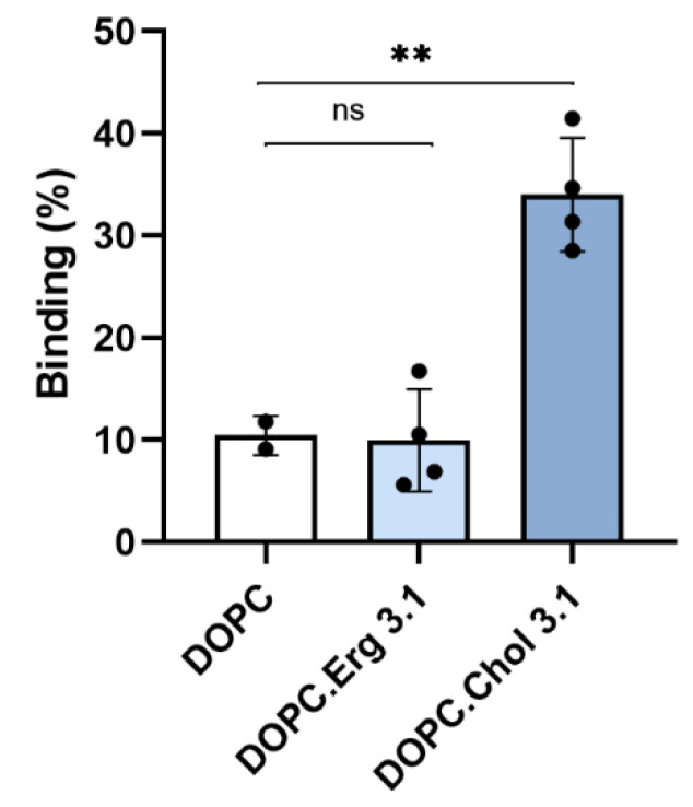
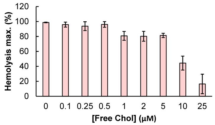
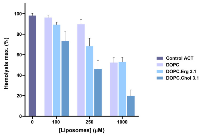
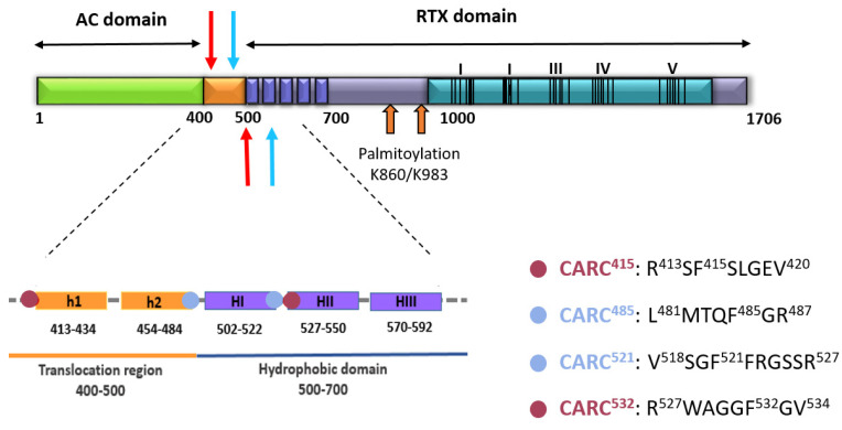
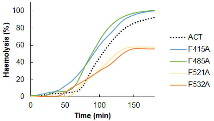
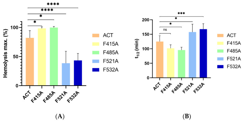
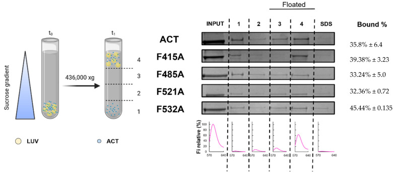
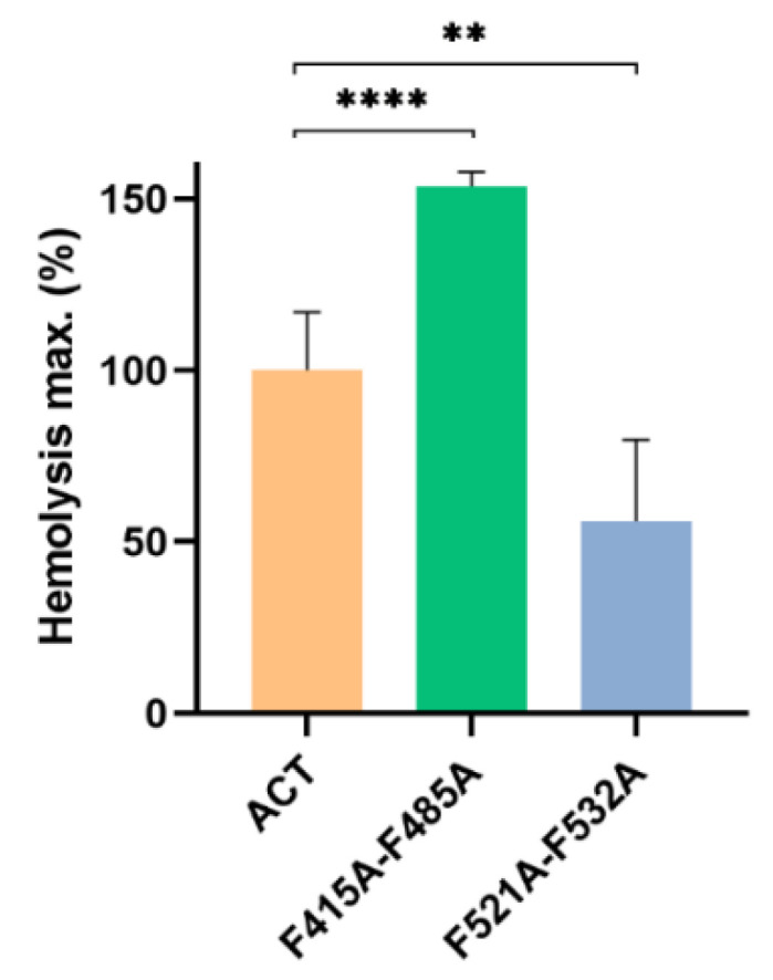
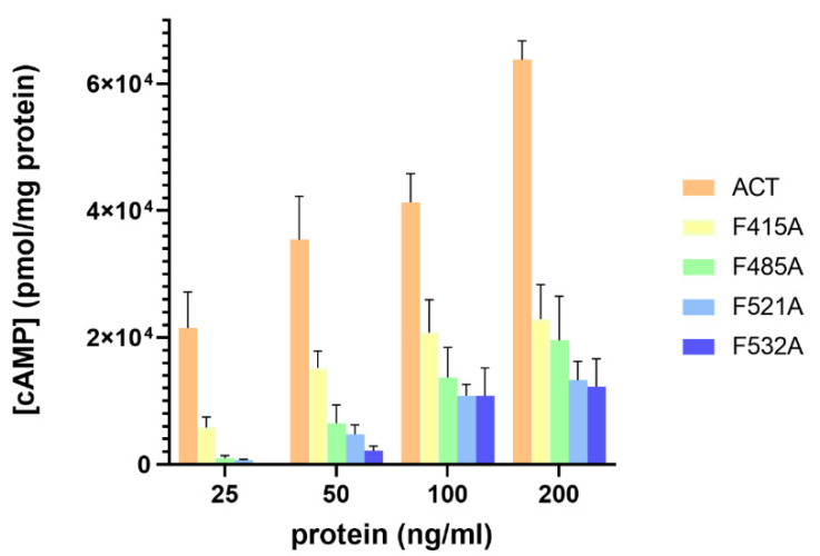
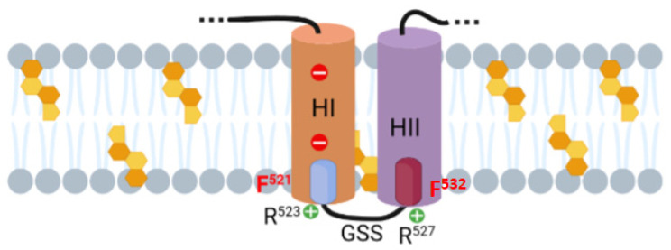
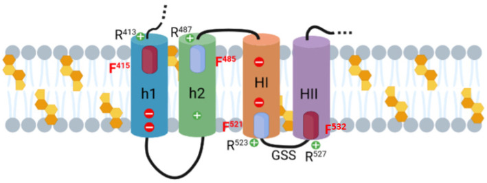
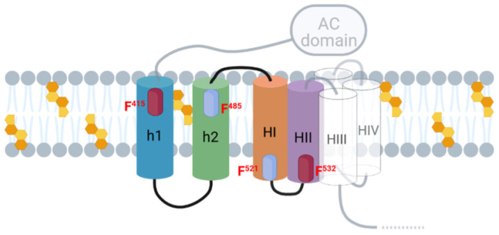
Adenylate Cyclase Toxin (ACT or CyaA) is one of the important virulence factors secreted by Bordetella pertussis, the bacterium causative of whooping cough. ACT debilitates host defenses by production of unregulated levels of cAMP into the cell cytosol upon delivery of its N-terminal domain with adenylate cyclase activity (AC domain) and by forming pores in the plasma membrane of macrophages. Binding of soluble toxin monomers to the plasma membrane of target cells and conversion into membrane-integrated proteins are the first and last step for these toxin activities; however, the molecular determinants in the protein or the target membrane that govern this conversion to an active toxin form are fully unknown. It was previously reported that cytotoxic and cytolytic activities of ACT depend on membrane cholesterol. Here we show that ACT specifically interacts with membrane cholesterol, and find in two membrane-interacting ACT domains, four cholesterol-binding motifs that are essential for AC domain translocation and lytic activities. We hypothesize that direct ACT interaction with membrane cholesterol through those four cholesterol-binding motifs drives insertion and stabilizes the transmembrane topology of several helical elements that ultimately build the ACT structure for AC delivery and pore-formation, thereby explaining the cholesterol-dependence of the ACT activities. The requirement for lipid-mediated stabilization of transmembrane helices appears to be a unifying mechanism to modulate toxicity in pore-forming toxins.

Keywords: RTX toxins; bacterial toxins; lipid-protein interactions; pore-forming toxins.


Conflict of interest statement

The authors declare that they have no conflict of interest with contents of this article. The funding sources had no involvement in the study design nor in the collection, analysis, and interpretation of data nor in the writing of the report or in the decision to submit the article for publication.

Figures

Figure 1
Figure 1
Binding of full length ACT to vesicles of different lipid composition. Membrane association of ACT to lipid bilayers of different lipid composition as measured by flotation assays using large unilamellar vesicles composed of DOPC, DOPC:Chol (1:1 molar ratio) or DOPC:Ergosterol (1:1 molar ratio). Details on the flotation assay methodology are provided in the Experimental Procedures section. The mean and standard deviations of three independent experiments are shown. Statistical differences are based on One-way ANOVA test with Dunnett’s T3 multiple comparisons; [ns] non-significant p ≥ 0.05 and ** p ≤ 0.01.
Figure 2
Figure 2
Effect of ACT preincubation with free cholesterol on ACT-induced haemolytic activity. ACT (100 nM) was preincubated for 30 min at RT in the presence of free cholesterol (0–25 µM). Then sheep erythrocytes at a density of 5 × 10⁸ cells/mL were added and the mixture was further incubated for 180 min at 37 °C. Haemolytic activity was measured as decrease of turbidity at 700 nm and expressed as haemolytic percentages (calculated as detailed in the Experimental Procedures section). Data represented in the figure correspond to the mean of three independent experiments ± SE.
Figure 3
Figure 3
Effect of ACT preincubation with liposomes of different lipid composition on ACT-induced haemolytic activity. ACT (50 nM) was preincubated for 30 min at RT in the presence of liposomes of different lipid compositions (DOPC, DOPC:Chol 3:1 and DOPC:Erg 3:1 molar ratio) at different total lipid concentrations (0–1000 µM). Then sheep erythrocytes at a density of 5 × 10⁸ cells/mL were added and the mixture was further incubated for 180 min at 37 °C. Haemolytic activity was measured as decrease of turbidity at 700 nm and expressed as haemolytic percentages (calculated as detailed in the Experimental Procedures section). Data represented in the figure correspond to the mean of three independent experiments ± SE.
Figure 4
Figure 4
Schematic drawing of the ACT polypeptide chain with the identified four potential cholesterol-recognition motifs. ACT is a 1706-residue-long polypeptide that consists of an N-terminal adenylate cyclase enzyme domain (AC domain, residues 1 to ≈400) (in green) that is fused by an “AC to Hly linker segment” of about 100 residues (in orange) so-called translocation region (residues ≈400–500) to a pore-forming RTX haemolysin (Hly) moiety of approximately 1200 residues (different blue tones). The RTX haemolysin moiety in turn, contains a hydrophobic pore-forming domain comprising residues 500 to 700, constituted by five alpha-helices (dark blue-coloured five cylinders), an acylated domain between residues 800 and 1000, where the posttranslational acylation at two lysine residues (K860 and K983, two orange arrows) occurs, a typical calcium-binding repeats domain (in light blue) organized in five blocks (I to V) and a C-terminal secretion signal (last ≈100 residues). Two predicted α-helices in the translocation region, namely, h1 and h2 (in orange), and three of the five predicted amphipathic/hydrophobic helices of the pore-forming domain, namely HI, HII and HIII (in dark blue) have been depicted below with greater detail. Blue or red spots have been used in the scheme below to specify the location of each one of the four potential cholesterol-recognition motifs identified in this study (CARC415, CRAC485, CRAC521 and CARC532). Sequences of each one of the motifs are specified on the right side of the scheme. The respective N-terminal leucine/valine or arginine, the central phenylalanine (F415, F485, F521 and F532) and the C-terminal arginine or valine residues of the predicted CRAC and CARC motifs are specified. The four motifs are also indicated with blue and red arrows in the schematic drawing of the ACT structure.
Figure 5
Figure 5
Effect of Ala substitutions in the central Phe residues of the potential cholesterol-binding sites CRAC415, CARC485, CRAC521 and CARC532 on the kinetics of the ACT-induced haemolysis. Raw traces of the kinetics recorded from a representative experiment of the haemolysis induced by intact ACT (50 nM) or by each one of the four mutant toxins (50 nM). A suspension of sheep erythrocytes (5 × 10⁸ cells/mL) was incubated with each protein for 180 min at 37 °C, recording the scattering changes measured at 700 nm at every second. Then the haemolysis percentage was calculated as detailed in the Experimental Procedures section and depicted in the figure. The traces shown correspond to a representative experiment from three experiments performed independently.
Figure 6
Figure 6
Effect of Ala substitutions in the central Phe residues of the potential cholesterol-binding sites CRAC415, CARC485, CRAC521 and CARC532 on the (A) maximum haemolytic percentage, and (B) t1/2 of the ACT-induced haemolysis. Haemolysis induced by 50 nM of intact ACT or by each one of the four mutant toxins was assayed with a suspension of sheep erythrocytes (5 × 10⁸ cells/mL) incubated with each protein for 180 min at 37 °C. Data represented in the figure correspond to the mean of three independent experiments ± SE. p-values for the plot in the left subpanel * p  = 0.0444 (ACT/F415A); * p  = 0.0262 (ACT/F485A); **** p < 0.0001; p-values for the plot in the right subpanel * p  = 0.0202 (ACT/F485A); * p  = 0.047 (ACT/F521A); *** p = 0.0008.
Figure 7
Figure 7
Quantification of the binding of ACT or ACT mutants to lipid bilayers. Membrane partitioning as measured by flotation assays using large unilamellar vesicles composed of DOPC:Chol (3:1 molar ratio). Details on the flotation assay methodology are provided in the section of Experimental Procedures. Bound % data correspond to the mean of three independent experiments ± SE.
Figure 8
Figure 8
Effect of double Ala substitutions in the central Phe residues of the potential cholesterol-binding sites F415A-F485A and F521A-F532A on the maximum haemolytic percentage. Haemolysis induced by 50 nM of intact ACT or by each one of the mutant toxins was assayed with a suspension of sheep erythrocytes (5 × 10⁸ cells/mL) incubated with each protein for 180 min at 37 °C. Data represented in the figure correspond to the mean of three independent experiments ± SE. The one-way ANOVA (Brown–Forsythe test) with Dunnett multiple comparisons test was used to determine whether there is a significant difference between the mean values of our independent groups (** if p ≤ 0.01 and **** if p ≤ 0.0001).
Figure 9
Figure 9
Point Ala substitutions in the central Phe residue of the potential cholesterol-binding sites CRAC415, CARC485, CRAC521 and CARC532 prominently decrease AC domain translocation. Translocation of AC domain was assessed by determining the intracellular concentration of cAMP (pmol/mg protein), generated in J774A.1 cells (1 × 10⁵ cells/mL) suspended in 20 mM Tris-HCl, pH = 8.0 buffer, supplemented with 150 mM NaCl and 2 mM CaCl2. Cells were treated for 30 min at 37 °C with different concentrations (25–200 ng/mL) of intact ACT, or the corresponding mutant toxin. Data represent the mean ± SD of at least three independent experiments.
Figure 10
Figure 10
Schematic representation of the proposed membrane topology for HI and HII helices of the pore-forming domain of ACT. Proposed topology for the HI and HII helices as predicted by the algorithm Philius (https://topcons.cbr.su.se/, accessed on 20 February 2021); the two cholesterol recognition motifs identified in this study, CARC532 and CRAC521 motifs, are represented by blue and purple cylinders, respectively, and the central aromatic residue of each site (F521 and F532) is highlighted in red. Cholesterol molecules are represented by orange-coloured penta-hexagonal figures. More details are explained in the Discussion section. Original picture created with BioRender.com.
Figure 11
Figure 11
Schematic model of the membrane topology for the h1-h2-HI-HII helices. The figure shows a scheme of the membrane topology for the h1, h2 (TR region) and HI-HII (pore-forming domain) helices as proposed here. Assuming that HI and HII would adopt Nout→Cin and Cout→Nin topology, respectively (as explained in the text), then, h2 and h1 topology would be Cout→Nin and Nout→Cin, respectively, with the difference that in this case the CARC415 and CRAC485 motifs would be facing the extracellular side, and so h1 and h2 would bind cholesterol in the exofacial, side of the membrane. The four cholesterol-binding sites identified in this study, the CRAC415 and CRAC532 motifs (purple cylinders) and the CARC485 and CARC521 motifs (blue cylinders) and their respective central phenylalanine residues F415, F485, F521 and F532 (in red) are highlighted. Other residues cited in the text, in the Discussion section, have been included. Cholesterol molecules are represented by orange-coloured penta-hexagonal figures. Original picture created with BioRender.com.
Figure 12
Figure 12
Schematic model of the membrane topology for the translocation region and the pore-forming domain. The figure shows a scheme of the putative membrane topology for the h1, h2 (TR region) and HI-HV (pore-forming domain) helices involved in AC translocation and in building the ACT pore structure as proposed here. Original picture created with BioRender.com.

Similar articles

Cited by

References

    1. Parker M.W., Feil S.C. Pore-forming protein toxins: From structure to function. Prog. Biophys. Mol. Biol. 2005;88:91–142. doi: 10.1016/j.pbiomolbio.2004.01.009. - DOI - PubMed
    1. Dal Peraro M., van der Goot F.G. Pore-forming toxins: Ancient, but never really out of fashion. Nat. Rev. Microbiol. 2016;14:77–92. doi: 10.1038/nrmicro.2015.3. - DOI - PubMed
    1. Gilbert R.J.C., Dalla Serra M., Froelich C.J., Wallace M.I., Anderluh G. Membrane pore formation at protein-lipid interfaces. Trends Biochem. Sci. 2014;39:510–516. doi: 10.1016/j.tibs.2014.09.002. - DOI - PubMed
    1. Tilley S.J., Saibil H.R. The mechanism of pore formation by bacterial toxins. Curr. Opin. Struct. Biol. 2006;16:230–236. doi: 10.1016/j.sbi.2006.03.008. - DOI - PubMed
    1. Janmey P.A., Kinnunen P.K. Biophysical properties of lipids and dynamic membranes. Trends Cell Biol. 2006;16:538–546. doi: 10.1016/j.tcb.2006.08.009. - DOI - PubMed

MeSH terms

LinkOut - more resources