. 2022 Aug 5;23(15):8738.
doi: 10.3390/ijms23158738.

Cobalt Ferrite Magnetic Nanoparticles for Tracing Mesenchymal Stem Cells in Tissue: A Preliminary Study

Affiliations

Cobalt Ferrite Magnetic Nanoparticles for Tracing Mesenchymal Stem Cells in Tissue: A Preliminary Study

Željka Večerić-Haler et al. Int J Mol Sci. .

Abstract

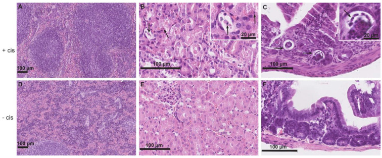
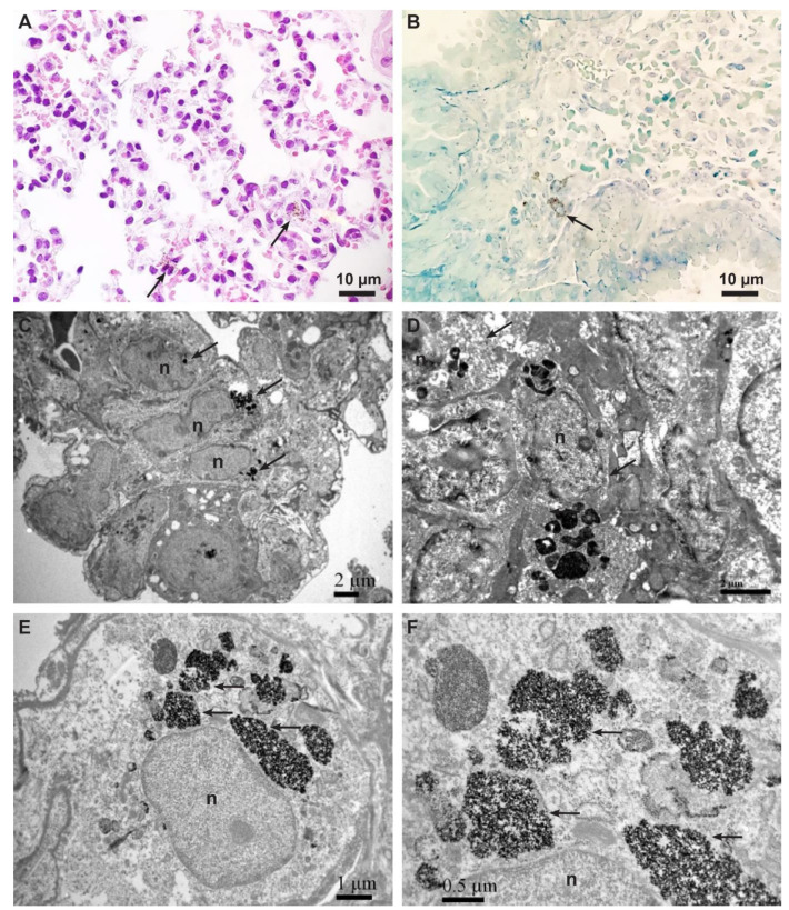
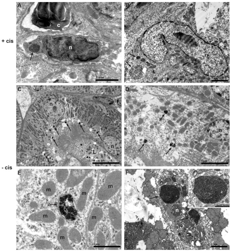
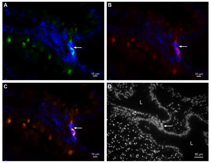
Therapy with mesenchymal stem cells (MSCs) is promising in many diseases. Evaluation of their efficacy depends on adequate follow-up of MSCs after transplantation. Several studies have shown that MSCs can be labeled and subsequently visualized with magnetic nanoparticles (NPs). We investigated the homing of MSCs labeled with magnetic cobalt ferrite NPs in experimentally induced acute kidney injury in mice. To explore the homing of MSCs after systemic infusion into mice, we developed a pre-infusion strategy for optimal tracing and identification of MSCs with polyacrylic acid-coated cobalt ferrite (CoFe2O4) NPs by light and transmission electron microscopy (TEM) in various organs of mice with cisplatin-induced acute kidney injury and control mice. By correlative microscopy, we detected MSCs labeled with NPs in the lungs, spleen, kidney, and intestine of cisplatin-treated mice and in the lungs and spleen of control mice. Our results confirm that labeling MSCs with metal NPs did not affect the ultrastructure of MSCs and their ability to settle in various organs. This study demonstrates the usefulness of cobalt ferrite NPs in ex vivo visualization of MSCs and offers correlative microscopy as a useful method in routine histopathology laboratories for tracing MSCs in paraffin-embedded tissue.

Keywords: markers; mesenchymal stem cells; metal nanoparticles; tissue injury.


Conflict of interest statement

The authors declare no conflict of interest.

Figures

Figure 1
Figure 1
Representative micrographs of histopathologic changes of the spleen, kidney, and intestine in cisplatin-treated mice receiving MSCs labeled with NPs (+cis) (AC) compared to control mice receiving MSCs labeled with NPs without cisplatin treatment (−cis) (DF). (A) In the spleen, cisplatin-induced widening of the red pulp and blurring of the boundaries between white and red pulp is evident. (B) In the renal cortex, numerous apoptotic tubular epithelial cells (arrows) are found. In the upper right corner is an insert with apoptotic epithelial cells at higher magnification. (C) In the intestine, necrotic epithelial cells are present particularly in crypts (arrows). In the upper right corner is an insert with necrotic epithelial cells at higher magnification. (DF) Normal parenchyma of the spleen, kidney, and intestine of control mice receiving MSCs labeled with NPs. Scale bars: 100 µm and 20 µm.
Figure 2
Figure 2
Representative micrographs of MSCs with internalized CoFe2O4. (A) Light micrograph of hematoxylin and eosin staining showing scattered cells containing brown particles inside the cytoplasm (arrows). (B) Micrograph of a semi-thin section stained with Azure II. Note the cell full of brown particles inside the cytoplasm (arrow). (C) TEM micrograph of three MSCs (arrows) containing electron-dense NPs in endosomes in the lungs of control mouse. Scale bar: 2 µm. (D) TEM micrograph of two MSCs (arrows) containing electron-dense NPs in endosomes in the spleen of cisplatin-treated mouse. Scale bar: 2 µm. (E) TEM micrograph of MSC containing electron-dense NPs in numerous endosomes (arrows) in the lungs of cisplatin−treated mouse. Scale bar: 1 µm. (F) Higher magnification of endosomes (from (E)) fully loaded with electron−dense NPs (arrows). Scale bar: 0.5 µm. Legend: n−nucleus.
Figure 3
Figure 3
Representative TEM micrographs of intracellular localization of electron−dense NPs in the kidney and intestine of cisplatin-treated mouse (+cis) (A,B) and in the kidney and liver of control mouse (−cis) (CF). (A) Electron-dense NPs (arrow) in endosome of MSC located in interstitium between peritubular capillary and tubular basement membrane. Scale bar: 2 µm. (B) Three endosomes containing electron−dense NPs (arrows) inside the cytoplasm of MSC in intestine. Scale bar: 2 µm. (C) Several small endosomes with electron-dense NPs (arrows) in apical cytoplasm of proximal tubular epithelial cells. Scale bar: 10 µm. (D) Endosomes with electron-dense NPs (arrows) in basal and apical cytoplasm of proximal tubular epithelial cell. Scale bar: 5 µm. (E) Higher magnification of endosome containing NPs (arrow) in proximal tubular epithelial cell. Scale bar: 1 µm. (F) Electron-dense NPs in endosomes (arrows) of Kupffer cell. Scale bar: 2 µm. Note a high magnification view of large endosome fully loaded with electron-dense NPs in the upper right corner of the figure. Scale bar: 1 µm. Legend: n−nucleus; c−cytoplasm of neutrophilic granulocyte; L−lumen of proximal tubule; m−mitochondria.
Figure 4
Figure 4
Representative images of immunofluorescent labeling against CD44 and CD105 in lungs. Two MSCs with positive immunoreaction against CD44 (green fluorescence) (A) and CD105 (red fluorescence) (B). The same two MSCs are orange−colored due to merged green fluorescence of CD44 and red fluorescence of CD105 confirming the colocalization of both surface markers (C). MSCs were located in the interstitium between alveoli ((D) nuclear staining only). Nuclei are stained with DAPI. Arrow denotes two MSCs. L−lumen of the alveolus.

Similar articles

Cited by

References

    1. Baksh D., Song L., Tuan R.S. Adult mesenchymal stem cells: Characterization, differentiation, and application in cell and gene therapy. J. Cell. Mol. Med. 2004;8:301–316. doi: 10.1111/j.1582-4934.2004.tb00320.x. - DOI - PMC - PubMed
    1. Rodriguez-Fuentes D.E., Fernández-Garza L.E., Samia-Meza J.A., Barrera-Barrera S.A., Caplan A.I., Barrera-Saldaña H.A. Mesenchymal Stem Cells Current Clinical Applications: A Systematic Review. Arch. Med. Res. 2021;52:93–101. doi: 10.1016/j.arcmed.2020.08.006. - DOI - PubMed
    1. Gnecchi M., Danieli P., Malpasso G., Ciuffreda M.C. Paracrine Mechanisms of Mesenchymal Stem Cells in Tissue Repair. Methods Mol. Biol. 2016;1416:123–146. - PubMed
    1. Pittenger M.F., Discher D.E., Péault B.M., Phinney D.G., Hare J.M., Caplan A.I. Mesenchymal stem cell perspective: Cell biology to clinical progress. NPJ Regen. Med. 2019;4:22. doi: 10.1038/s41536-019-0083-6. - DOI - PMC - PubMed
    1. Musial-Wysocka A., Kot M., Majka M. The Pros and Cons of Mesenchymal Stem Cell-Based Therapies. Cell Transpl. 2019;28:801–812. doi: 10.1177/0963689719837897. - DOI - PMC - PubMed

LinkOut - more resources