. 2022 Aug 6;23(15):8746.
doi: 10.3390/ijms23158746.

Efficacy of Nerve-Derived Hydrogels to Promote Axon Regeneration Is Influenced by the Method of Tissue Decellularization

Affiliations

Efficacy of Nerve-Derived Hydrogels to Promote Axon Regeneration Is Influenced by the Method of Tissue Decellularization

Vijay Kumar Kuna et al. Int J Mol Sci. .

Abstract

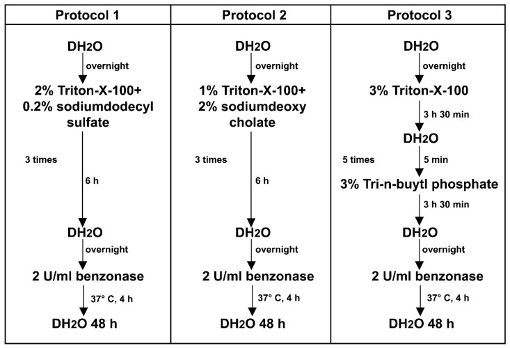
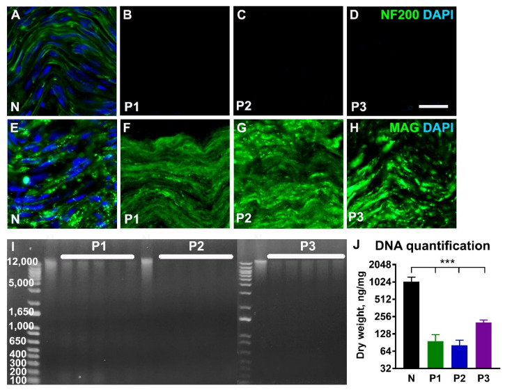
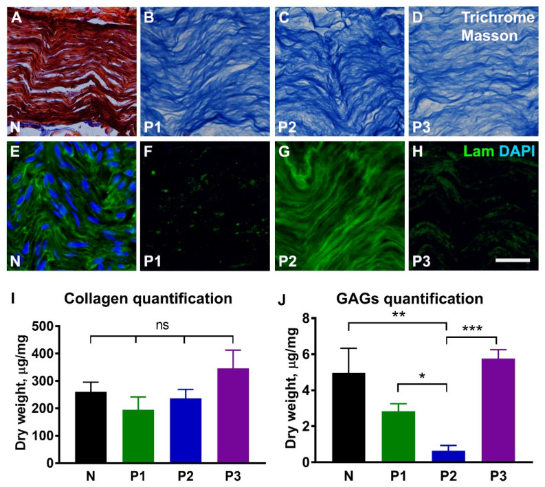
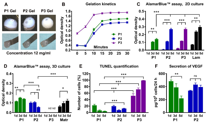
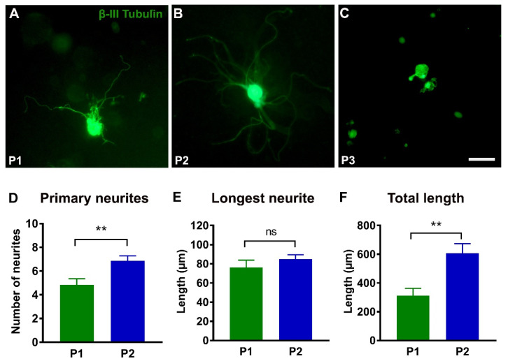
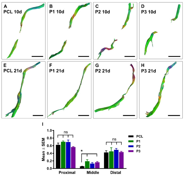
Injuries to large peripheral nerves are often associated with tissue defects and require reconstruction using autologous nerve grafts, which have limited availability and result in donor site morbidity. Peripheral nerve-derived hydrogels could potentially supplement or even replace these grafts. In this study, three decellularization protocols based on the ionic detergents sodium dodecyl sulfate (P1) and sodium deoxycholate (P2), or the organic solvent tri-n-butyl phosphate (P3), were used to prepare hydrogels. All protocols resulted in significantly decreased amounts of genomic DNA, but the P2 hydrogel showed the best preservation of extracellular matrix proteins, cytokines, and chemokines, and reduced levels of sulfated glycosaminoglycans. In vitro P1 and P2 hydrogels supported Schwann cell viability, secretion of VEGF, and neurite outgrowth. Surgical repair of a 10 mm-long rat sciatic nerve gap was performed by implantation of tubular polycaprolactone conduits filled with hydrogels followed by analyses using diffusion tensor imaging and immunostaining for neuronal and glial markers. The results demonstrated that the P2 hydrogel considerably increased the number of axons and the distance of regeneration into the distal nerve stump. In summary, the method used to decellularize nerve tissue affects the efficacy of the resulting hydrogels to support regeneration after nerve injury.

Keywords: MRI; biosynthetic conduit; decellularized nerve tissue; diffusion tensor imaging; nerve-derived hydrogel; peripheral nerve injury.


Conflict of interest statement

The authors declare no conflict of interest. The funders had no role in the design of the study; in the collection, analyses, or interpretation of data; in the writing of the manuscript; or in the decision to publish the results.

Figures

Figure 1
Figure 1
Decellularization protocols. Schematic diagram showing different steps of the nerve tissue processing, using protocols P1, P2, and P3.
Figure 2
Figure 2
Characterization of the decellularized nerve tissue. Representative longitudinal sections from normal nerve (N) and decellularized nerves following treatment with protocols P1, P2, and P3 and immunostained for axonal neurofilaments (NF 200, (AD)) and myelin-associated glycoprotein (MAG, (EH)). Cell nuclei are counterstained with DAPI. The agarose gel electrophoresis in ((I), n = 5) and histogram in ((J), n = 5) shows significant reduction of the genomic DNA following treatments. Error bars show the S.E.M. p < 0.001 is indicated by ***. Scale bar, 25 µm.
Figure 3
Figure 3
Extracellular matrix in the decellularized nerve tissue. Representative longitudinal sections from normal nerve (N) and decellularized nerves following treatment with protocols P1, P2, and P3, and stained for collagen (AD) and laminin (EH). The red colour in (A) corresponds to the cell cytoplasm in normal nerve. Histograms in (I,J) show quantitative analysis of insoluble fibrillary collagen (n = 5) and sulfated glycosaminoglycans (GAGs, n = 5). Note strong laminin immunostaining following treatment with P2 protocol. Error bars show the S.E.M. p-values are indicated as follows: * p ≤ 0.05, ** p ≤ 0.01, and *** p ≤ 0.001; ns, not significantly different. Scale bar, 25 µm.
Figure 4
Figure 4
Characterization of the nerve-derived hydrogels. Images and histogram in (A,B) show appearance and handling of the 12 mg/mL P1, P2, and P3 hydrogels after solidification, and their gelation kinetics (n = 5). Histograms in (C,D) demonstrate Schwann cell proliferation and viability at 1, 3, and 5 days in the wells coated with P1, P2, or P3 hydrogels (2D culture, n = 6), or following suspension of the cells into hydrogels (3D culture, n = 3). Laminin (Lam in C) and Matrigel (Matr in D) are used as control substrates. Histograms in (E,F) show the proportion of TUNEL-positive Schwann cells in hydrogels (n = 3) and production of growth factor VEGF (n = 3) in 3D culture. Error bars show the S.E.M., nd, not detected. p-values are indicated as follows: * p ≤ 0.05, ** p ≤ 0.01 and *** p ≤ 0.001; ns, not significantly different. Scale bar, 25 µm.
Figure 5
Figure 5
Neurite outgrowth from dorsal root ganglion (DRG) neurons. (AC): Representative images of the DRG neurons stained for βIII tubulin after 76 h in the P1, P2, and P3 hydrogels. Note that neurons in P3 hydrogel have no neurite outgrowth. (DF): Histograms showing the number of primary neurites (P1, n = 26 and P2, n = 48), the length of the longest neurite (P1, n = 26 and P2, n = 48), and the total length (n = 10) of all neurites in P1 and P2 hydrogels. Error bars show the S.E.M. p < 0.01 is indicated by **. ns, not significantly different. Scale bar, 50 µm.
Figure 6
Figure 6
Diffusion tensor imaging of regenerating axons in the conduits. Representative axial DTI images at 10 days (AD) and 21 days (EH) after sciatic nerve injury and repair with empty PCL conduit or PCL conduit filled with P1, P2, or P3 hydrogel. Histogram in (I) shows quantification of the fractional anisotropy measured in proximal, middle, and distal ROIs at 21 days after nerve injury and repair (PCL, n = 5, P1, n = 5, P2, n = 7, and P3, n = 7). p < 0.05 is indicated by * (PCL versus P1 and P3 hydrogels); ns, not significantly different. Scale bar, 5 mm.
Figure 7
Figure 7
The effects of hydrogels implantation on regeneration into the distal nerve stump. Representative longitudinal sections demonstrate neurofilament-labelled axons alone (NF in A,D,G,J) or in combination with S100-stained Schwann cells (S100 in B,E,H,K)) in the empty PCL conduit (PCL) and in PCL conduits filled with hydrogels (P1, P2, and P3) at 3 weeks following nerve injury and repair. Boxed areas in (A,D,G,J) are enlarged in (C,F,I,L). Note that regenerating axons reached the distal nerve stump in all experimental groups. Histograms in (M,N) show the number of regeneration axons and the length of the longest axon in the distal nerve stump (PCL, n = 5, P1, n = 5, P2, n = 7, and P3, n = 7). Error bars represent the S.E.M. p-values are indicated as follows: * p ≤ 0.05, ** p ≤ 0.01, and *** p ≤ 0.001. Scale bars: 750 µm in the images with conduit reconstructions and 100 µm in the insertions from boxed areas.

Similar articles

Cited by

References

    1. Palispis W.A., Gupta R. Surgical repair in humans after traumatic nerve injury provides limited functional neural regeneration in adults. Exp. Neurol. 2017;290:106–114. doi: 10.1016/j.expneurol.2017.01.009. - DOI - PubMed
    1. Dahlin L.B., Wiberg M. Nerve injuries of the upper extremity and hand. EFORT Open Rev. 2017;2:158–170. doi: 10.1302/2058-5241.2.160071. - DOI - PMC - PubMed
    1. Kouyoumdjian J.A. Peripheral nerve injuries: A retrospective survey of 456 cases. Muscle Nerve. 2006;34:785–788. doi: 10.1002/mus.20624. - DOI - PubMed
    1. Wang M.L., Rivlin M., Graham J.G., Beredjiklian P.K. Peripheral nerve injury, scarring, and recovery. Connect. Tissue Res. 2019;60:3–9. doi: 10.1080/03008207.2018.1489381. - DOI - PubMed
    1. Ruijs A.C., Jaquet J.B., Kalmijn S., Giele H., Hovius S.E. Median and ulnar nerve injuries: A meta-analysis of predictors of motor and sensory recovery after modern microsurgical nerve repair. Plast. Reconstr. Surg. 2005;116:484–494. doi: 10.1097/01.prs.0000172896.86594.07. - DOI - PubMed

Grants and funding

LinkOut - more resources