

PEBP4 Directs the Malignant Behavior of Hepatocellular Carcinoma Cells via Regulating mTORC1 and mTORC2
- PMID: 35955931
- PMCID: PMC9369291
- DOI: 10.3390/ijms23158798
PEBP4 Directs the Malignant Behavior of Hepatocellular Carcinoma Cells via Regulating mTORC1 and mTORC2
Abstract
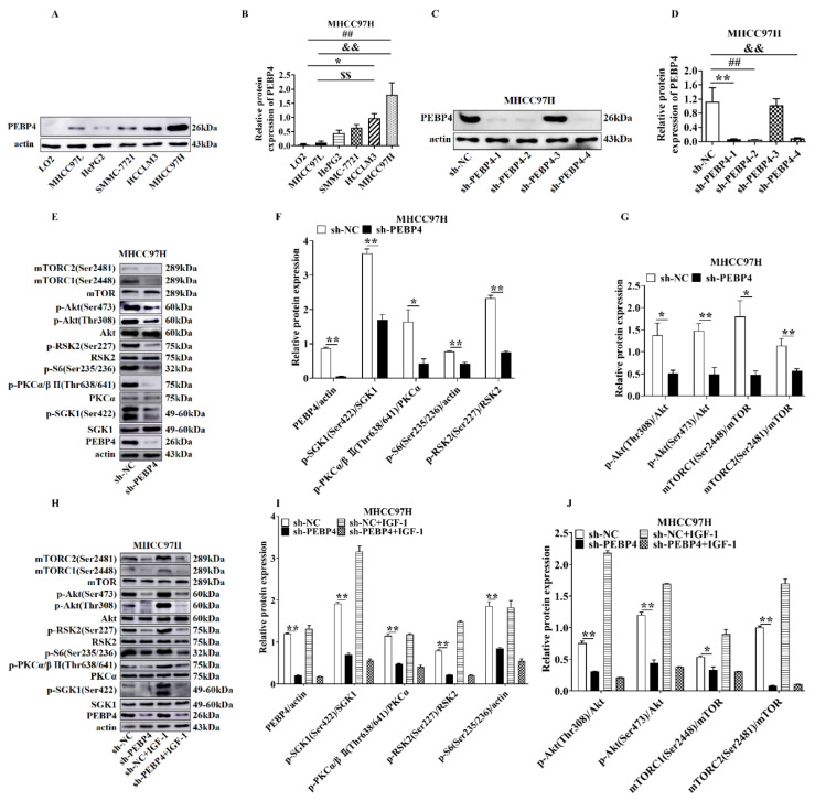
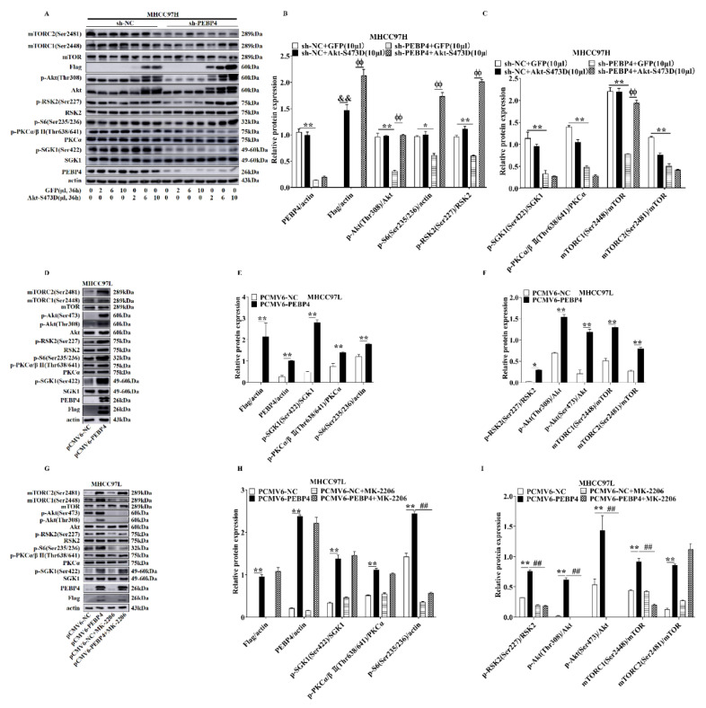
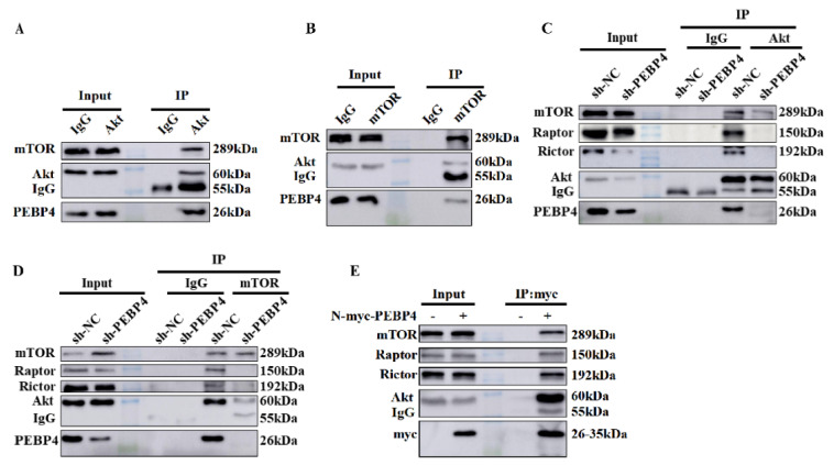
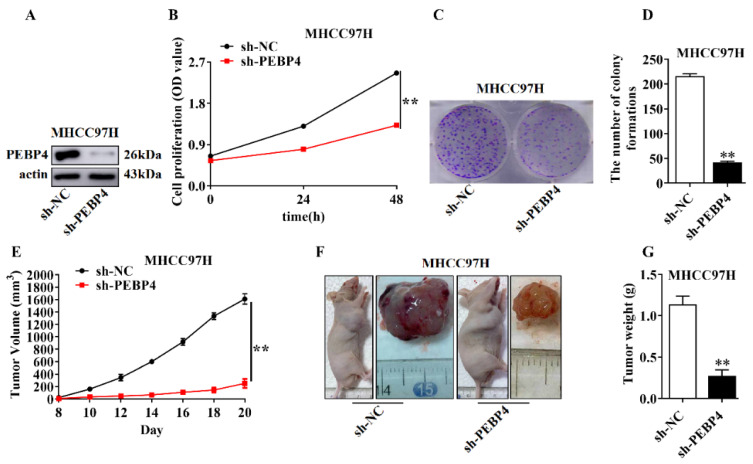
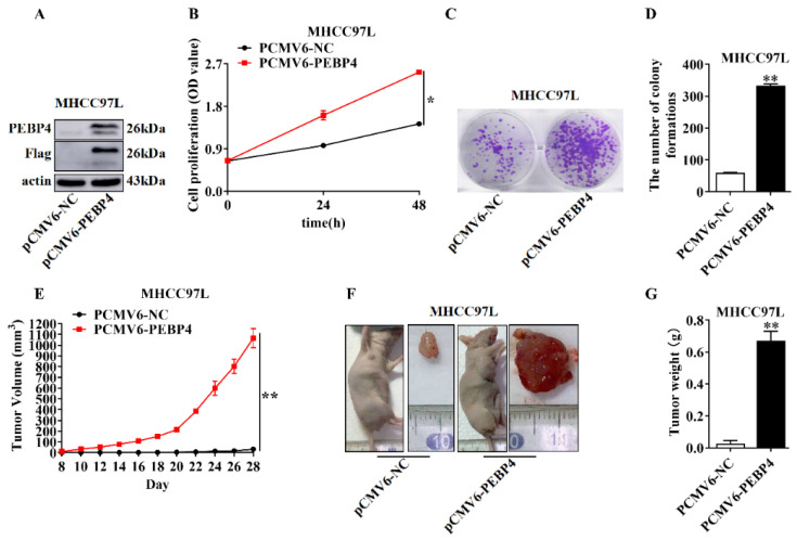
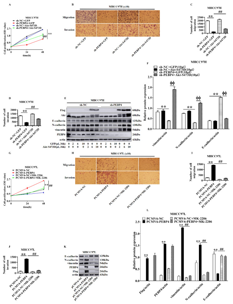
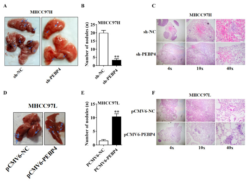
Phosphatidylethanolamine binding protein 4 (PEBP4) is an understudied multifunctional small protein. Previous studies have shown that the expression of PEBP4 is increased in many cancer specimens, which correlates to cancer progression. The present study explored the mechanism by which PEBP4 regulates the growth and progression of hepatocellular carcinoma cells. Thus, we showed that knockdown of PEBP4 in MHCC97H cells, where its expression was relatively high, diminished activities of serine/threonine protein kinase B (PKB, also known as Akt), mammalian target of rapamycin complex 1(mTORC1), and mTORC2, events that were not restored by insulin-like growth factor 1 (IGF-1). Conversely, overexpression of PEBP4 in MHCC97L cells with the low endogenous level yielded opposite effects. Furthermore, physical association of PEBP4 with Akt, mTORC1, and mTORC2 was observed. Interestingly, introduction of AktS473D mutant, bypassing phosphorylation by mTORC2, rescued mTORC1 activity, but without effects on mTORC2 signaling. In contrast, the effect of PEBP4 overexpression on the activity of mTORC1 but not that of mTORC2 was suppressed by MK2206, a specific inhibitor of Akt. In conjunction, PEBP4 knockdown-engendered reduction of cell proliferation, migration and invasion was partially rescued by Akt S473D while increases in these parameters induced by overexpression of PEBP4 were completely abolished by MK2206, although the expression of epithelial mesenchymal transition (EMT) markers appeared to be fully regulated by the active mutant of Akt. Finally, knockdown of PEBP4 diminished the growth of tumor and metastasis, whereas they were enhanced by overexpression of PEBP4. Altogether, our study suggests that increased expression of PEBP4 exacerbates malignant behaviors of hepatocellular cancer cells through cooperative participation of mTORC1 and mTORC2.
Keywords: Akt; HCC; PEBP4; mTORC1; mTORC2.
Conflict of interest statement
The work described has not been submitted elsewhere for publication, and all the authors listed have approved the manuscript. There are no conflicts of interest associated with this work.
Figures
Similar articles
-
TSC/mTORC1 mediates mTORC2/AKT1 signaling in c-MYC-induced murine hepatocarcinogenesis via centromere protein M.J Clin Invest. 2024 Sep 26;134(22):e174415. doi: 10.1172/JCI174415. J Clin Invest. 2024. PMID: 39325536 Free PMC article.
-
Inhibition of mTORC2 Induces Cell-Cycle Arrest and Enhances the Cytotoxicity of Doxorubicin by Suppressing MDR1 Expression in HCC Cells.Mol Cancer Ther. 2015 Aug;14(8):1805-15. doi: 10.1158/1535-7163.MCT-15-0029. Epub 2015 May 29. Mol Cancer Ther. 2015. PMID: 26026051 Free PMC article.
-
FilGAP regulates tumor growth in Glioma through the regulation of mTORC1 and mTORC2.Sci Rep. 2023 Dec 8;13(1):20956. doi: 10.1038/s41598-023-47892-1. Sci Rep. 2023. PMID: 38065968 Free PMC article.
-
Disentangling the signaling pathways of mTOR complexes, mTORC1 and mTORC2, as a therapeutic target in glioblastoma.Adv Biol Regul. 2022 Jan;83:100854. doi: 10.1016/j.jbior.2021.100854. Epub 2021 Dec 6. Adv Biol Regul. 2022. PMID: 34996736 Review.
-
Diverse signaling mechanisms of mTOR complexes: mTORC1 and mTORC2 in forming a formidable relationship.Adv Biol Regul. 2019 May;72:51-62. doi: 10.1016/j.jbior.2019.03.003. Epub 2019 Apr 11. Adv Biol Regul. 2019. PMID: 31010692 Review.
Cited by
-
Metformin Suppresses Stemness of Non-Small-Cell Lung Cancer Induced by Paclitaxel through FOXO3a.Int J Mol Sci. 2023 Nov 22;24(23):16611. doi: 10.3390/ijms242316611. Int J Mol Sci. 2023. PMID: 38068934 Free PMC article.
-
PEBP4 deficiency aggravates LPS-induced acute lung injury and alveolar fluid clearance impairment via modulating PI3K/AKT signaling pathway.Cell Mol Life Sci. 2024 Mar 13;81(1):133. doi: 10.1007/s00018-024-05168-5. Cell Mol Life Sci. 2024. PMID: 38472560 Free PMC article.
-
Deep learning classification of uveal melanoma based on histopathological images and identification of a novel indicator for prognosis of patients.Biol Proced Online. 2023 Jun 2;25(1):15. doi: 10.1186/s12575-023-00207-0. Biol Proced Online. 2023. PMID: 37268878 Free PMC article.
-
Advances in Molecular Research of Oncogenes.Int J Mol Sci. 2023 Apr 13;24(8):7222. doi: 10.3390/ijms24087222. Int J Mol Sci. 2023. PMID: 37108381 Free PMC article.
References
-
- Baka R., Eckersall D., Horvatic A., Gelemanovic A., Mrljak V., McLaughlin M., Athanasiou L.V., Papaioannou N., Stylianaki I., Hanh H.Q., et al. Quantitative proteomics of cerebrospinal fluid using tandem mass tags in dogs with recurrent epileptic seizures. J. Proteom. 2021;231:103997. doi: 10.1016/j.jprot.2020.103997. - DOI - PubMed
MeSH terms
Substances
Grants and funding
LinkOut - more resources
Full Text Sources
Medical
Miscellaneous
