

Facile Microwave Assisted Synthesis of Silver Nanostars for Ultrasensitive Detection of Biological Analytes by SERS
- PMID: 35955966
- PMCID: PMC9369225
- DOI: 10.3390/ijms23158830
Facile Microwave Assisted Synthesis of Silver Nanostars for Ultrasensitive Detection of Biological Analytes by SERS
Abstract
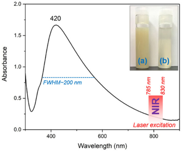
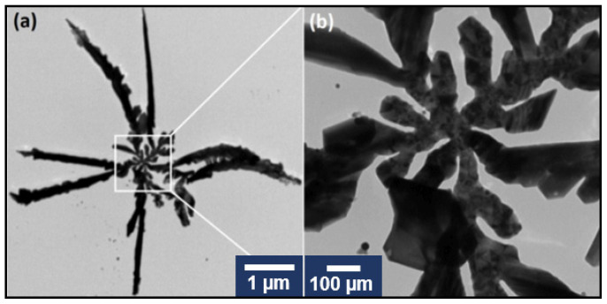
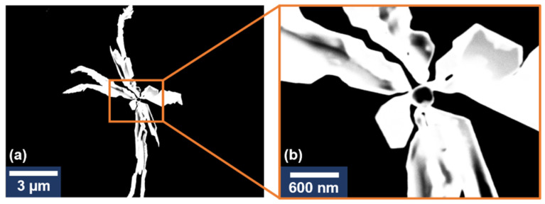
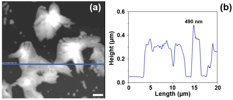
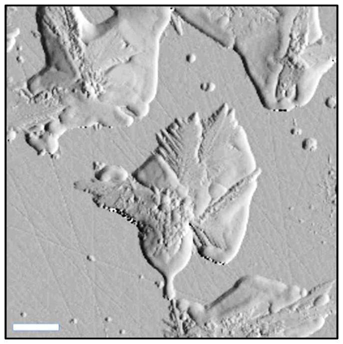
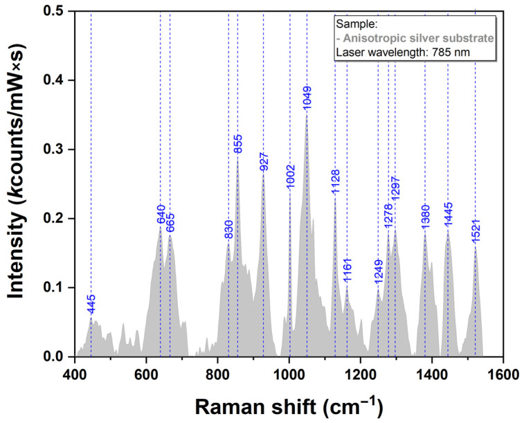
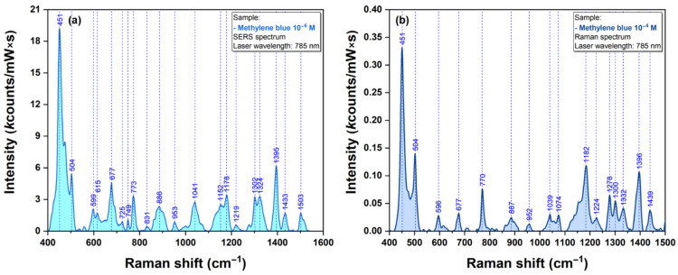
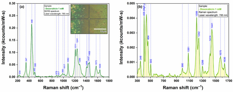
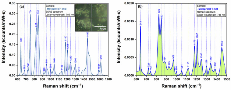
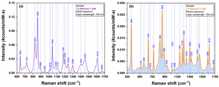
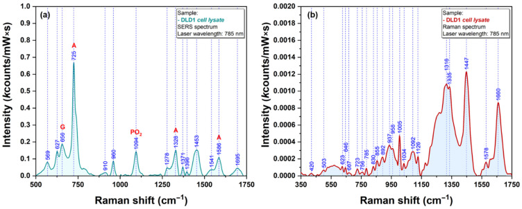
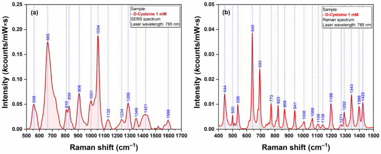
We report a very simple, rapid and reproducible method for the fabrication of anisotropic silver nanostars (AgNS) that can be successfully used as highly efficient SERS substrates for different bioanalytes, even in the case of a near-infra-red (NIR) excitation laser. The nanostars have been synthesized using the chemical reduction of Ag+ ions by trisodium citrate. This is the first research reporting the synthesis of AgNS using only trisodium citrate as a reducing and stabilizing agent. The key elements of this original synthesis procedure are rapid hydrothermal synthesis of silver nanostars followed by a cooling down procedure by immersion in a water bath. The synthesis was performed in a sealed bottom flask homogenously heated and brought to a boil in a microwave oven. After 60 s, the colloidal solution was cooled down to room temperature by immersion in a water bath at 35 °C. The as-synthesized AgNS were washed by centrifugation and used for SERS analysis of test molecules (methylene blue) as well as biological analytes: pharmaceutical compounds with various Raman cross sections (doxorubicin, atenolol & metoprolol), cell lysates and amino acids (methionine & cysteine). UV-Vis absorption spectroscopy, (Scanning) Transmission Electron Microscopy ((S)TEM) and Atomic Force Microscopy (AFM) have been employed for investigating nanostars' physical properties.
Keywords: SERS; anisotropic silver nanostars; bioanalytes; cell lysates; pharmaceutical compounds.
Conflict of interest statement
The authors declare no conflict of interest.
Figures
Similar articles
-
Long-term stable silver subsurface ion-exchanged glasses for SERS applications.Chemphyschem. 2011 Jun 20;12(9):1683-8. doi: 10.1002/cphc.201100098. Epub 2011 May 27. Chemphyschem. 2011. PMID: 21626643
-
Ultra-trace SERS detection of cocaine and heroin using bimetallic gold-silver nanostars (BGNS-Ag).Anal Chim Acta. 2023 Apr 22;1251:340956. doi: 10.1016/j.aca.2023.340956. Epub 2023 Feb 10. Anal Chim Acta. 2023. PMID: 36925275
-
Fabrication of surface-enhanced Raman spectroscopy substrates using silver nanoparticles produced by laser ablation in liquids.Spectrochim Acta A Mol Biomol Spectrosc. 2023 Aug 5;296:122694. doi: 10.1016/j.saa.2023.122694. Epub 2023 Apr 5. Spectrochim Acta A Mol Biomol Spectrosc. 2023. PMID: 37030254 Review.
-
Porous Silicon Covered with Silver Nanoparticles as Surface-Enhanced Raman Scattering (SERS) Substrate for Ultra-Low Concentration Detection.Appl Spectrosc. 2015 Dec;69(12):1417-24. doi: 10.1366/14-07729. Appl Spectrosc. 2015. PMID: 26556231
-
[Green synthesis of silver nanoparticles and their application in SERS].Guang Pu Xue Yu Guang Pu Fen Xi. 2013 Jul;33(7):1816-9. Guang Pu Xue Yu Guang Pu Fen Xi. 2013. PMID: 24059181 Chinese.
Cited by
-
Multifunctional Nanomaterials: Synthesis, Properties, and Applications 2.0.Int J Mol Sci. 2023 Apr 21;24(8):7619. doi: 10.3390/ijms24087619. Int J Mol Sci. 2023. PMID: 37108782 Free PMC article.
-
A New Detection Method of Oral and Oropharyngeal Squamous Cell Carcinoma Based on Multivariate Analysis of Surface Enhanced Raman Spectra of Salivary Exosomes.J Pers Med. 2023 Apr 28;13(5):762. doi: 10.3390/jpm13050762. J Pers Med. 2023. PMID: 37240933 Free PMC article.
-
Silver-Based Surface Plasmon Sensors: Fabrication and Applications.Int J Mol Sci. 2023 Feb 18;24(4):4142. doi: 10.3390/ijms24044142. Int J Mol Sci. 2023. PMID: 36835553 Free PMC article. Review.
References
-
- Parak W.J., Gerion D., Pellegrino T., Zanchet D., Micheel C., Williams S.C., Boudreau R., Le Gros M.A., Larabell C.A., Alivisatos A.P. Biological applications of colloidal nanocrystals. Nanotechnology. 2003;14:R15–R27. doi: 10.1088/0957-4484/14/7/201. - DOI
-
- Garcia-Leis A., Garcia-Ramos J.V., Sanchez-Cortes S. Silver Nanostars with High SERS Performance. J. Phys. Chem. C. 2013;117:7791–7795. doi: 10.1021/jp401737y. - DOI
MeSH terms
Substances
Grants and funding
LinkOut - more resources
Full Text Sources
Miscellaneous
