

Large Amplitude Iris Fluttering Detected by Consecutive Anterior Segment Optical Coherence Tomography Images in Eyes with Intrascleral Fixation of an Intraocular Lens
- PMID: 35956211
- PMCID: PMC9369625
- DOI: 10.3390/jcm11154596
Large Amplitude Iris Fluttering Detected by Consecutive Anterior Segment Optical Coherence Tomography Images in Eyes with Intrascleral Fixation of an Intraocular Lens
Abstract
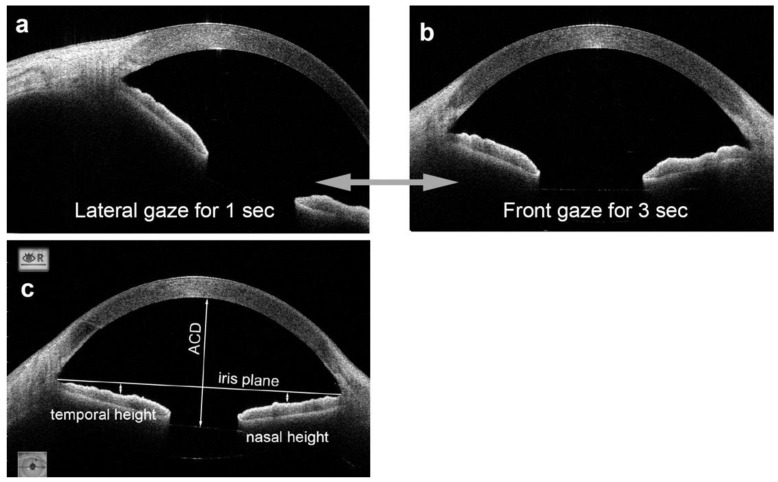
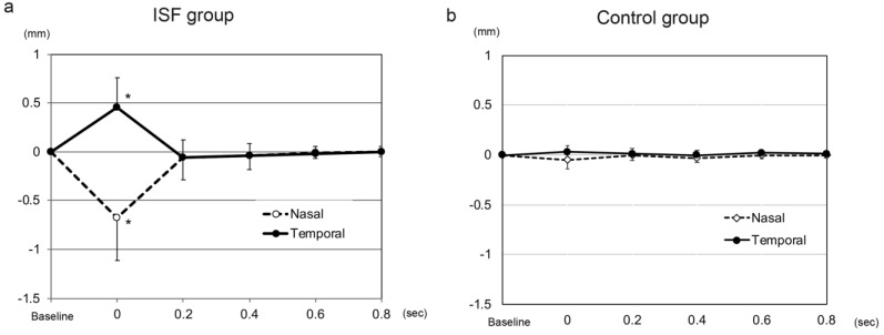
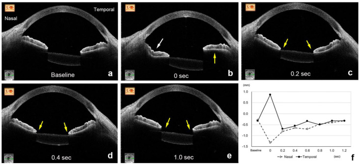
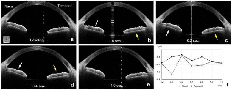
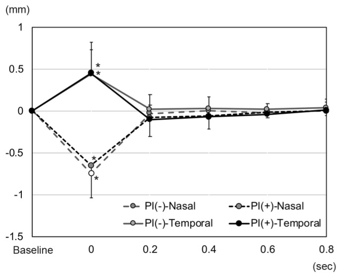
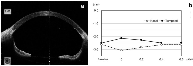
Saccadic eye movements induce movements of the aqueous and vitreous humor and iris fluttering. To evaluate iris fluttering during eye movements, anterior segment optical coherence tomography (AS-OCT) was used in 29 eyes with pars plana vitrectomy (PPV) and intrascleral fixation of an intraocular lens (ISF group) and 15 eyes with PPV and an IOL implantation into lens capsular bag (control group). The height of the iris from the iris plane (the line between the anterior chamber angles) was compared every 0.2 s after the eye had moved from a temporal to the primary position (time 0). The height of the nasal iris in the ISF group decreased to −0.68 ± 0.43 mm at 0 s (p < 0.001) and returned to −0.06 ± 0.23 mm at 0.2 s. The height of the temporal iris increased to 0.45 ± 0.31 mm at 0 s (p < 0.001) and returned to −0.06 ± 0.18 mm at 0.2 s. The height of the nasal iris at 0 s in the ISF group was significantly lower, and that of the temporal iris was significantly higher than the control (−0.05 ± 0.09 mm, 0.03 ± 0.06 mm, p < 0.001, respectively). Iris fluttering can act as a check valve for aqueous and vitreous humor movements and can be quantified by consecutive AS-OCT images. Large amplitude iris fluttering in eyes with intrascleral fixation is important because it can lead to a reverse pupillary block.
Keywords: anterior segment optical coherence tomography; intraocular lens; intrascleral fixation; iris capture; peripheral iridectomy; reverse pupillary block.
Conflict of interest statement
M.I.: research grants from Alcon Laboratories, Inc., and personal fees (lecture fees) from Alcon Laboratories, Inc., Novartis Pharma K.K., Bayer AG, Carl Zeiss Meditec AG, Novartis Pharma K.K., Santen Pharmaceutical Co., Ltd., and Senju Pharmaceutical Co., Ltd. AMO, Logitec and Design, outside the submitted work. T.K.: research grants from Ellex, and personal fees (lecture fees) from Alcon Laboratories, Inc., Novartis Pharma K.K., Bayer AG, Carl Zeiss Meditec AG, Novartis Pharma K.K., Santen Pharmaceutical Co., Ltd., and Senju Pharmaceutical Co., Ltd. AMO., outside the submitted work. A.H.: research grants from Santen Pharmaceutical Co., Ltd., personal fees (lecture fees) from Santen Pharmaceutical Co., Ltd., Alcon Laboratories, Inc., Novartis Pharma K.K., Bayer AG, Sanwagkagaku, KOWA, Senju Pharmaceutical Co., Ltd., outside the submitted work.
Figures
Similar articles
-
Evaluations of bridging sutures in preventing iris capture in eyes with intrascleral fixation of implanted intraocular lens.Graefes Arch Clin Exp Ophthalmol. 2023 Feb;261(2):427-434. doi: 10.1007/s00417-022-05816-1. Epub 2022 Aug 31. Graefes Arch Clin Exp Ophthalmol. 2023. PMID: 36042055
-
Anterior segment optical coherence tomography findings of reverse pupillary block after scleral-fixated sutured posterior chamber intraocular lens implantation.J Cataract Refract Surg. 2009 Sep;35(9):1540-7. doi: 10.1016/j.jcrs.2009.04.030. J Cataract Refract Surg. 2009. PMID: 19683150
-
Combined Keratoplasty, Pars Plana Vitrectomy, and Flanged Intrascleral Intraocular Lens Fixation to Restore Vision in Complex Eyes With Coexisting Anterior and Posterior Segment Problems.Cornea. 2018 Nov;37 Suppl 1:S78-S85. doi: 10.1097/ICO.0000000000001716. Cornea. 2018. PMID: 30216334
-
Sutureless intrascleral fixation using different three-piece posterior chamber intraocular lenses: a literature review of surgical techniques in cases of insufficient capsular support and a retrospective multicentre study.Acta Ophthalmol. 2020 May;98(3):224-236. doi: 10.1111/aos.14307. Epub 2019 Dec 1. Acta Ophthalmol. 2020. PMID: 31788964 Review.
-
[Intraocular lens explantation and retroiridal iris claw lens implantation via the pars plana : Video article].Ophthalmologe. 2020 Nov;117(11):1133-1137. doi: 10.1007/s00347-020-01246-8. Ophthalmologe. 2020. PMID: 33034738 Review. German.
Cited by
-
Secondary intrascleral intraocular lens (IOL) fixation with capsule preservation for IOL dislocation following mature cataract surgery with incomplete capsulorhexis: A case report.Medicine (Baltimore). 2025 Jun 20;104(25):e43030. doi: 10.1097/MD.0000000000043030. Medicine (Baltimore). 2025. PMID: 40550021 Free PMC article.
-
Secondary Intrascleral Intraocular Lens Fixation With Lens Capsule Preservation for Aphakic Eyes in Patients With Pseudoexfoliation Syndrome: A Case Series.Cureus. 2024 Oct 2;16(10):e70688. doi: 10.7759/cureus.70688. eCollection 2024 Oct. Cureus. 2024. PMID: 39372382 Free PMC article.
-
Comparative Functional and Morphological Data of Different IOL Dislocation Treatment Methods.J Clin Med. 2025 Feb 21;14(5):1462. doi: 10.3390/jcm14051462. J Clin Med. 2025. PMID: 40094913 Free PMC article.
References
LinkOut - more resources
Full Text Sources
Research Materials
Miscellaneous
