

Pinus mugo Essential Oil Impairs STAT3 Activation through Oxidative Stress and Induces Apoptosis in Prostate Cancer Cells
- PMID: 35956786
- PMCID: PMC9369512
- DOI: 10.3390/molecules27154834
Pinus mugo Essential Oil Impairs STAT3 Activation through Oxidative Stress and Induces Apoptosis in Prostate Cancer Cells
Abstract
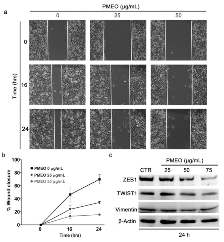
Essential oils (EOs) and their components have been reported to possess anticancer properties and to increase the sensitivity of cancer cells to chemotherapy. The aim of this work was to select EOs able to downregulate STAT3 signaling using Western blot and RT-PCR analyses. The molecular mechanism of anti-STAT3 activity was evaluated through spectrophotometric and fluorometric analyses, and the biological effect of STAT3 inhibition was analyzed by flow cytometry and wound healing assay. Herein, Pinus mugo EO (PMEO) is identified as an inhibitor of constitutive STAT3 phosphorylation in human prostate cancer cells, DU145. The down-modulation of the STAT3 signaling cascade decreased the expression of anti-proliferative as well as anti-apoptotic genes and proteins, leading to the inhibition of cell migration and apoptotic cell death. PMEO treatment induced a rapid drop in glutathione (GSH) levels and an increase in reactive oxygen species (ROS) concentration, resulting in mild oxidative stress. Pretreatment of cells with N-acetyl-cysteine (NAC), a cell-permeable ROS scavenger, reverted the inhibitory action of PMEO on STAT3 phosphorylation. Moreover, combination therapy revealed that PMEO treatment displayed synergism with cisplatin in inducing the cytotoxic effect. Overall, our data highlight the importance of STAT3 signaling in PMEO cytotoxic activity, as well as the possibility of developing adjuvant therapy or sensitizing cancer cells to conventional chemotherapy.
Keywords: STAT3; apoptosis; essential oil; oxidative stress.
Conflict of interest statement
The authors declare no conflict of interest.
Figures


Similar articles
-
Mild oxidative stress induces S-glutathionylation of STAT3 and enhances chemosensitivity of tumoural cells to chemotherapeutic drugs.Free Radic Biol Med. 2013 Dec;65:1322-1330. doi: 10.1016/j.freeradbiomed.2013.09.015. Epub 2013 Oct 1. Free Radic Biol Med. 2013. PMID: 24095958
-
2'-Hydroxycinnamaldehyde inhibits proliferation and induces apoptosis via signal transducer and activator of transcription 3 inactivation and reactive oxygen species generation.Cancer Sci. 2019 Jan;110(1):366-378. doi: 10.1111/cas.13852. Epub 2018 Nov 20. Cancer Sci. 2019. PMID: 30375708 Free PMC article.
-
Isoalantolactone induces apoptosis through ROS-mediated ER stress and inhibition of STAT3 in prostate cancer cells.J Exp Clin Cancer Res. 2018 Dec 12;37(1):309. doi: 10.1186/s13046-018-0987-9. J Exp Clin Cancer Res. 2018. PMID: 30541589 Free PMC article.
-
8-Epi-xanthatin induces the apoptosis of DU145 prostate carcinoma cells through signal transducer and activator of transcription 3 inhibition and reactive oxygen species generation.Phytother Res. 2021 Mar;35(3):1508-1520. doi: 10.1002/ptr.6918. Epub 2020 Nov 8. Phytother Res. 2021. PMID: 33164240
-
Thymoquinone induces oxidative stress-mediated apoptosis through downregulation of Jak2/STAT3 signaling pathway in human melanoma cells.Food Chem Toxicol. 2021 Nov;157:112604. doi: 10.1016/j.fct.2021.112604. Epub 2021 Oct 7. Food Chem Toxicol. 2021. PMID: 34627931
Cited by
-
Alpha-pinene ameliorates liver fibrosis by suppressing oxidative stress, inflammation, and the TGF-β/Smad3 signaling pathway.Iran J Basic Med Sci. 2025;28(4):451-460. doi: 10.22038/ijbms.2025.81693.17678. Iran J Basic Med Sci. 2025. PMID: 39968080 Free PMC article.
-
Chemical Composition, In Vitro Antitumor Effect, and Toxicity in Zebrafish of the Essential Oil from Conyza bonariensis (L.) Cronquist (Asteraceae).Biomolecules. 2023 Sep 24;13(10):1439. doi: 10.3390/biom13101439. Biomolecules. 2023. PMID: 37892120 Free PMC article.
-
Effect of Different Soil Treatments on Production and Chemical Composition of Essential Oils Extracted from Foeniculum vulgare Mill., Origanum vulgare L. and Thymus vulgaris L.Plants (Basel). 2023 Jul 31;12(15):2835. doi: 10.3390/plants12152835. Plants (Basel). 2023. PMID: 37570990 Free PMC article.
-
Antioxidant Activity of Essential Oils from Pinaceae Species.Antioxidants (Basel). 2024 Feb 26;13(3):286. doi: 10.3390/antiox13030286. Antioxidants (Basel). 2024. PMID: 38539820 Free PMC article. Review.
-
Essential Oils from Mediterranean Plants Inhibit In Vitro Monocyte Adhesion to Endothelial Cells from Umbilical Cords of Females with Gestational Diabetes Mellitus.Int J Mol Sci. 2023 Apr 13;24(8):7225. doi: 10.3390/ijms24087225. Int J Mol Sci. 2023. PMID: 37108387 Free PMC article.
References
-
- Cocchiola R., Rubini E., Altieri F., Chichiarelli S., Paglia G., Romaniello D., Carissimi S., Giorgi A., Giamogante F., Macone A., et al. STAT3 Post-Translational Modifications Drive Cellular Signaling Pathways in Prostate Cancer Cells. Int. J. Mol. Sci. 2019;20:1815. doi: 10.3390/ijms20081815. - DOI - PMC - PubMed
-
- Butturini E., de Prati A.C., Chiavegato G., Rigo A., Cavalieri E., Darra E., Mariotto S. Mild oxidative stress induces S-glutathionylation of STAT3 and enhances chemosensitivity of tumoural cells to chemotherapeutic drugs. Free Radic. Biol. Med. 2013;65:1322–1330. doi: 10.1016/j.freeradbiomed.2013.09.015. - DOI - PubMed
-
- Butturini E., Cavalieri E., de Prati A.C., Darra E., Rigo A., Shoji K., Murayama N., Yamazaki H., Watanabe Y., Suzuki H., et al. Two naturally occurring terpenes, dehydrocostuslactone and costunolide, decrease intracellular GSH content and inhibit STAT3 activation. PLoS ONE. 2011;6:e20174. doi: 10.1371/journal.pone.0020174. - DOI - PMC - PubMed
MeSH terms
Substances
LinkOut - more resources
Full Text Sources
Medical
Miscellaneous
