. 2022 Aug 5;12(15):2691.
doi: 10.3390/nano12152691.

Cellular Mechanosensitivity: Validation of an Adaptable 3D-Printed Device for Microindentation

Affiliations

Cellular Mechanosensitivity: Validation of an Adaptable 3D-Printed Device for Microindentation

Giulio Capponi et al. Nanomaterials (Basel). .

Abstract

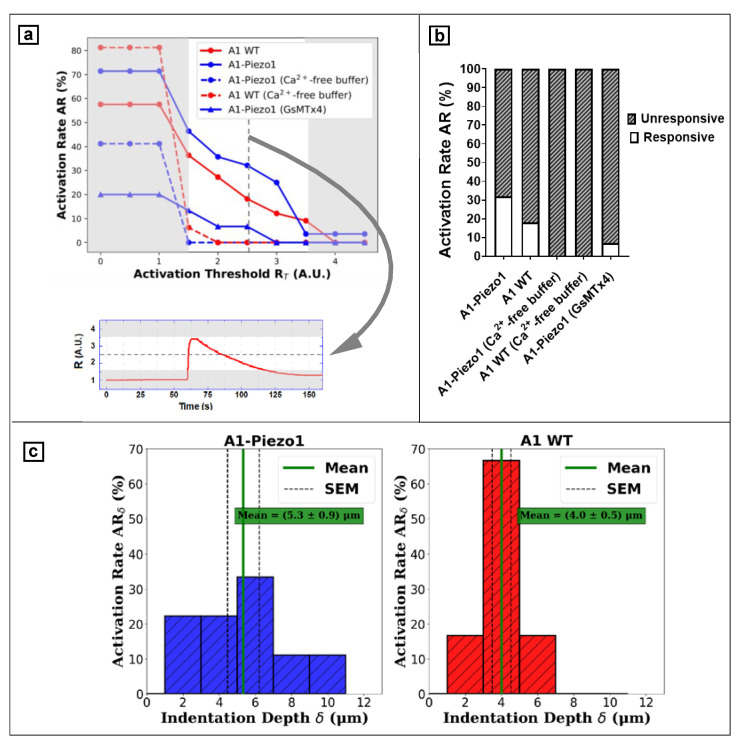
Mechanotransduction refers to the cellular ability to sense mechanical stimuli from the surrounding environment and convert them into biochemical signals that regulate cellular physiology and homeostasis. Mechanosensitive ion channels (MSCs), especially ones of Piezo family (Piezo1 and Piezo2), play a crucial role in mechanotransduction. These transmembrane proteins directly react to mechanical cues by triggering the onset of an ionic current. The relevance of this mechanism in driving physiology and pathology is emerging, and there is a growing need for the identification of an affordable and reliable assay to measure it. Setting up a mechanosensitivity assay requires exerting a mechanical stimulus on single cells while observing the downstream effects of channels opening. We propose an open-hardware approach to stimulate single adherent cells through controlled microindentation, using a 3D-printed actuation platform. We validated the device by measuring the mechanosensitivity of a neural mice cell line where the expression level and activity of Piezo1 were genetically and pharmacologically manipulated. Moreover, this extremely versatile device could be integrated with different read-out technologies, offering a new tool to improve the understanding of mechanotransduction in living cells.

Keywords: 3D printing; mechanobiology; mechanosensitivity; mechanotransduction; piezo1.


Conflict of interest statement

The authors declare no conflict of interest.

Figures

Figure 1
Figure 1
Translation motion stage integrated with microscope set-up for fluorescence calcium imaging. (a) Colour-coded CAD of motion translation stage, 3D printed in few parts: a base (purple), a main body (grey), the moving platform (blue) and the actuating screws with knobs (red). (b) Cells plated on a glass coverslip held by a steel chamber (1) positioned upon objective of the inverted microscope. The stage (2) enables the driving of Z-axis displacements of the glass probe (3) toward the glass coverslip for single-cell indentation. A micropipette holder (4) is used to hold a glass probe and it is connected to the moving platform of the stage. Vertical movements for indentation are automated by 28-BYJ48 step motor (5). A Newport Agilis AG-LS25 linear piezo motor (6) has no role in the indentation session; it only drives large vertical movements of the probe for initially positioning the steel chamber upon the objective, avoiding chamber edges that would damage the probe tip.
Figure 2
Figure 2
Schematic picture of the single-cell microindentation experiment. (a) The perturbation of the cell membrane provokes the opening of the Piezo1 channels, inducing an increase of the intracellular Ca2+ concentration, which is monitored by the ratiometric fura2-based fluorescence microscopy. (b) Timeline of glass probe position Z: the needle tip is initially positioned 20 μm (h0) above the adhesion plane; cell indentation takes place upon an area of maximum thickness Tmax corresponding to the nucleus zone. At instant t0, the probe begins vertically move downward by a distance D; at the instant ti, the cellular membrane is indented until a depth δ. Since the moving speed of the motion stage is v = 3 μm/s, the time interval Δt = ti − t0 depends on the probe displacement D, which is Δt = 4 s and D = 12 μm in the figure.
Figure 3
Figure 3
Calibration of the translation stage: step size is evaluated by interferometric measurement. Actuate 20 steps forward and 20 steps backward with nominal displacement of 1 μm per step along Z direction, repeating this displacement sequence 3 times to quantify the backlash and bidirectional positioning accuracy of Z-axis. (a) Calibration set-up: 3 fixed IR lasers (1) and three retroreflectors (2) jointed to the moving platform of the stage (3). The interferometer registers actual displacements of the translation stage for three main directions (X, Y and Z) depending on the geometry (in the picture: Y-axis geometry). (b) The actual displacements of every step in Z-direction are evaluated for positive (Z+) and negative (Z−) displacements. The red line states the mean of single displacement (1 μm), the green area is limiting steps within one standard deviation and the orange area represents the confidence interval of 95%. (c) Three sequences of displacements along the Z-axis: actual positions of retroreflectors jointed to the translation stage (red points) are strongly affected by backlash (blue points): the bidirectional systematic error is E = 3.2 μm. Backlash correction is actuated by overshooting steps to reduce deviation from target positions (black points). (d) Conduct 3 sequences of displacements along the Z-axis with backlash correction: the systematic bidirectional error is E = 0.5 μm.
Figure 4
Figure 4
Cellular morphometry via digital holographic microscopy. (a) A 3D phase image reconstruction of A1-Piezo1 cells. The scale (at left) relates the phase shift to the thickness (in μm). (b) Assessment of the average maximal vertical thickness for A1 WT (n = 45) and A1-Piezo1 (n = 65) shows no significant morphological differences.
Figure 5
Figure 5
Single-cell microindentation of Piezo1-A1 cells: the changes of intracellular Ca2+ concentration are monitored using ratiometric fluorescence microscopy; examples of a responsive (ae) and an unresponsive cell (fj) are reported. Cell-indenter glass probe is initially positioned above (h0 = 20 μm) cell adhesion plane. Thus, deeper and deeper vertical displacements are actuated (a,f) until contact with cell membrane. Fluorescence intensity in respect to the excitation wavelengths of 340 nm and 380 nm is averaged over the intracellular area, and displayed in a plot of the fluorescence ratio R (b,g) or a channels plot (c,h). Both responsive and unresponsive cells are indented until membrane rupture occurs, resulting in a plateau in the R plot. Before the disruptive indentation, the cell presents a transient peak in R signal (b) with tFWHM~40 s (responsive). On the other hand, when no increase in the trace of R is observed before the plateau, i.e., the membrane rupture, cell is classified as unresponsive (g). The initial positioning of the probe requires a tip position visualisation: bright-field microscopy is used to monitor the shadow of the tip (d,i), which can be moved above cell area through horizontal displacements of the probe. Fluorescence ratio R is imaged (Supplementary Videos S1–S2) on five representative color-coded frames displaying the peak of a responsive cell € and the plateau of a ruptured unresponsive cell (j).
Figure 6
Figure 6
Data analysis and device validation. Assessment of cell responsivity requires setting a proper fluorescence ratio threshold RT. Among activated cells, average indentation depth δ obtained from each group provides a calibration parameter. (a) Relationship between activation rate AR and the activation threshold RT used for classification; extreme values of threshold RT (grey area) were excluded to avoid misleading values of AR, and the median RT = 2.5 was selected for evaluating inter-group AR. (b) AR among different A1 cell groups. (c) Histogram showing the depth of indentation δ associated to the response event rate ARδ for A1-Piezo1 and A1 WT.

Similar articles

Cited by

References

    1. Liu H., Hu J., Zheng Q., Feng X., Zhan F., Wang X., Xu G., Hua F. Piezo1 Channels as Force Sensors in Mechanical Force-Related Chronic Inflammation. Front. Immunol. 2022;13:816149. doi: 10.3389/fimmu.2022.816149. - DOI - PMC - PubMed
    1. Chen X., Wanggou S., Bodalia A., Zhu M., Dong W., Fan J.J., Yin W.C., Min H.K., Hu M., Draghici D., et al. A Feedforward Mechanism Mediated by Mechanosensitive Ion Channel PIEZO1 and Tissue Mechanics Promotes Glioma Aggression. Neuron. 2018;100:799–815.e7. doi: 10.1016/j.neuron.2018.09.046. - DOI - PubMed
    1. Solis A.G., Bielecki P., Steach H.R., Sharma L., Harman C.C.D., Yun S., de Zoete M.R., Warnock J.N., To S.D.F., York A.G., et al. Mechanosensation of cyclical force by PIEZO1 is essential for innate immunity. Nature. 2019;573:69–74. doi: 10.1038/s41586-019-1485-8. - DOI - PMC - PubMed
    1. Ranade S.S., Qiu Z., Woo S.H., Hur S.S., Murthy S.E., Cahalan S.M., Xu J., Mathur J., Bandell M., Coste B., et al. Piezo1, a mechanically activated ion channel, is required for vascular development in mice. Proc. Natl. Acad. Sci. USA. 2014;111:10347–10352. doi: 10.1073/pnas.1409233111. - DOI - PMC - PubMed
    1. Alcaino C., Knutson K.R., Treichel A.J., Yildiz G., Strege P.R., Linden D.R., Li J.H., Leiter A.B., Szurszewski J.H., Farrugia G., et al. A population of gut epithelial enterochromaffin cells is mechanosensitive and requires Piezo2 to convert force into serotonin release. Proc. Natl. Acad. Sci. USA. 2018;115:E7632–E7641. doi: 10.1073/pnas.1804938115. - DOI - PMC - PubMed

LinkOut - more resources