. 2022 Aug 4;22(15):5819.
doi: 10.3390/s22155819.

Assessment of a Therapeutic X-ray Radiation Dose Measurement System Based on a Flexible Copper Indium Gallium Selenide Solar Cell

Affiliations

Assessment of a Therapeutic X-ray Radiation Dose Measurement System Based on a Flexible Copper Indium Gallium Selenide Solar Cell

Dong-Seok Shin et al. Sensors (Basel). .

Abstract

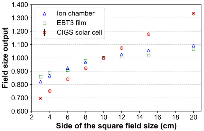
Several detectors have been developed to measure radiation doses during radiotherapy. However, most detectors are not flexible. Consequently, the airgaps between the patient surface and detector could reduce the measurement accuracy. Thus, this study proposes a dose measurement system based on a flexible copper indium gallium selenide (CIGS) solar cell. Our system comprises a customized CIGS solar cell (with a size 10 × 10 cm2 and thickness 0.33 mm), voltage amplifier, data acquisition module, and laptop with in-house software. In the study, the dosimetric characteristics, such as dose linearity, dose rate independence, energy independence, and field size output, of the dose measurement system in therapeutic X-ray radiation were quantified. For dose linearity, the slope of the linear fitted curve and the R-square value were 1.00 and 0.9999, respectively. The differences in the measured signals according to changes in the dose rates and photon energies were <2% and <3%, respectively. The field size output measured using our system exhibited a substantial increase as the field size increased, contrary to that measured using the ion chamber/film. Our findings demonstrate that our system has good dosimetric characteristics as a flexible in vivo dosimeter. Furthermore, the size and shape of the solar cell can be easily customized, which is an advantage over other flexible dosimeters based on an a-Si solar cell.

Keywords: copper indium gallium selenide solar cell; flexible dosimeter; radiation therapy.


Conflict of interest statement

The authors declare no conflict of interest.

Figures

Figure 1
Figure 1
A commercialized copper indium gallium selenide (CIGS) solar cell and its structure. The area comprising the CIGS cell is the active area that converts therapeutic X-ray radiation to electrical signals. TCO and CdS denote transparent conductive oxide and cadmium sulfide, respectively.
Figure 2
Figure 2
Configuration and data acquisition process of our measurement system based on a copper indium gallium selenide (CIGS) solar cell. In this system, therapeutic X-ray radiation is converted to electrical signals by the CIGS solar cell, and the electrical signals were digitalized through data acquisition (DAQ) module. The digitalized signals were visualized in real-time using a laptop with in-house software.
Figure 3
Figure 3
Experimental setup for quantifications/evaluations for dosimetric characteristics of measurement system based on a copper indium gallium selenide (CIGS) solar cell. A bolus (flexible/water equivalent material) was used instead of a solid water phantom to reduce airgaps between the solid water phantom and bolus. LINAC and dmax denote linear accelerator and depth at maximum dose, respectively.
Figure 4
Figure 4
Relationship between the signals of the copper indium gallium selenide (CIGS) solar cell and doses delivered using 6 MV photon beams. Total signal intensity is the sum of the obtained signals from the CIGS solar cell during irradiation. The normalizations were performed based on the signal corresponding to a dose of 1200 cGy. Error bars denote the standard deviations between three repeated measurements.
Figure 5
Figure 5
The signals obtained from the copper indium gallium selenide (CIGS) solar cell according to changes in dose rate from 100 to 600 MU/min at intervals of 100 MU/min. Total signal intensity is the sum of the obtained signals from the CIGS solar cell during irradiation. All the signals were normalized based on the signal corresponding to a dose rate of 600 MU/min. Error bars denote the standard deviations (numbers above the error bars) between three repeated measurements.
Figure 6
Figure 6
The signals obtained from the copper indium gallium selenide (CIGS) solar cell according to energy changes of the photon beam (6, 10, and 15 MV). Total signal intensity is the sum of the obtained signals from the CIGS solar cell during irradiation. All the signals were normalized based on the signal corresponding to the photon beam energy of 6 MV. Error bars denote the standard deviations (numbers above the error bars) between three repeated measurements.
Figure 7
Figure 7
Field size output factors of copper indium gallium selenide (CIGS) solar cell, ion chamber, and film for various field sizes (3 × 3, 4 × 4, 6 × 6, 8 × 8, 10 × 10, 12 × 12, 15 × 15, and 20 × 20 cm2) in 6 MV photon beams. The normalization was performed based on the output factor corresponding to a field size of 10 × 10 cm2. Error bars denote the standard deviations between three repeated measurements.
Figure 8
Figure 8
The copper indium gallium selenide (CIGS) solar cell signals under flat and bent conditions, respectively, in 6 MV photon beams. Zero curvature means a flat condition. The normalization was performed based on the signal corresponding to the flat CIGS solar cell. Error bars denote the standard deviations between three repeated measurements.
Figure 9
Figure 9
A simple example of in vivo dosimetry using our system based on copper indium gallium selenide (CIGS) solar cell. The CIGS solar cell was attached to the surface of the anthropomorphic phantom in the bent condition in this example.

Similar articles

Cited by

References

    1. Berlangieri A., Elliott S., Wasiak J., Chao M., Foroudi F. Use of magnetic resonance image-guided radiotherapy for breast cancer: A scoping review. J. Med. Radiat. Sci. 2022;69:122–133. doi: 10.1002/jmrs.545. - DOI - PMC - PubMed
    1. Bryce-Atkinson A., De Jong R., Marchant T., Whitfield G., Aznar M.C., Bel A., van Herk M. Low dose cone beam CT for paediatric image-guided radiotherapy: Image quality and practical recommendations. Radiother. Oncol. 2021;163:68–75. doi: 10.1016/j.radonc.2021.07.027. - DOI - PubMed
    1. De Ridder M., Raaijmakers C.P.J., Pameijer F.A., de Bree R., Reinders F.C.J., Doornaert P.A.H., Terhaard C.H.J., Philippens M.E.P. Target Definition in MR-Guided Adaptive Radiotherapy for Head and Neck Cancer. Cancers. 2022;14:3027. doi: 10.3390/cancers14123027. - DOI - PMC - PubMed
    1. Christiansen R.L., Dysager L., Hansen C.R., Jensen H.R., Schytte T., Nyborg C.J., Bertelsen A.S., Agergaard S.N., Mahmood F., Hansen S., et al. Online adaptive radiotherapy potentially reduces toxicity for high-risk prostate cancer treatment. Radiother. Oncol. 2022;167:165–171. doi: 10.1016/j.radonc.2021.12.013. - DOI - PubMed
    1. Williams M.V. Radiotherapy Near Misses, Incidents and Errors: Radiotherapy Incident at Glasgow. Clin. Oncol. 2007;19:1–3. doi: 10.1016/j.clon.2006.12.004. - DOI - PubMed

LinkOut - more resources