

MiR-340-5p alleviates neuroinflammation and neuronal injury via suppressing STING in subarachnoid hemorrhage
- PMID: 35957622
- PMCID: PMC9480905
- DOI: 10.1002/brb3.2687
MiR-340-5p alleviates neuroinflammation and neuronal injury via suppressing STING in subarachnoid hemorrhage
Abstract
Background: Subarachnoid hemorrhage (SAH) is a severe acute neurological disorder. SAH causes neuroinflammation and leads to early brain injury (EBI) and secondary injury. MicroRNAs are crucial regulators in a variety of neurological diseases. This study was performed to decipher how miR-340-5p functions in SAH.
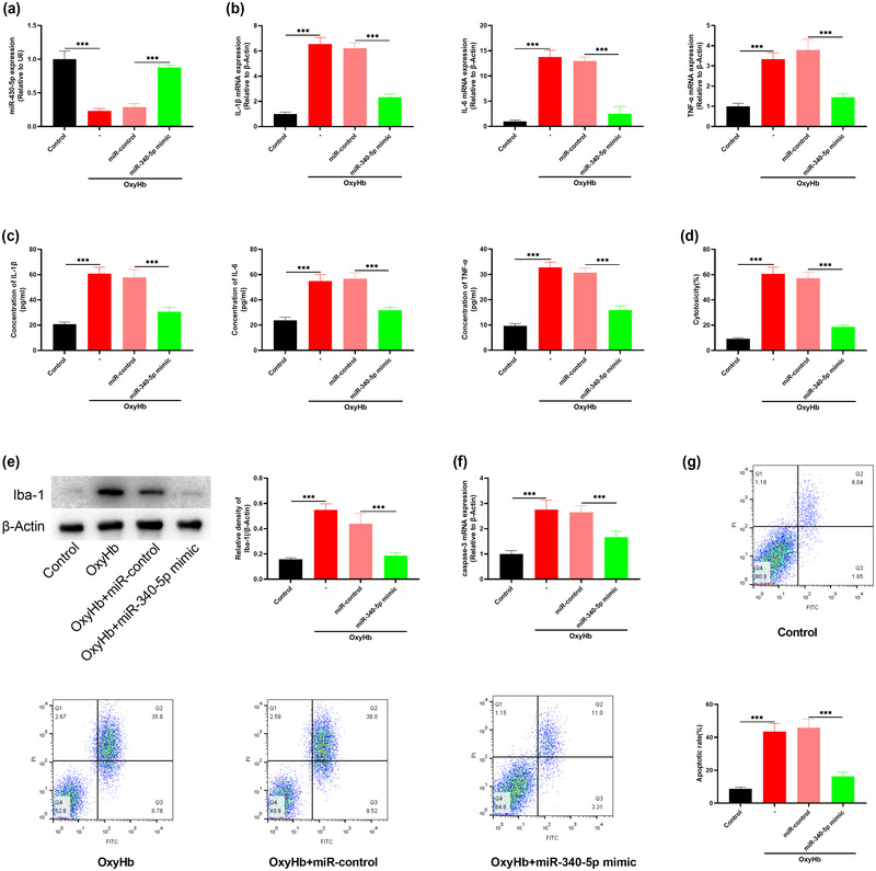
Methods: An experimental mouse model with SAH was established by the intravascular perforation, and the in vitro SAH model was constructed by exposing cocultured primary neurons and microglia to oxyhemoglobin. After overexpression of miR-340-5p in mice, the neurobehavioral disorders were evaluated by Garcia test; brain edema was evaluated by wet-dry method; blood-brain barrier (BBB) damage was detected with Evan's blue staining; levels of inflammatory cytokines were detected with enzyme-linked immunosorbent assay. After miR-340-5p was transfected in to microglia, Iba-1 expression was detected by Western blot, and neuronal apoptosis were detected with flow cytometry. The targeting relationship between miR-340-5p and STING was verified by dual-luciferase reporter gene assay and RNA immunoprecipitation assay.
Results: MiR-340-5p was significantly inhibited in the brain tissues of mice with SAH and microglia of SAH model, and neurological impairment, brain edema, BBB injury, and neuroinflammation were significantly alleviated in mice after overexpressing miR-340-5p. STING was identified as a target of miR-340-5p, and STING overexpression could counteract the effects of miR-340-5p overexpression on neurons.
Conclusion: MiR-340-5p can attenuate EBI caused by SAH-induced neuroinflammation by inhibiting STING.
Keywords: STING; early brain injury; miR-340-5p; neuroinflammation; subarachnoid hemorrhage.
© 2022 The Authors. Brain and Behavior published by Wiley Periodicals LLC.
Conflict of interest statement
The authors declare that they have no competing interests.
Figures


Similar articles
-
Up-regulation of circARF3 reduces blood-brain barrier damage in rat subarachnoid hemorrhage model via miR-31-5p/MyD88/NF-κB axis.Aging (Albany NY). 2021 Sep 12;13(17):21345-21363. doi: 10.18632/aging.203468. Epub 2021 Sep 12. Aging (Albany NY). 2021. PMID: 34511434 Free PMC article.
-
Propofol alleviates M1 polarization and neuroinflammation of microglia in a subarachnoid hemorrhage model in vitro, by targeting the miR-140-5p/TREM-1/NF-κB signaling axis.Eur J Histochem. 2024 Sep 17;68(3):4034. doi: 10.4081/ejh.2024.4034. Eur J Histochem. 2024. PMID: 39287134 Free PMC article.
-
RU.521 mitigates subarachnoid hemorrhage-induced brain injury via regulating microglial polarization and neuroinflammation mediated by the cGAS/STING/NF-κB pathway.Cell Commun Signal. 2023 Sep 28;21(1):264. doi: 10.1186/s12964-023-01274-2. Cell Commun Signal. 2023. PMID: 37770901 Free PMC article.
-
Tenascin-C in brain injuries and edema after subarachnoid hemorrhage: Findings from basic and clinical studies.J Neurosci Res. 2020 Jan;98(1):42-56. doi: 10.1002/jnr.24330. Epub 2018 Sep 22. J Neurosci Res. 2020. PMID: 30242870 Review.
-
The blood-brain barrier and the neurovascular unit in subarachnoid hemorrhage: molecular events and potential treatments.Fluids Barriers CNS. 2022 Apr 11;19(1):29. doi: 10.1186/s12987-022-00312-4. Fluids Barriers CNS. 2022. PMID: 35410231 Free PMC article. Review.
Cited by
-
Novel insight into cGAS-STING pathway in ischemic stroke: from pre- to post-disease.Front Immunol. 2023 Oct 17;14:1275408. doi: 10.3389/fimmu.2023.1275408. eCollection 2023. Front Immunol. 2023. PMID: 37915571 Free PMC article. Review.
-
Targeting Non-Coding RNA for CNS Injuries: Regulation of Blood-Brain Barrier Functions.Neurochem Res. 2023 Jul;48(7):1997-2016. doi: 10.1007/s11064-023-03892-1. Epub 2023 Feb 14. Neurochem Res. 2023. PMID: 36786944 Review.
-
A Mouse Systems Genetics Approach Reveals Common and Uncommon Genetic Modifiers of Hepatic Lysosomal Enzyme Activities and Glycosphingolipids.Int J Mol Sci. 2023 Mar 3;24(5):4915. doi: 10.3390/ijms24054915. Int J Mol Sci. 2023. PMID: 36902345 Free PMC article.
-
The pivotal role of microglia in injury and the prognosis of subarachnoid hemorrhage.Neural Regen Res. 2025 Jul 1;20(7):1829-1848. doi: 10.4103/NRR.NRR-D-24-00241. Epub 2024 Jul 10. Neural Regen Res. 2025. PMID: 38993136 Free PMC article.
-
The STING Signaling: A Novel Target for Central Nervous System Diseases.Cell Mol Neurobiol. 2025 Apr 7;45(1):33. doi: 10.1007/s10571-025-01550-4. Cell Mol Neurobiol. 2025. PMID: 40195137 Free PMC article. Review.
References
-
- Kamp, M. A. , Steiger, H. J. , & van Lieshout, J. H. (2020). Experimental aneurysmal subarachnoid hemorrhage: Tiding over. Translational Stroke Research, 11(1), 1–3. - PubMed
-
- Helbok, R. , Schiefecker, A. J. , Beer, R. , Dietmann, A. , Antunes, A. P. , Sohm, F. , Fischer, M. , Hackl, W. O. , Rhomberg, P. , Lackner, P. , Pfausler, B. , Thomé, C. , Humpel, C. , & Schmutzhard, E. (2015). Early brain injury after aneurysmal subarachnoid hemorrhage: A multimodal neuromonitoring study. Critical Care (London, England), 19(1), 75. - PMC - PubMed
-
- Dou, Y. , Shen, H. , Feng, D. , Li, H. , Tian, X. , Zhang, J. , Wang, Z. , & Chen, G. (2017). Tumor necrosis factor receptor‐associated factor 6 participates in early brain injury after subarachnoid hemorrhage in rats through inhibiting autophagy and promoting oxidative stress. Journal of Neurochemistry, 142(3), 478–492. - PubMed
-
- Liu, W. , Li, R. , Yin, J. , Guo, S. , Chen, Y. , Fan, H. , Li, G. , Li, Z. , Li, X. , Zhang, X. , He, X. , & Duan, C. (2019). Mesenchymal stem cells alleviate the early brain injury of subarachnoid hemorrhage partly by suppression of Notch1‐dependent neuroinflammation: Involvement of Botch. Journal of Neuroinflammation, 16(1), 8. - PMC - PubMed
MeSH terms
Substances
LinkOut - more resources
Full Text Sources
Research Materials
