

Detection of Antiviral Tissue Responses and Increased Cell Stress in the Pancreatic Islets of Newly Diagnosed Type 1 Diabetes Patients: Results From the DiViD Study
- PMID: 35957810
- PMCID: PMC9360491
- DOI: 10.3389/fendo.2022.881997
Detection of Antiviral Tissue Responses and Increased Cell Stress in the Pancreatic Islets of Newly Diagnosed Type 1 Diabetes Patients: Results From the DiViD Study
Abstract
Aims/hypothesis: The Diabetes Virus Detection (DiViD) study has suggested the presence of low-grade enteroviral infection in pancreatic tissue collected from six of six live adult patients newly diagnosed with type 1 diabetes. The present study aimed to compare the gene and protein expression of selected virally induced pathogen recognition receptors and interferon stimulated genes in islets from these newly diagnosed type 1 diabetes (DiViD) subjects vs age-matched non-diabetic (ND) controls.
Methods: RNA was extracted from laser-captured islets and Affymetrix Human Gene 2.0 ST arrays used to obtain gene expression profiles. Lists of differentially expressed genes were subjected to a data-mining pipeline searching for enrichment of canonical pathways, KEGG pathways, Gene Ontologies, transcription factor binding sites and other upstream regulators. In addition, the presence and localisation of specific viral response proteins (PKR, MxA and MDA5) were examined by combined immunofluorescent labelling in sections of pancreatic tissue.
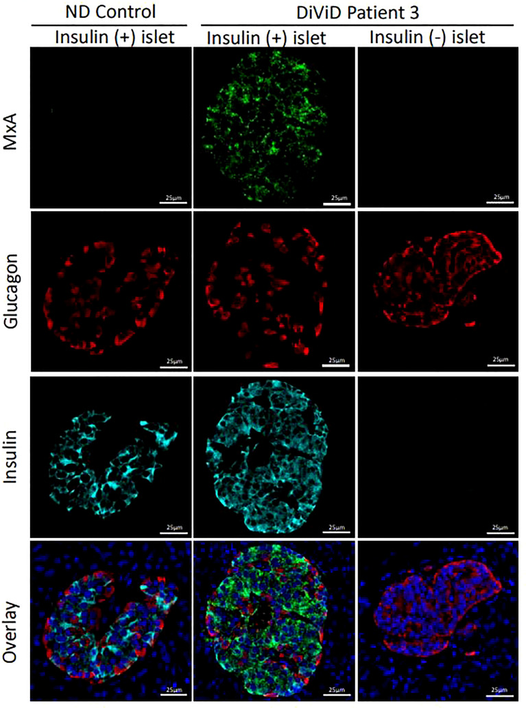
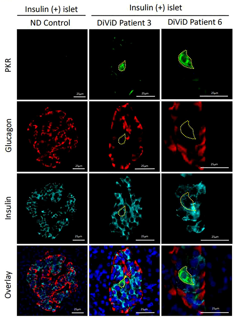
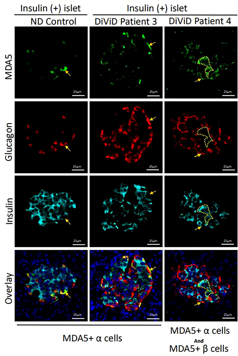
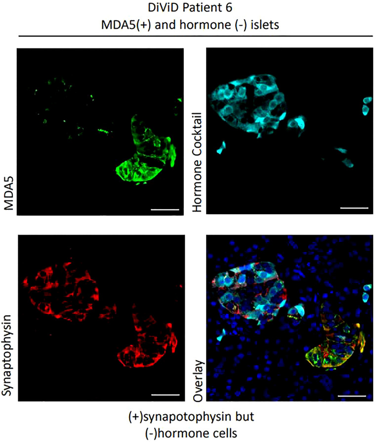
Results: The data analysis and data mining process revealed a significant enrichment of gene ontologies covering viral reproduction and infectious cycles; peptide translation, elongation and initiation, as well as oxidoreductase activity. Enrichment was identified in the KEGG pathways for oxidative phosphorylation; ribosomal and metabolic activity; antigen processing and presentation and in canonical pathways for mitochondrial dysfunction, oxidative phosphorylation and EIF2 signaling. Protein Kinase R (PKR) expression did not differ between newly diagnosed type 1 diabetes and ND islets at the level of total RNA, but a small subset of β-cells displayed markedly increased PKR protein levels. These PKR+ β-cells correspond to those previously shown to contain the viral protein, VP1. RNA encoding MDA5 was increased significantly in newly diagnosed type 1 diabetes islets, and immunostaining of MDA5 protein was seen in α- and certain β-cells in both newly diagnosed type 1 diabetes and ND islets, but the expression was increased in β-cells in type 1 diabetes. In addition, an uncharacterised subset of synaptophysin positive, but islet hormone negative, cells expressed intense MDA5 staining and these were more prevalent in DiViD cases. MxA RNA was upregulated in newly diagnosed type 1 diabetes vs ND islets and MxA protein was detected exclusively in newly diagnosed type 1 diabetes β-cells.
Conclusion/interpretation: The gene expression signatures reveal that pathways associated with cellular stress and increased immunological activity are enhanced in islets from newly diagnosed type 1 diabetes patients compared to controls. The increases in viral response proteins seen in β-cells in newly diagnosed type 1 diabetes provide clear evidence for the activation of IFN signalling pathways. As such, these data strengthen the hypothesis that an enteroviral infection of islet β-cells contributes to the pathogenesis of type 1 diabetes.
Keywords: MDA5; MxA; PKR; Type 1 diabetes; biopsy; enterovirus; gene expression; pancreas.
Copyright © 2022 Krogvold, Leete, Mynarek, Russell, Gerling, Lenchik, Mathews, Richardson, Morgan and Dahl-Jørgensen.
Conflict of interest statement
The authors declare that the research was conducted in the absence of any commercial or financial relationships that could be construed as a potential conflict of interest.
Figures
Similar articles
-
Expression of the enteroviral capsid protein VP1 in the islet cells of patients with type 1 diabetes is associated with induction of protein kinase R and downregulation of Mcl-1.Diabetologia. 2013 Jan;56(1):185-93. doi: 10.1007/s00125-012-2745-4. Epub 2012 Oct 14. Diabetologia. 2013. PMID: 23064357
-
Pancreas Whole Tissue Transcriptomics Highlights the Role of the Exocrine Pancreas in Patients With Recently Diagnosed Type 1 Diabetes.Front Endocrinol (Lausanne). 2022 Apr 13;13:861985. doi: 10.3389/fendo.2022.861985. eCollection 2022. Front Endocrinol (Lausanne). 2022. PMID: 35498413 Free PMC article.
-
Detection of a low-grade enteroviral infection in the islets of langerhans of living patients newly diagnosed with type 1 diabetes.Diabetes. 2015 May;64(5):1682-7. doi: 10.2337/db14-1370. Epub 2014 Nov 24. Diabetes. 2015. PMID: 25422108
-
Enterovirus infection and type 1 diabetes: unraveling the crime scene.Clin Exp Immunol. 2019 Jan;195(1):15-24. doi: 10.1111/cei.13223. Epub 2018 Nov 13. Clin Exp Immunol. 2019. PMID: 30307605 Free PMC article. Review.
-
Molecular Footprints of the Immune Assault on Pancreatic Beta Cells in Type 1 Diabetes.Front Endocrinol (Lausanne). 2020 Sep 15;11:568446. doi: 10.3389/fendo.2020.568446. eCollection 2020. Front Endocrinol (Lausanne). 2020. PMID: 33042023 Free PMC article.
Cited by
-
The role of the interferon/JAK-STAT axis in driving islet HLA-I hyperexpression in type 1 diabetes.Front Endocrinol (Lausanne). 2023 Oct 6;14:1270325. doi: 10.3389/fendo.2023.1270325. eCollection 2023. Front Endocrinol (Lausanne). 2023. PMID: 37867531 Free PMC article. Review.
-
Transcriptional changes in multiple endocrine organs from lethal cases of COVID-19.J Mol Med (Berl). 2023 Aug;101(8):973-986. doi: 10.1007/s00109-023-02334-3. Epub 2023 May 29. J Mol Med (Berl). 2023. PMID: 37246981 Free PMC article.
-
LncRNA ARGI Contributes to Virus-Induced Pancreatic β Cell Inflammation Through Transcriptional Activation of IFN-Stimulated Genes.Adv Sci (Weinh). 2023 Sep;10(25):e2300063. doi: 10.1002/advs.202300063. Epub 2023 Jun 29. Adv Sci (Weinh). 2023. PMID: 37382191 Free PMC article.
-
Beta cell dysfunction occurs independently of insulitis in type 1 diabetes pathogenesis.bioRxiv [Preprint]. 2025 Mar 1:2024.12.29.630665. doi: 10.1101/2024.12.29.630665. bioRxiv. 2025. PMID: 39763971 Free PMC article. Preprint.
-
A Monovalent Mt10-CVB3 Vaccine Prevents CVB4-Accelerated Type 1 Diabetes in NOD Mice.Vaccines (Basel). 2022 Dec 29;11(1):76. doi: 10.3390/vaccines11010076. Vaccines (Basel). 2022. PMID: 36679922 Free PMC article.
References
-
- Harris HF. A Case of Diabetes Quickly Following Mumps. Boston Med Surg J (1899) CXL:465–9. doi: 10.1056/NEJM189905181402001 - DOI
Publication types
MeSH terms
Substances
Grants and funding
LinkOut - more resources
Full Text Sources
Medical
Research Materials