. 2022 Jul 25:12:913498.
doi: 10.3389/fonc.2022.913498. eCollection 2022.

Sema3d Restrained Hepatocellular Carcinoma Progression Through Inactivating Pi3k/Akt Signaling via Interaction With FLNA

Affiliations

Sema3d Restrained Hepatocellular Carcinoma Progression Through Inactivating Pi3k/Akt Signaling via Interaction With FLNA

Yiming Li et al. Front Oncol. .

Abstract

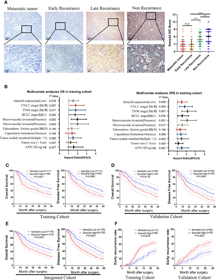
Hepatocellular carcinoma (HCC) is one of the most lethal malignant tumors worldwide due to the high incidence rate of metastasis and recurrence. Semaphorin 3d (Sema3d) has been shown to play a critical role in vascular development during early embryogenesis and several forms of cancer progression via regulating cell migration. However, the function of Sema3d in hepatocellular carcinoma (HCC) remains elusive. This study aimed to explore the function and mechanisms of Sema3d in HCC. In our study, Sema3d expression was significantly downregulated in HCC tissues and cell lines. Downregulated Sema3d was closely correlated with aggressive clinicopathological features and poor clinical outcomes in HCC patients. Moreover, overexpression of Sema3d in HCCLM3 cells was significantly inhibited and knockdown of Sema3d in PLC/PRF/5 cells promoted proliferation, migration, invasion, and epithelial-mesenchymal transition (EMT) of HCC cells in vitro and tumor growth, EMT, and metastasis in vivo. Furthermore, the RNA sequencing and gene set enrichment analysis (GSEA) indicated that these phenotypic and functional changes in Sema3d-interfered HCC cells were mediated by the Pi3k/Akt signaling pathway, and co-IP-combined mass spectrometry indicated Sema3d might interact with FLNA. Finally, we proved that Sema3d exerted its tumor-restraining effect by interacting with FLNA to inactivate the Pi3k/Akt signaling pathway and remodel the cytoskeleton. Our data showed that Sema3d restrained hepatocellular carcinoma proliferation, invasion, and metastasis through inactivating Pi3k/Akt via interaction with FLNA, which may serve as a novel prognostic predictor and a potential therapeutic target for HCC patients.

Keywords: EMT; Pi3k/Akt signaling; Sema3d; cancer progression; hepatocellular carcinoma; prognosis; semaphorin 3d.


Conflict of interest statement

The authors declare that the research was conducted in the absence of any commercial or financial relationships that could be construed as a potential conflict of interest.

Figures

Figure 1
Figure 1
Sema3d is downregulated in HCC cell lines and tissues. (A) Sema3d expression in Gene Expression Omnibus (GEO) datasets (GSE22058, GSE102079, GSE56372) and TCGA database. (B) Sema3d mRNA expression level was detected by qRT-PCR in 50 HCC and paired ANLT samples (upper panel), 8 HCC cell lines, and primary human hepatocytes (PHHs) (lower panel). (C) Sema3d protein expression was examined by Western blot in PHHs and HCC cell lines, representative normal liver tissue, paired HCC tissues, and ANLTs. (D) Sema3d mRNA expression in HCC tissues of 3 clinical subtypes (SLHCC, SHCC, and NHCC) was detected by qRT-PCR. (E) Representative IHC images of Sema3d expression in HCC tissues and corresponding ANLTs. ** p < 0.01; *** p < 0.001. n.s, no significance.
Figure 2
Figure 2
Downregulated Sema3d is associated with HCC’s poor prognosis. (A) Representative IHC staining for Sema3d in metastatic tumor (n = 34), early recurrence of HCC ( < 2 years, n = 142), late recurrence of HCC ( > 2 years, n = 66), and nonrecurrence of HCC (5 years, n = 92). IHC staining score of Sema3d in the integrated cohorts (composed of training and validation cohorts). p-values were calculated by the Mann–Whitney U test. ** p < 0.01; *** p < 0.001. n.s, no significance. (B) Multivariate analysis revealed that low Sema3d expression was an independent risk factor for overall survival (OS) and disease-free survival (DFS) in the training cohort. (C) Survival analysis for the training cohort reveals the Sema3d-low group had worse OS and DFS. (D) Survival analysis for the validation cohort verified that the Sema3d-low group had worse OS and DFS. (E) Survival analysis for integrated cohort (composed of training and validation cohorts) verified that the Sema3d-low group had worse OS and DFS. (F) Kaplan–Meier curves for the cumulative early recurrence ( < 2 years) rate of HCC patients based on Sema3d expression in training and validation cohorts.
Figure 3
Figure 3
Sema3d restrains HCC migration, invasion, proliferation, and metastasis in vitro and in vivo. (A) Wound-healing assay detected the migratory PLC/PRF/5shSema3d and HCCLM3Sema3d, as well as the corresponding control cells (n = 6 for each group). (B) A Transwell invasion assay was performed to detect the invasive capacities of HCC cells (n = 6 for each group). (C) MTT assay detected the proliferation capacities of Sema3d-interfered HCC cells (n = 6 for each group). (D) In subcutaneous tumors derived from PLC/PRF/5shSema3d and HCCLM3Sema3d cells and their control cells, tumor volumes were calculated with the formula (length × width2)/2. (E) At week 6, representative bioluminescent images monitored by IVIS and orthotopic xenograft tumors were shown, as well as the volume of orthotopic tumors and the growth curves constructed by the quantified bioluminescent signal (n = 6 for each group). (F) Representative bioluminescent images and HE staining images of lung tissue and a number of pulmonary metastasis nodules in different groups were shown (upper panel). ** p < 0.01; *** p < 0.001.
Figure 4
Figure 4
Sema3d restrains HCC progression via inactivating Pi3k/Akt signaling. (A) RNA-seq and KEGG analysis of HCCLM3Sema3d cells compared with HCCLM3NC cells revealed the potential signaling pathway regulated by Sema3d in HCC cells. (B) GSEA was performed between the Sema3d high- and low-expression patients in TCGA dataset. (C) Representative IHC images of Sema3d and p-AKT and their expression correlations were analyzed by Spearman’s rank correlation tests in 40 HCC tissue samples. (D) Expression of Pi3k and Akt and their phosphorylation level in Sema3d-interfered HCC cells were detected by Western blot. (E) EdU staining assay detected the proliferation for Sema3d-interfered HCC cells with/without Wortmannin treatment. (F) MTT assay detected the proliferation of ema3d-interfered HCC cells, and their control cells with/without Wortmannin treatment. (G) Wound healing and Transwell invasion assays detected the migration and invasion of Sema3d-interfered HCC cells with/without Wortmannin treatment. ** p < 0.01; *** p < 0.001. n.s, no significance.
Figure 5
Figure 5
Sema3d directly interacts with FLNA and affects cytoskeleton remodeling. (A) Coimmunoprecipitation (co-IP) was performed to extract Sema3d direct combined proteins from HCCLM3Sema3d cells, which were then identified using LC-MS/MS. The top 20 combined proteins sorted by Score Sequest are shown in the diagram. (B) co-IP was performed to determine that Sema3d and FLNA could directly interact with each other. (C) Double immunofluorescence staining showed that Sema3d affected the cellular expression of FLNA (green) and induced cytoskeleton remodeling (F-actin, red). (D) Protein expression levels were analyzed by Western blot and showed that Sema3d regulated the phosphorylation level of FLNA and the amount of nuclear C-FLNA. (E) Immunofluorescence staining revealed that Sema3d promotes C-FLNA translocated from the cytoplasm to the nucleus.
Figure 6
Figure 6
Downregulation of Sema3d promotes EMT in HCC. (A) Correlation of FLNA and vimentin mRNA expression levels was analyzed from TCGA. (B) Representative image of Sema3d, E-cadherin, and vimentin expression level in HCC tissue detected by IHC. (C) EMT marker proteins in PLC/PRF/5shSema3d and HCCLM3Sema3d cells and their corresponding control cells were detected by Western blot. (D) EMT marker proteins in Sema3d-interfered PLC/PRF/5 and HCCLM3 cells detected by IF. (E) IHC was performed to detect the FLNA and EMT marker proteins in consecutive sections of a liver orthotopic xenograft tumor derived from Sema3d-interfered PLC/PRF/5 and HCCLM3 cells.
Figure 7
Figure 7
Sema3d suppressed Pi3k/AKT signaling and restrained HCC proliferation, metastasis, and EMT through FLNA. (A) Protein expression level was analyzed by Western blot and showed that Sema3d regulated the phosphorylation level of Pi3k and AKT and expression of EMT markers through FLNA. (B) F-actin and EMT marker proteins in Seme3d- and FLNA-interfered HCC cells were detected by IF. (C) Wound healing, Transwell invasion, and EdU staining assays detected the migration, invasion, and proliferation of HCCLM3Sema3d ectopic expression of FLNA. (D) Wound healing, Transwell invasion, and EdU staining assays detected the migration, invasion, and proliferation of PLC/PRF/5shSema3d knockdown FLNA. (E) MTT assay detected a proliferation of HCCLM3Sema3d ectopic expression of FLNA and PLC/PRF/5shSema3d interference of FLNA compared with the corresponding control cells. (F) Sema3d restrained hepatocellular carcinoma proliferation, metastases, EMT, and cytoskeleton remodeling through inactivating Pi3k/Akt via interaction with FLNA. ** p < 0.01; *** p < 0.001. n.s, no significance.

Similar articles

Cited by

References

    1. Bray F, Ferlay J, Soerjomataram I, Siegel RL, Torre LA, Jemal A. Global Cancer Statistics 2018: GLOBOCAN Estimates of Incidence and Mortality Worldwide for 36 Cancers in 185 Countries. CA Cancer J Clin (2018) 68(6):394–424. doi: 10.3322/caac.21492 [published Online First: 2018/09/13 - DOI - PubMed
    1. Forner A, Reig M, Bruix J. Hepatocellular Carcinoma. Lancet (2018) 391(10127):1301–14. doi: 10.1016/S0140-6736(18)30010-2 - DOI - PubMed
    1. Tsilimigras DI, Mehta R, Paredes AZ, Moris D, Sahara K, Bagante F, et al. . Overall Tumor Burden Dictates Outcomes for Patients Undergoing Resection of Multinodular Hepatocellular Carcinoma Beyond the Milan Criteria. Ann Surg (2020) 272(4):574–81. doi: 10.1097/SLA.0000000000004346 - DOI - PubMed
    1. Yang LY, Fang F, Ou DP, Wu W, Zeng ZJ, Wu F. Solitary Large Hepatocellular Carcinoma: A Specific Subtype of Hepatocellular Carcinoma With Good Outcome After Hepatic Resection. Ann Surg (2009) 249(1):118–23. doi: 10.1097/SLA.0b013e3181904988 [published Online First: 2008/12/25 - DOI - PubMed
    1. Franssen B, Alshebeeb K, Tabrizian P, Marti J, Pierobon ES, Lubezky N, et al. . Differences in Surgical Outcomes Between Hepatitis B- and Hepatitis C-Related Hepatocellular Carcinoma: A Retrospective Analysis of a Single North American Center. Ann Surg (2014), 260(4). doi: 10.1097/SLA.0000000000000917 - DOI - PubMed