

Research progress on the application of optical coherence tomography in the field of oncology
- PMID: 35957903
- PMCID: PMC9358962
- DOI: 10.3389/fonc.2022.953934
Research progress on the application of optical coherence tomography in the field of oncology
Abstract
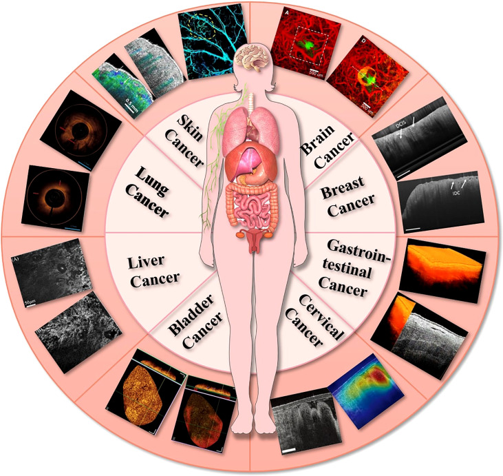
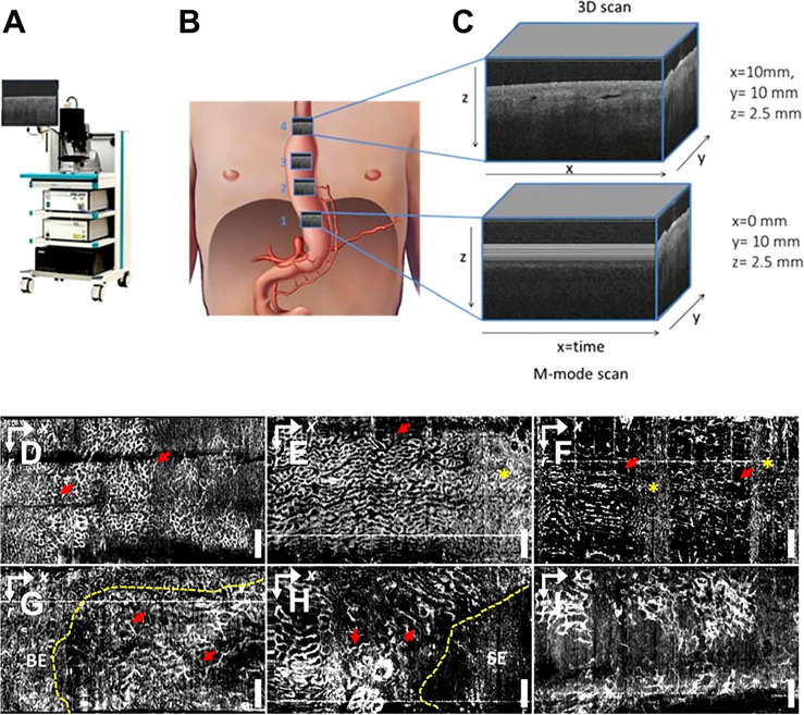
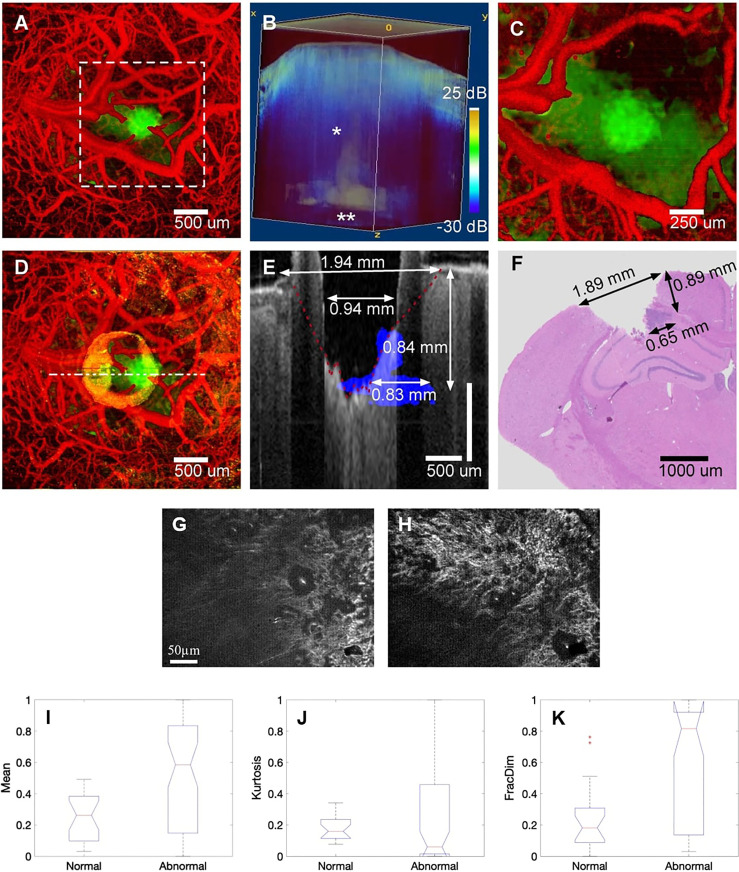
Optical coherence tomography (OCT) is a non-invasive imaging technique which has become the "gold standard" for diagnosis in the field of ophthalmology. However, in contrast to the eye, nontransparent tissues exhibit a high degree of optical scattering and absorption, resulting in a limited OCT imaging depth. And the progress made in the past decade in OCT technology have made it possible to image nontransparent tissues with high spatial resolution at large (up to 2mm) imaging depth. On the one hand, OCT can be used in a rapid, noninvasive way to detect diseased tissues, organs, blood vessels or glands. On the other hand, it can also identify the optical characteristics of suspicious parts in the early stage of the disease, which is of great significance for the early diagnosis of tumor diseases. Furthermore, OCT imaging has been explored for imaging tumor cells and their dynamics, and for the monitoring of tumor responses to treatments. This review summarizes the recent advances in the OCT area, which application in oncological diagnosis and treatment in different types: (1) superficial tumors:OCT could detect microscopic information on the skin's surface at high resolution and has been demonstrated to help diagnose common skin cancers; (2) gastrointestinal tumors: OCT can be integrated into small probes and catheters to image the structure of the stomach wall, enabling the diagnosis and differentiation of gastrointestinal tumors and inflammation; (3) deep tumors: with the rapid development of OCT imaging technology, it has shown great potential in the diagnosis of deep tumors such in brain tumors, breast cancer, bladder cancer, and lung cancer.
Keywords: cancer imaging; imaging technique; oncological diseases; optical coherence tomography; tumor diagnoses.
Copyright © 2022 Yang, Chen, Ling, Wang, Wang, Zhang, Zhao, Zhao and Mao.
Conflict of interest statement
The authors declare that the research was conducted in the absence of any commercial or financial relationships that could be construed as a potential conflict of interest.
Figures
Similar articles
-
Methods and applications of full-field optical coherence tomography: a review.J Biomed Opt. 2022 May;27(5):050901. doi: 10.1117/1.JBO.27.5.050901. J Biomed Opt. 2022. PMID: 35596250 Free PMC article. Review.
-
Review of optical coherence tomography in oncology.J Biomed Opt. 2017 Dec;22(12):1-23. doi: 10.1117/1.JBO.22.12.121711. J Biomed Opt. 2017. PMID: 29274145 Free PMC article. Review.
-
Applications of optical coherence tomography in dermatology.J Dermatol Sci. 2005 Nov;40(2):85-94. doi: 10.1016/j.jdermsci.2005.07.006. Epub 2005 Aug 31. J Dermatol Sci. 2005. PMID: 16139481 Review.
-
[Progress of the application of optical coherence tomography in gastrointestinal tumor surgery].Zhonghua Wei Chang Wai Ke Za Zhi. 2017 Jun 25;20(6):716-720. Zhonghua Wei Chang Wai Ke Za Zhi. 2017. PMID: 28643319 Chinese.
-
Optical Coherence Tomography for Ophthalmology Imaging.Adv Exp Med Biol. 2021;3233:197-216. doi: 10.1007/978-981-15-7627-0_10. Adv Exp Med Biol. 2021. PMID: 34053029
Cited by
-
Emerging Trends in Point-of-Care Technology Development for Oncology in Low- and Middle-Income Countries.JCO Glob Oncol. 2025 Jun;11:e2500142. doi: 10.1200/GO-25-00142. Epub 2025 Jun 18. JCO Glob Oncol. 2025. PMID: 40532137 Free PMC article. Review.
-
Recent advances in non-invasive in vivo tracking of cell-based cancer immunotherapies.Biomater Sci. 2025 Apr 8;13(8):1939-1959. doi: 10.1039/d4bm01677g. Biomater Sci. 2025. PMID: 40099377 Review.
-
Cutting Edge Microscopic Intraoperative Tissue Assessment for Guidance in Oncologic Surgery: A Systematic Review of the Role of Optical Coherence Tomography.Ann Surg Oncol. 2025 Mar;32(3):2191-2205. doi: 10.1245/s10434-024-16632-8. Epub 2024 Dec 8. Ann Surg Oncol. 2025. PMID: 39648239
-
Optical imaging for screening and early cancer diagnosis in low-resource settings.Nat Rev Bioeng. 2024 Jan;2(1):25-43. doi: 10.1038/s44222-023-00135-4. Epub 2023 Dec 22. Nat Rev Bioeng. 2024. PMID: 39301200 Free PMC article.
-
Recent Advances in Melanoma Diagnosis and Prognosis Using Machine Learning Methods.Curr Oncol Rep. 2023 Jun;25(6):635-645. doi: 10.1007/s11912-023-01407-3. Epub 2023 Mar 31. Curr Oncol Rep. 2023. PMID: 37000340 Free PMC article. Review.
References
Publication types
LinkOut - more resources
Full Text Sources
Miscellaneous