Review
. 2022 Jul 26:13:921470.
doi: 10.3389/fphys.2022.921470. eCollection 2022.

The O2-sensitive brain stem, hyperoxic hyperventilation, and CNS oxygen toxicity

Affiliations
Review

The O2-sensitive brain stem, hyperoxic hyperventilation, and CNS oxygen toxicity

Jay B Dean et al. Front Physiol. .

Abstract

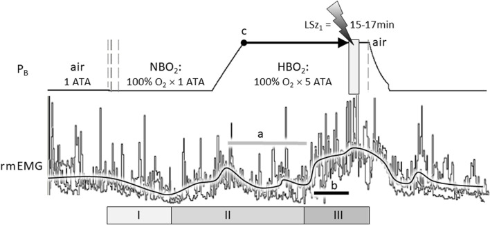
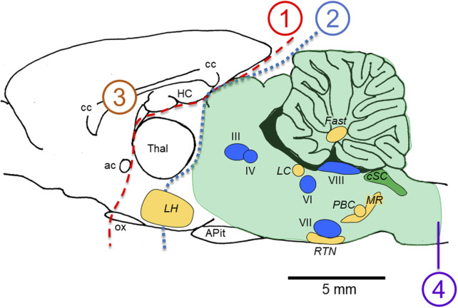
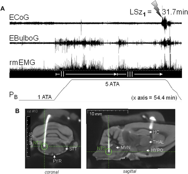
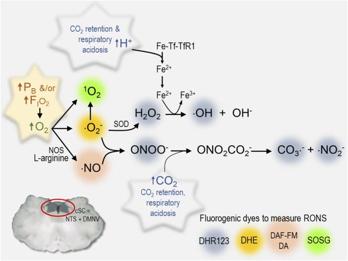
Central nervous system oxygen toxicity (CNS-OT) is a complex disorder that presents, initially, as a sequence of cardio-respiratory abnormalities and nonconvulsive signs and symptoms (S/Sx) of brain stem origin that culminate in generalized seizures, loss of consciousness, and postictal cardiogenic pulmonary edema. The risk of CNS-OT and its antecedent "early toxic indications" are what limits the use of hyperbaric oxygen (HBO2) in hyperbaric and undersea medicine. The purpose of this review is to illustrate, based on animal research, how the temporal pattern of abnormal brain stem responses that precedes an "oxtox hit" provides researchers a window into the early neurological events underlying seizure genesis. Specifically, we focus on the phenomenon of hyperoxic hyperventilation, and the medullary neurons presumed to contribute in large part to this paradoxical respiratory response; neurons in the caudal Solitary complex (cSC) of the dorsomedial medulla, including putative CO2 chemoreceptor neurons. The electrophysiological and redox properties of O2-/CO2-sensitive cSC neurons identified in rat brain slice experiments are summarized. Additionally, evidence is summarized that supports the working hypothesis that seizure genesis originates in subcortical areas and involves cardio-respiratory centers and cranial nerve nuclei in the hind brain (brainstem and cerebellum) based on, respectively, the complex temporal pattern of abnormal cardio-respiratory responses and various nonconvulsive S/Sx that precede seizures during exposure to HBO2.

Keywords: CO2-chemosensitive; O2-sensing; cardiorespiration; hyperbaric oxygen therapy; hyperoxia; seizure; undersea medicine.


Conflict of interest statement

The authors declare that the research was conducted in the absence of any commercial or financial relationships that could be construed as a potential conflict of interest.

Figures

FIGURE 1
FIGURE 1
The compound hyperoxic ventilatory response (HO2VR) has three distinct phases (I-III). These experiments were done in a male Sprague-Dawley rat that was exposed to hyperbaric oxygen (HBO2: 5 ATA) three times, once every 7 days, until onset of seizure (lightning bolt). The latency time to first seizure (LSz1) was measured from time reaching maximum depth (5 ATA, time = 0, indicated by “c”) until onset of increased cortical electroencephalogram activity and visible seizure (not shown). In this animal, LSz1 ranged from 15 to 17min over 3 “dives”; the grey box at the lightning bolt defines the range of LSz1 values for 3 dives. Phase I HO2VR—breathing decreases and is attributed to inhibition of peripheral low O2 chemoreceptions (i.e., peripheral chemoreceptor physiological denervation). Phase I, if it occurs, is transient and lasts a few minutes. Phase II HO2VR—continued breathing of NBO2 followed by HBO2 stimulates respiration; however, alveolar hyperventilation blows off CO2 and blunts or attenuates breathing in the second half of phase II (horizontal grey line “a”). Phase III HO2VR—continued breathing of HBO2 builds up oxidative stress centrally and stimulates neurons, presumably O2-/RONS-/CO2-sensitive neurons in the cSC and other regions. In this example, phase III began from ∼5 to 7 min prior to Sz (length of black line “b” in phase III is equal to 5 min). The vertical dashed lines on the barometric pressure (PB) trace (left) indicate when the rat’s breathing atmosphere was changed from 1 ATA air to 100% O2 at room pressure (NBO2) or, after seizure, from 100% O2 at 5 ATA (HBO2) to air (right) followed by decompression. The three sets of respiratory traces (rmEMG, respiratory muscle electromyogram) were superimposed and aligned on the y-axis to emphasize the consistent temporal pattern of the HO2VR over all 3 dives. Likewise, the black line averaging the breathing responses was drawn manually by eye to emphasize the three phases of the HO2VR. Refer to the text for further details on the HO2VR. Figure 1 was adapted from (Pilla et al., 2013).
FIGURE 2
FIGURE 2
Locations of lesions in animal models that do not (1–3) versus do (4) abolish the seizures of CNS-OT. Removing the forebrain (lesions 1 and 2) and bisecting the corpus callosum (lesion 3) do not abolish seizure genesis when breathing HBO2. A medullary pyramidal tractotomy does abolish seizure, however (lesion 4). Thus, the critical areas involved in seizure genesis must reside between lesions 2 and 4; that is, in the hind brain; i.e., brain stem plus cerebellum (light green shaded region). Nuclei postulated to function as “oxtox trigger nuclei” based on the early abnormal cardio-respiratory responses and nonconvulsive S/Sx that precede HBO2-seizures are indicated, including CO2-chemosensitive areas (golden yellow nuclei) and cranial nerve nuclei (blue nuclei). While neurons in CO2-chemosenitive areas are reported to be stimulated during exposure to hypercapnic acidosis, to date, only CO2-sensitive neurons in the cSC (green nuclei) are reported to be stimulated by exposure to hyperoxia and chemical oxidants in control O2. CO2-excited neurons in the other chemosensitive areas of the mCNS have not been studied yet to determine their sensitivity to increased oxygenation and cellular oxidation. The sagittal view presented is 0.40 mm lateral to midline in the rat brain, which was adapted from Figure 164 in the brain atlas of Paxinos and Watson (Paxinos and Watson 2007). The relative locations of cranial nerve nuclei III, IV, VI, VII, VIII and X and chemosensitive nuclei indicated come from sagittal views passing 0.18 through 2.4 mm lateral to midline (Figures 163–171 in the rat brain atlas). Recall that CN X is the dorsal motor nucleus of vagus, which with the nucleus tractus solitarius comprises the cSC. The purpose of the Figure 2 is to emphasize, based on lesion studies, the region of the mCNS that likely contains neurons involved in seizure genesis in CNS-OT; that is, the rostro-caudal distribution of nuclei postulated to function as “oxtox trigger nuclei”. Their locations presented here do not to accurately convey their true medial-to-lateral distributions. Abbreviations used: III, oculomotor nucleus; IV, trochlear nucleus; VI, abducens nucleus; VII, facial nucleus; VIII, vestibular-cochlear nuclei; ac, anterior commissure; APit, anterior pituitary; cc, corpus callosum; cSC, caudal Solitary Complex (= NTS and DMNV/CN X); Fast, fastigial nucleus of the cerebellum; HC, hippocampus; LC, locus coeruleus; LH, lateral hypothalamus; MR, medullary raphe; ox, optic chiasm; PBC, pre-Bötzinger Complex; RTN, retrotrapezoid nucleus; Thal, thalamus.
FIGURE 3
FIGURE 3
Increasing inspired oxygen from 1 to 5 ATA stimulates neural activity in the DCM and breathing and again prior to corticomotor seizure in a freely behaving male Sprague-Dawley rat. (A) Radio telemetry recordings of cortical (ECoG), medullary (EBulboG), and respiratory (rmEMG) activities during exposure to normobaric hyperoxia (1 ATA) and HBO2 (>1 to 5 ATA); entire record shown is 54.4min in duration. EBulboG activity increased during compression on 100% O2 and remained intermittently active until onset of seizure (lightning bolt). The LSz1 measured from 3 ATA to onset of ictal ECoG activity and visible seizure was 31.7 min. EBulboG activity increased 33.6 min before increased ECoG activity. EBulboG activity also correlated with increased ventilation during exposure to HBO2. Respiration did not decrease initially when breathing 1 ATA O2, thus there was no phase I HO2VR as sometimes is the case (see text). Phase II HO2VR is evident and quite long while phase III waxes and wanes until surging ∼4–5min prior to Sz. (B) Composite CT-MRI image showing location of deep tungsten electrode in the CDM, which was on the border of the NTS and the parvicellular reticular nucleus. HC, hippocampus; HYPO, hypothalamus; MVN, medial vestibular nucleus; NTS, nucleus tractus solitarius; PYR, pyramids; STT, spinotrigeminal tract; THAL, thalamus.
FIGURE 4
FIGURE 4
Summary of various RONS measured in cSC cells that increase during exposure to hyperoxia. Each reactive species is color coded to identify the fluorogenic dye used to measure its production in rat brain slices (1O2, singlet oxygen) SOSG, Singlet Oxygen Sensor Green, 2.5 μM (Roberts et al., 2017) (⋅O2 , superoxide radical) DHE, dihydroethidium, 2.5 μM (Ciarlone and Dean 2016b; Hinojo et al., 2021) (⋅NO, nitric oxide) DAF-FM DA, 4-amino-5-methylamino-2′,7′-difluorofluorescein diacetate, 5 μM) (Matott et al., 2014; Ciarlone and Dean 2016b); an aggregate of RONS including hydrogen peroxide (H2O2), hydroxyl radical (⋅OH), peroxynitrite (ONOO), carbonate (⋅CO3 ), and nitrogen dioxide radicals (⋅NO2 ): DHR123, dihydrorhodamine 123, 10 μM) (Ciarlone and Dean 2016c). Other abbreviations used include the following: cSC, caudal solitary complex; DMNV, dorsal motor nucleus of vagus; Fe-Tf-TfR1, transferrin + iron; Fe2+, ferrous iron; Fe3+, ferric iron; FIO2, fractional concentration of inspired oxygen; NOS, nitric oxide synthase; NTS, nucleus tractus solitarius; ONO2CO2 , nitrosoperoxocarboxylate; PB, barometric pressure; RONS, reactive oxygen and nitrogen species; SOD, superoxide dismutase.

Similar articles

Cited by

References

    1. Accorsi-Mendonca D., Machado B. H. (2013). Synaptic transmission of baro- and chemoreceptors afferents in the NTS second order neurons. Aut. Neurosci. 175, 3–8. 10.1016/j.autneu.2012.12.002 - DOI - PubMed
    1. Aitken P. G., Breese G. R., Dudek F. F., Edwards F., Espanol M. T., Larkman P. M., et al. (1995). Preparative methods for brain slices: A discussion. J. Neurosci. Methods 59, 139–149. 10.1016/0165-0270(94)00204-t - DOI - PubMed
    1. Ari C., Koutnik A. P., DeBlasi J., Landon C. S., Rogers C. Q., Vallas J., et al. (2019). Delaying latency to hyperbaric oxygen-induced CNS oxygen toxicity seizures by combinations of exogenous ketone supplements. Physiol. Rep. 7, e13961. 10.14814/phy2.13961 - DOI - PMC - PubMed
    1. Arias-Mayenco I., González-Rodríguez P., Torres-Torrelo H., Gao L., Fernández-Agüera M. C., Bonilla-Henao V., et al. (2018). Acute O2 sensing: Role of coenzyme QH2/Q ratio and mitochondrial ROS compartmentalization. Cell Metab. 28 (1), 145–158. 10.1016/j.cmet.2018.05.009 - DOI - PubMed
    1. Atochin D., Demchenko I., Astern J., Boso A., Piantadosi C., Huang P., et al. (2003). Contributions of endothelial and neuronal nitric oxide synthases to cerebrovascular responses to hyperoxia. J. Cereb. Blood Flow. Metab. 23 (10), 1219–1226. 10.1097/01.WCB.0000089601.87125.E4 - DOI - PubMed

LinkOut - more resources