. 2022 Aug 1:2022:7275765.
doi: 10.1155/2022/7275765. eCollection 2022.

Heat Shock Protein 70 Mediates the Protective Effect of Naringenin on High-Glucose-Induced Alterations of Endothelial Function

Affiliations

Heat Shock Protein 70 Mediates the Protective Effect of Naringenin on High-Glucose-Induced Alterations of Endothelial Function

Zhihan Zhang et al. Int J Endocrinol. .

Abstract

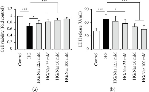
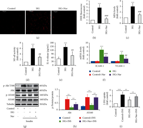
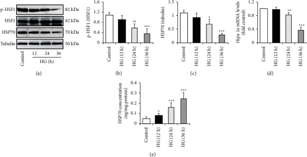
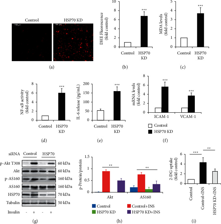
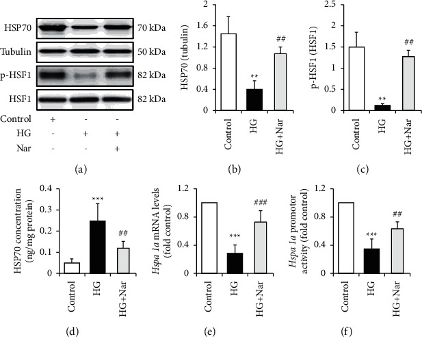
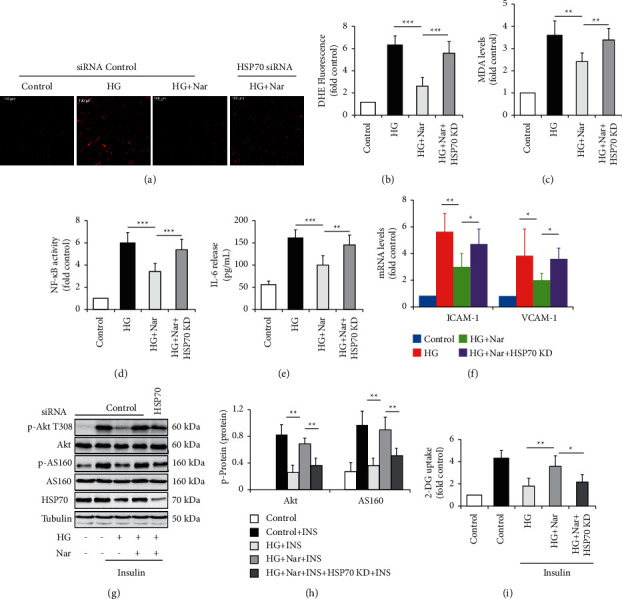
Endothelial dysfunction plays a pivotal role in the development and progression of diabetic vascular complications. Naringenin (Nar) is a flavanone bioactive isolated from citrus fruits known to have in vitro and in vivo antidiabetic properties. However, whether Nar affects endothelial function remains unclear in diabetes or under high-glucose (HG) condition. Using an in vitro model of hyperglycemia in human umbilical vein endothelial cells (HUVECs), we found that Nar administration markedly attenuated HG-induced alterations of endothelial function, evidenced by the mitigation of oxidative stress and inflammation, the reduction of cell adhesion molecular expressions, and the improvement of insulin resistance. We also found that HG exposure significantly reduced the levels of intracellular heat shock protein 70 (iHSP70 or iHSPA1A) and the release of HSP70 from HUVECs. HSP70 depletion mimicked and clearly diminished the protective effects of Nar on HG-induced alterations of endothelial function. In addition, Nar treatment significantly enhanced iHSP70 protein levels through a transcription-dependent manner. These results demonstrated that Nar could protect HUVECs against HG-induced alterations of endothelial function through upregulating iHSP70 protein levels. These findings are also helpful in providing new therapeutic strategies that are promising in the clinical use of Nar for the treatment of diabetes and diabetic complications.


Conflict of interest statement

The authors declare that they have no conflicts of interest regarding the publication of this paper.

Figures

Figure 1
Figure 1
Effect of Nar on HG-induced cell damage. HUVECs were exposed to NG (5.5 mM glucose) or HG (30 mM glucose) media with or without the indicated concentration of Nar for 72 h. (a) Effect of Nar on cell viability. (b) Effect of Nar on LDH concentration in the culture media. Data are expressed as means ± SD (n = 4). P < 0.05 and ∗∗∗P < 0.001 versus the indicated group.
Figure 2
Figure 2
Effect of Nar on HG-induced alterations of endothelial function. HUVECs were exposed to NG (5.5 mM glucose) or HG (30 mM glucose) media with or without the 50 mM of Nar for 36 h. To observe the effect of Nar on insulin signaling, the cells were treated with or without 100 nM of insulin for 10 min. (a) Effect of Nar on ROS formation by DHE staining. (b) Quantification of relative DHE fluorescence in (a). (c) Effect of Nar on intracellular MDA concentration. (d) Effect of Nar on NF-κB activity. (e) Effect of Nar on IL-6 concentration in the culture media. (f) Effect of Nar on mRNA levels of cell adhesion molecules ICAM-1 and VCAM-1. (g) Effect of Nar on insulin-stimulated phosphorylation of Akt and its downstream AS160. (h) Quantification of phosphorylated Akt and AS160 in (g). (i) Effect of Nar on insulin-stimulated 2-DG uptake. Data are expressed as means ± SD (n = 3). ∗∗P < 0.01 and ∗∗∗P < 0.001 versus control (NG) group or the indicated group. ##P < 0.01 and ###P < 0.001 versus high-glucose (HG) group.
Figure 3
Figure 3
Effect of HG on iHSP70 levels. HUVECs were exposed to NG (5.5 mM glucose) or HG (30 mM glucose) media for the indicated times. (a) Effect of HG on HSF1 phosphorylation and iHSP70 levels. (b) Quantification of HSF1 phosphorylation in (a). (c) Quantification of iHSP70 levels in (a). (d) Effect of HG on Hspa1a mRNA levels. (e) Effect of HG on HSP70 concentration in the culture media. Data are expressed as means ± SD (n = 3). P < 0.05, ∗∗P < 0.01, and ∗∗∗P < 0.001 versus control (NG) group.
Figure 4
Figure 4
Effect of HSP70 knockdown (KD) on endothelial function. The siRNA technique was used to silence HSP70 in HUVECs. The cells were then grown in NG (5.5 mM glucose) media for 36 h. To observe the effect of Nar on insulin signaling, the cells were treated with or without 100 nM of insulin for 10 min. (a) Effect of HSP70 KD on ROS formation by DHE staining. (b) Quantification of relative DHE fluorescence in (a). (c) Effect of HSP70 KD on intracellular MDA concentration. (d) Effect of HSP70 KD on NF-κB activity. (e) Effect of HSP70 KD on IL-6 concentration in the culture media. (f) Effect of HSP70 KD on mRNA levels of cell adhesion molecules ICAM-1 and VCAM-1. (g) Effect of HSP70 KD on insulin-stimulated phosphorylation of Akt and its downstream AS160. (h) Quantification of phosphorylated Akt and AS160 in (g). (i) Effect of HSP70 KD on insulin-stimulated 2-DG uptake. Data are expressed as means ± SD (n = 3). ∗∗P < 0.01 and ∗∗∗P < 0.001 versus siRNA control group or the indicated group.
Figure 5
Figure 5
Effect of Nar on HG-reduced HSP70 levels. HUVECs were exposed to NG (5.5 mM glucose) or HG (30 mM glucose) media with or without the 50 mM of Nar for 36 h. (a). Effect of Nar on iHSP70 levels and HSF1 phosphorylation. (b) Quantification of iHSP70 levels in (a). (c) Quantification of HSF1 phosphorylation in (a). (d) Effect of Nar on HSP70 concentration in the culture media. (e) Effect Nar on Hspa1a mRNA levels. (f) Effect of Nar on Hspa1a promotor activity. Data are expressed as means ± SD (n = 3). ∗∗P < 0.01 and ∗∗∗P < 0.001 versus control (NG) group. ##P < 0.01 and ###P < 0.001 versus high-glucose (HG) group.
Figure 6
Figure 6
HSP70 knockdown (KD) attenuated the protective effect of Nar on HG-induced alterations of endothelial function. The siRNA technique was used to silence HSP70 in HG-treated HUVECs. The cells were then exposed to NG (5.5 mM glucose) or HG (30 mM glucose) media with or without the 50 mM of Nar for 36 h. To observe the effect of Nar on insulin signaling, the cells were treated with or without 100 nM of insulin for 10 min. (a) Effect of HSP70 KD on ROS formation by DHE staining. (b) Quantification of relative DHE fluorescence in (a). (c) Effect of HSP70 KD on intracellular MDA concentration. (d) Effect of HSP70 KD on NF-κB activity. (e) Effect of HSP70 KD on IL-6 concentration in the culture media. (f) Effect of HSP70 KD on mRNA levels of cell adhesion molecules ICAM-1 and VCAM-1. (g) Effect of HSP70 KD on insulin-stimulated phosphorylation of Akt and its downstream AS160. (h) Quantification of phosphorylated Akt and AS160 in (g). (i) Effect of HSP70 KD on insulin-stimulated 2-DG uptake. Data are expressed as means ± SD (n = 3). P < 0.05, ∗∗P < 0.01, and ∗∗∗P < 0.001 versus the indicated group.

Similar articles

Cited by

References

    1. Shi Y., Vanhoutte P. M. Macro- and microvascular endothelial dysfunction in diabetes. Journal of Diabetes . 2017;9(5):434–449. - PubMed
    1. Wołoszyn-Durkiewicz A., Myśliwiec M. The prognostic value of inflammatory and vascular endothelial dysfunction biomarkers in microvascular and macrovascular complications in type 1 diabetes. Pediatric Endocrinology, Diabetes and Metabolism . 2019;25(1):28–35. - PubMed
    1. Poredos P., Poredos A. V., Gregoric I. Endothelial dysfunction and its clinical implications. Angiology . 2021;72(7):604–615. doi: 10.1177/0003319720987752. - DOI - PubMed
    1. Maruhashi T., Higashi Y. Pathophysiological association between diabetes mellitus and endothelial dysfunction. Antioxidants . 2021;10(8):p. 1306. doi: 10.3390/antiox10081306. - DOI - PMC - PubMed
    1. Takeda Y., Matoba K., Sekiguchi K., et al. Endothelial dysfunction in diabetes. Biomedicines . 2020;8(7):p. 182. doi: 10.3390/biomedicines8070182. - DOI - PMC - PubMed

LinkOut - more resources