Case Reports
. 2022 Jul 31:39:101715.
doi: 10.1016/j.rmcr.2022.101715. eCollection 2022.

Natural course of pulmonary hyalinizing granuloma over a decade

Affiliations
Case Reports

Natural course of pulmonary hyalinizing granuloma over a decade

Kohei Fujita et al. Respir Med Case Rep. .

Abstract

Background: Pulmonary hyalinizing granuloma (PHG) is a very rare pulmonary disease characterized by multiple fibrosclerotic inflammatory lung nodules. The disease is supposedly caused by an unusual immune response.

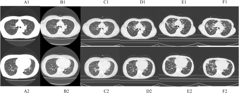
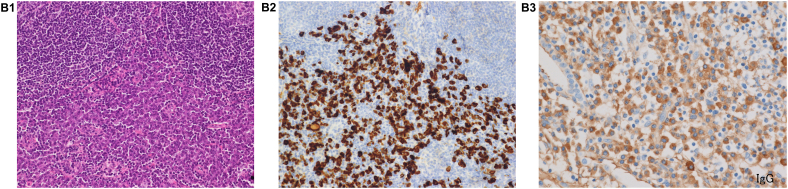
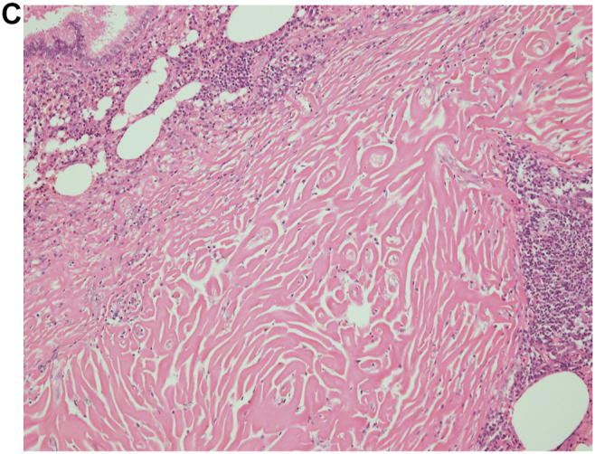
Case presentation: We present a case involving a 53-year-old female with a history of lumpectomy surgery due to invasive ductal carcinoma who was admitted for slowly progressive pulmonary nodules. The patient's elevated serum IgG4 level and the pathological findings obtained in surgical biopsy indicated IgG4-related lung disease. The nodules continued to enlarge despite administration of corticosteroid therapy, and we performed a second surgical biopsy to obtain a correct diagnosis. The pathological findings obtained in the second biopsy were different and consistent with the features of PHG.

Conclusions: In this report, the radiological follow-up data obtained after lumpectomy surgery demonstrate the very early stage of PHG and the following radiological changes over a decade, and the two surgical biopsies support us to realize the pathological change from previous diagnosed disease before PHG.

Keywords: IgG4-related lung disease; Interstitial pneumonia; Pulmonary hyalinizing granuloma.


Conflict of interest statement

The authors have no conflicts of interest to declare.

Figures

Fig. 1
Fig. 1
Chest computed tomography scans in the upper and lower lobes showing slowly progressive diffuse centrilobular nodules. (A, B, C, D, E and F were obtained in 2008, 2010, 2012, 2014, 2016, and 2018, respectively. 1: Upper area of the lung, 2: Lower area of the lung).
Fig. 2
Fig. 2
(A), View of the first surgical biopsy in 2013, which shows lymphoplasmacyte infiltration into the interstitium, peribronchovascular sheath, and subpleura (20x). Inset: High-power view showing storiform fibrosis (100x). (B-1), HE staining in the first surgical biopsy (x200). (B-2), IgG4 staining in the first surgical biopsy (x200). (B-3), IgG staining in the first surgical biopsy (x200). An IgG4/IgG-ratio of 50%. (C), View of the second surgical biopsy in 2016, which shows lamellar hyalinized collagen bundles around bronchovascular bundles with an associated dense rim of plasma cells and lymphocytes (100x).
Fig. 2
Fig. 2
(A), View of the first surgical biopsy in 2013, which shows lymphoplasmacyte infiltration into the interstitium, peribronchovascular sheath, and subpleura (20x). Inset: High-power view showing storiform fibrosis (100x). (B-1), HE staining in the first surgical biopsy (x200). (B-2), IgG4 staining in the first surgical biopsy (x200). (B-3), IgG staining in the first surgical biopsy (x200). An IgG4/IgG-ratio of 50%. (C), View of the second surgical biopsy in 2016, which shows lamellar hyalinized collagen bundles around bronchovascular bundles with an associated dense rim of plasma cells and lymphocytes (100x).

References

    1. Lhote R., Haroche J., Duron L., et al. Pulmonary hyalinizing granuloma: a multicenter study of 5 new cases and review of the 135 cases of the literature. Immunol. Res. 2017 Feb;65(1):375–385. - PubMed
    1. Engleman P., Liebow A.A., Gmelich J., et al. Pulmonary hyalinizing granuloma. Am. Rev. Respir. Dis. 1977;115:997–1008. - PubMed
    1. Ussavarungsi K., Khoor A., Jolles H.I., et al. A 40-year-old woman with multiple pulmonary nodules. Pulmonary hyalinizing granuloma. Chest. 2014 Dec;146(6):e198–e203. - PubMed
    1. Düzgün N., Kurtipek E., Esme H., et al. Pulmonary hyalinizing granuloma mimicking metastatic lung cancer. Case Rep. Pulmonol. 2015;2015 - PMC - PubMed
    1. Na K.J., (1), Song S.Y., Kim J.H., et al. Subpleural pulmonary hyalinizing granuloma presenting as a solitary pulmonary nodule. J. Thorac. Oncol. 2007 Aug;2(8):777–779. - PubMed

Publication types

LinkOut - more resources