

Decreased low-density lipoprotein receptor-related protein 1 expression in pro-inflammatory monocytes is associated with subclinical atherosclerosis
- PMID: 35958411
- PMCID: PMC9360420
- DOI: 10.3389/fcvm.2022.949778
Decreased low-density lipoprotein receptor-related protein 1 expression in pro-inflammatory monocytes is associated with subclinical atherosclerosis
Abstract
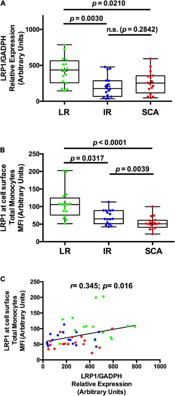
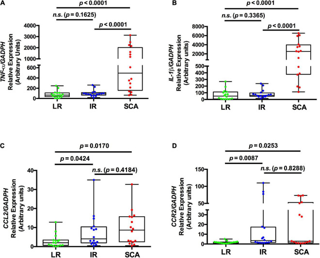
Subclinical atherosclerosis (SCA) occurs in asymptomatic individuals. Blood peripheral monocytes are involved in the development of atherosclerosis. Circulating monocytes acquire pro-inflammatory profiles, and they are involved in the early stages of atherosclerosis development. Low-density lipoprotein Receptor-related Protein 1 (LRP1) is expressed in monocytes, mainly in classical and intermediate subsets. Although LRP1 is highly expressed in macrophages and vascular smooth muscle cells (VSMCs) in atherosclerotic plaque formation, its expression in circulating monocytes has not been studied in SCA. The aim of this study was to characterize the LRP1 expression level in circulating monocytes of individuals with SCA and compared with individuals with low (LR) and intermediate (IR) risk of cardiovascular diseases, both without evidence of atherosclerotic lesions in carotid and coronary arteries. LRP1 and additional markers (CD11b, CD11c, and CD36) at cell surface of monocytes were analyzed by flow cytometry assays, whereas LRP1 and pro-inflammatory factors gene expressions were measured in isolated monocytes by quantitative RT-PCRs. Both LRP1 protein and LRP1 mRNA were significantly reduced in monocytes in SCA and IR respect to LR. Conversely, CD36, CD11b, and CD11c monocytic markers showed no significant changes between the different study groups. Finally, increased gene expressions of TNF-α and IL-1β were detected in monocytes of SCA, which were associated with decreased LRP1 expression at the cell surface in total monocytes. In summary, we propose that the decreased LRP1 expression at cell surface in total monocytes with pro-inflammatory profile is associated with the development of atherosclerosis in asymptomatic individuals.
Keywords: cardiovascular; cytokines; inflammation; lipids; lipoproteins; receptors.
Copyright © 2022 Albertini, Nicolas, Actis Dato, Ferrer, Tinti, Capra and Chiabrando.
Conflict of interest statement
The authors declare that the research was conducted in the absence of any commercial or financial relationships that could be construed as a potential conflict of interest.
Figures
Similar articles
-
Standardized flow cytometry assay for identification of human monocytic heterogeneity and LRP1 expression in monocyte subpopulations: decreased expression of this receptor in nonclassical monocytes.Cytometry A. 2014 Jul;85(7):601-10. doi: 10.1002/cyto.a.22455. Epub 2014 Mar 17. Cytometry A. 2014. PMID: 24639232
-
Circulating soluble low-density lipoprotein receptor-related protein 1 (sLRP1) concentration is associated with hypercholesterolemia: A new potential biomarker for atherosclerosis.Int J Cardiol. 2015 Dec 15;201:20-9. doi: 10.1016/j.ijcard.2015.07.085. Epub 2015 Aug 5. Int J Cardiol. 2015. PMID: 26285183
-
Adipocyte differentiation-related protein is induced by LRP1-mediated aggregated LDL internalization in human vascular smooth muscle cells and macrophages.J Lipid Res. 2007 Oct;48(10):2133-40. doi: 10.1194/jlr.M700039-JLR200. Epub 2007 Jul 9. J Lipid Res. 2007. PMID: 17620659
-
The Dual Role of Low-Density Lipoprotein Receptor-Related Protein 1 in Atherosclerosis.Front Cardiovasc Med. 2021 May 28;8:682389. doi: 10.3389/fcvm.2021.682389. eCollection 2021. Front Cardiovasc Med. 2021. PMID: 34124208 Free PMC article. Review.
-
Low-density lipoprotein receptor-related protein-1: role in the regulation of vascular integrity.Arterioscler Thromb Vasc Biol. 2014 Mar;34(3):487-98. doi: 10.1161/ATVBAHA.113.301924. Epub 2014 Feb 6. Arterioscler Thromb Vasc Biol. 2014. PMID: 24504736 Free PMC article. Review.
Cited by
-
Integrative bioinformatics frameworks for abdominal aortic aneurysm using GWAS meta-analysis, biological network construction, and structural modeling.Sci Rep. 2025 Jul 1;15(1):22331. doi: 10.1038/s41598-025-07989-1. Sci Rep. 2025. PMID: 40595262 Free PMC article.
-
Plasma Leptin Levels, Obstructive Sleep Apnea Syndrome, and Diabetes Are Associated with Obesity-Related Alterations of Peripheral Blood Monocyte Subsets.Immunohorizons. 2023 Mar 1;7(3):191-199. doi: 10.4049/immunohorizons.2300009. Immunohorizons. 2023. PMID: 36921085 Free PMC article.
-
The Peripheral Amyloid-β Nexus: Connecting Alzheimer's Disease with Atherosclerosis through Shared Pathophysiological Mechanisms.Neuromolecular Med. 2025 Mar 3;27(1):20. doi: 10.1007/s12017-025-08836-2. Neuromolecular Med. 2025. PMID: 40032716 Free PMC article. Review.
-
Mitigating atherosclerosis: Integrating vaccines with gene targets.Am Heart J Plus. 2025 Aug 6;57:100588. doi: 10.1016/j.ahjo.2025.100588. eCollection 2025 Sep. Am Heart J Plus. 2025. PMID: 40823653 Free PMC article. Review.
-
Multi-faceted role of LRP1 in the immune system.Front Immunol. 2023 Mar 20;14:1166189. doi: 10.3389/fimmu.2023.1166189. eCollection 2023. Front Immunol. 2023. PMID: 37020553 Free PMC article. Review.
References
-
- Albertini RA, Ferrer DG, Romagnoli PA, Tinti ME, Amigone JL, Capra R, et al. Association between cardiovascular disease risk scores and subclinical atherosclerosis prevalence in non-elderly adult patients from Argentina. Int J Cardiovasc Imaging. (2017) 33:1521–9. 10.1007/s10554-017-1152-9 - DOI - PubMed
-
- Bernal-Lopez MR, Llorente-Cortes V, Calleja F, Lopez-Carmona D, Mayas MD, Gomez-Huelgas R, et al. Effect of different degrees of impaired glucose metabolism on the expression of inflammatory markers in monocytes of patients with atherosclerosis. Acta Diabetol. (2013) 50:553–62. 10.1007/s00592-011-0337-2 - DOI - PubMed
LinkOut - more resources
Full Text Sources
Research Materials
Miscellaneous