

Design of functional biomaterials as substrates for corneal endothelium tissue engineering
- PMID: 35958516
- PMCID: PMC9362998
- DOI: 10.1093/rb/rbac052
Design of functional biomaterials as substrates for corneal endothelium tissue engineering
Abstract
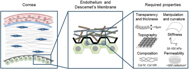
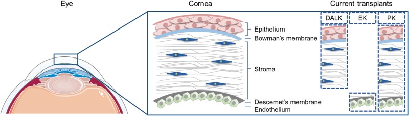
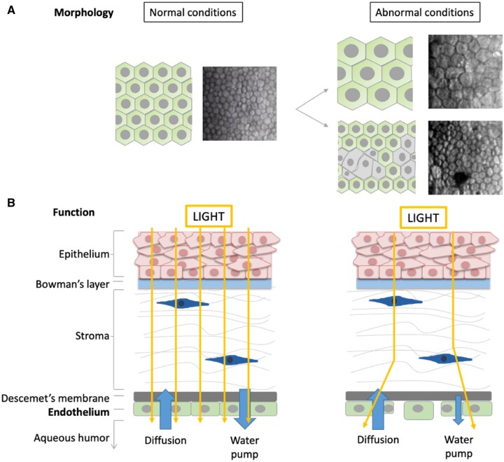
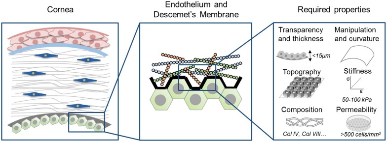
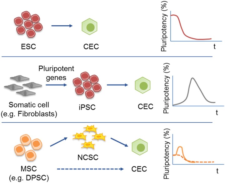
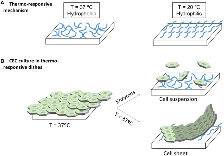
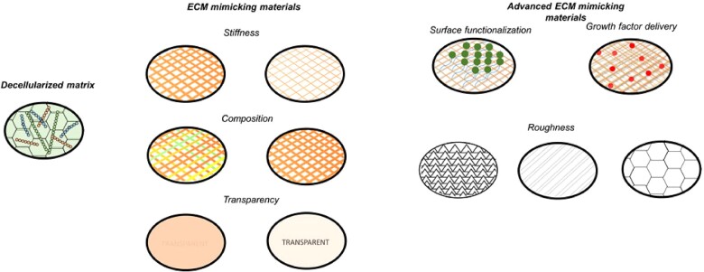
Corneal endothelium defects are one of the leading causes of blindness worldwide. The actual treatment is transplantation, which requires the use of human cadaveric donors, but it faces several problems, such as global shortage of donors. Therefore, new alternatives are being developed and, among them, cell therapy has gained interest in the last years due to its promising results in tissue regeneration. Nevertheless, the direct administration of cells may sometimes have limited success due to the immune response, hence requiring the combination with extracellular mimicking materials. In this review, we present different methods to obtain corneal endothelial cells from diverse cell sources such as pluripotent or multipotent stem cells. Moreover, we discuss different substrates in order to allow a correct implantation as a cell sheet and to promote an enhanced cell behavior. For this reason, natural or synthetic matrixes that mimic the native environment have been developed. These matrixes have been optimized in terms of their physicochemical properties, such as stiffness, topography, composition and transparency. To further enhance the matrixes properties, these can be tuned by incorporating certain molecules that can be delivered in a sustained manner in order to enhance biological behavior. Finally, we elucidate future directions for corneal endothelial regeneration, such as 3D printing, in order to obtain patient-specific substrates.
Keywords: biomaterials; cell therapy; corneal endothelium; tissue engineering.
© The Author(s) 2022. Published by Oxford University Press.
Figures
Similar articles
-
Corneal endothelium tissue engineering: An evolution of signaling molecules, cells, and scaffolds toward 3D bioprinting and cell sheets.J Cell Physiol. 2020 Oct 8. doi: 10.1002/jcp.30085. Online ahead of print. J Cell Physiol. 2020. PMID: 33090510 Review.
-
The Human Tissue-Engineered Cornea (hTEC): Recent Progress.Int J Mol Sci. 2021 Jan 28;22(3):1291. doi: 10.3390/ijms22031291. Int J Mol Sci. 2021. PMID: 33525484 Free PMC article. Review.
-
The Innovative Biomaterials and Technologies for Developing Corneal Endothelium Tissue Engineering Scaffolds: A Review and Prospect.Bioengineering (Basel). 2023 Nov 3;10(11):1284. doi: 10.3390/bioengineering10111284. Bioengineering (Basel). 2023. PMID: 38002407 Free PMC article. Review.
-
Stem cell-based therapeutic strategies for corneal epithelium regeneration.Tissue Cell. 2021 Feb;68:101470. doi: 10.1016/j.tice.2020.101470. Epub 2020 Nov 19. Tissue Cell. 2021. PMID: 33248403 Review.
-
Ex vivo expansion and characterization of human corneal endothelium for transplantation: a review.Stem Cell Res Ther. 2021 Oct 30;12(1):554. doi: 10.1186/s13287-021-02611-3. Stem Cell Res Ther. 2021. PMID: 34717745 Free PMC article. Review.
Cited by
-
Semiautomatic assessment of endothelial density and morphology in organ-cultured corneas - potential predictors for transplantation suitability and clinical outcome?Graefes Arch Clin Exp Ophthalmol. 2023 Sep;261(9):2593-2602. doi: 10.1007/s00417-023-06079-0. Epub 2023 Apr 28. Graefes Arch Clin Exp Ophthalmol. 2023. PMID: 37115267 Free PMC article.
-
Effects of ECM protein-coated surfaces on the generation of retinal pigment epithelium cells differentiated from human pluripotent stem cells.Regen Biomater. 2024 Aug 20;11:rbae091. doi: 10.1093/rb/rbae091. eCollection 2024. Regen Biomater. 2024. PMID: 39233867 Free PMC article.
-
Bioprinted Membranes for Corneal Tissue Engineering: A Review.Pharmaceutics. 2022 Dec 14;14(12):2797. doi: 10.3390/pharmaceutics14122797. Pharmaceutics. 2022. PMID: 36559289 Free PMC article. Review.
-
Advancements in Polymer Biomaterials as Scaffolds for Corneal Endothelium Tissue Engineering.Polymers (Basel). 2024 Oct 12;16(20):2882. doi: 10.3390/polym16202882. Polymers (Basel). 2024. PMID: 39458711 Free PMC article. Review.
-
The Effects of Biomimetic Surface Topography on Vascular Cells: Implications for Vascular Conduits.Adv Healthc Mater. 2024 Oct;13(27):e2400335. doi: 10.1002/adhm.202400335. Epub 2024 Jul 15. Adv Healthc Mater. 2024. PMID: 38935920 Free PMC article. Review.
References
-
- Mathews PM, Lindsley K, Aldave AJ, Akpek EK.. Etiology of global corneal blindness and current practices of corneal transplantation: a focused review. Cornea 2018;37:1198–6. - PubMed
-
- Gain P, Jullienne R, He Z, Aldossary M, Acquart S, Cognasse F, Thuret G.. Global survey of corneal transplantation and eye banking. JAMA Ophthalmol 2016;134:167–73. - PubMed
-
- Eye Bank Association of America, n.d. https://restoresight.org/members/publications/statistical-report/
Publication types
LinkOut - more resources
Full Text Sources
