Review
. 2022 Jul;11(4):380-388.
doi: 10.21037/acs-2021-bav-14.

Normal and abnormal development of the aortic valve and ascending aortic wall: a comprehensive overview of the embryology and pathology of the bicuspid aortic valve

Affiliations
Review

Normal and abnormal development of the aortic valve and ascending aortic wall: a comprehensive overview of the embryology and pathology of the bicuspid aortic valve

Nimrat Grewal et al. Ann Cardiothorac Surg. 2022 Jul.

Abstract

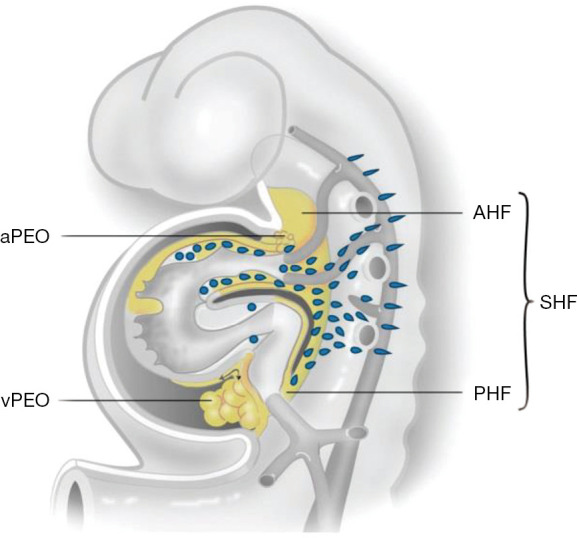
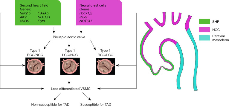
A bicuspid aortic valve (BAV) is the most prevalent congenital cardiac anomaly, in which the valve has only two leaflets, instead of the normal three. Patients with a BAV have an increased risk of aneurysm formation and the development of an aortic dissection. Vascular smooth muscle cells in both the non- and dilated aortic wall are characterized by a maturation defect in all BAV patients, as compared to patients with a tricuspid aortic valve, which can contribute to inherent developmental susceptibility. Besides structural abnormalities of the vascular wall, a turbulent blood flow, caused by bicuspid valve geometry, could expedite the pathological process in the aortic wall, leading to aortopathy. Although the risk for aortopathy is significant, not all BAV patients experience (acute) aortic complications in their lifespan, highlighting the complexity of the pathogenetic process. Recent studies have focused on the embryonic development of semilunar valves and the ascending aortic wall. Their findings highlight that a defect in the embryogenesis could not only explain the development of a malformed aortic valve but also the increased risk for ascending aorta and arch pathology. This review presents an overview of the normal and abnormal development of the aortic valve and the aortic wall: a common defect in early embryogenesis causes the development of a BAV and associated aortopathy.

Keywords: Bicuspid aortic valve (BAV); aortopathy; embryology; neural crest cells; pathology.


Conflict of interest statement

Conflicts of Interest: The authors have no conflicts of interest to declare.

Figures

Figure 1
Figure 1
The developing heart tube. During embryogenesis the SHF contributes to the arterial pole which includes the great arteries and the right ventricle from the AHF. From the PHF, second heart field cells enter the venous pole. The myocardium and intrapericardial part of the aorta are covered by epicardial cells that are provided by a proepicardial organ of the vPEO and aPEO, respectively. Not shown in this figure: neural crest cells migrate from the neural tube primarily to the arterial pole of the heart [adapted from Grewal et al. 2014 (5)]. SHF, second heart field; AHF, anterior population; PHF, posterior heart field; vPEO, venous pole; aPEO, arterial pole.
Figure 2
Figure 2
Developmental origin of the BAV and aortic wall. Progenitor neural crest and second heart field cells contribute to the normal development of an aortic valve and the vascular smooth muscle cells in the ascending aortic wall. Mutations in genes related to the neural crest and second heart lead to the development of a BAV and a maturation defect of vascular smooth muscle cells in the aortic root, ascending aorta and arch. Type 1 RCC-NCC and type 1 RCC-LCC have been attributed to defects in the neural crest and second heart field cell signalling respectively. RCC-NCC: fusion between the RCC and NCC. LCC-NCC: fusion between the LCC and NCC. RCC-LCC: fusion between the RCC and LCC. TAD: reduced glutathione. BAV, bicuspid aortic valve; RCC, right coronary cusp; NCC, non-coronary cusp; LCC, left coronary cusp; VSMC, vascular smooth muscle cell; SHF, second heart field.
Video
Video
Normal and abnormal development of the aortic valve and ascending aortic wall: an overview of the embryology and pathology of the bicuspid aortic valve

Similar articles

Cited by

References

    1. Tzemos N, Therrien J, Yip J, et al. Outcomes in adults with bicuspid aortic valves. JAMA 2008;300:1317-25. 10.1001/jama.300.11.1317 - DOI - PubMed
    1. Neri E, Barabesi L, Buklas D, et al. Limited role of aortic size in the genesis of acute type A aortic dissection. Eur J Cardiothorac Surg 2005;28:857-63. 10.1016/j.ejcts.2005.10.013 - DOI - PubMed
    1. Pape LA, Tsai TT, Isselbacher EM, et al. Aortic diameter >or = 5.5 cm is not a good predictor of type A aortic dissection: observations from the International Registry of Acute Aortic Dissection (IRAD). Circulation 2007;116:1120-7. 10.1161/CIRCULATIONAHA.107.702720 - DOI - PubMed
    1. Gittenberger-de Groot AC, Bartelings MM, Deruiter MC, et al. Basics of cardiac development for the understanding of congenital heart malformations. Pediatr Res 2005;57:169-76. 10.1203/01.PDR.0000148710.69159.61 - DOI - PubMed
    1. Grewal N, DeRuiter MC, Jongbloed MR, et al. Normal and abnormal development of the aortic wall and valve: correlation with clinical entities. Neth Heart J 2014;22:363-9. 10.1007/s12471-014-0576-2 - DOI - PMC - PubMed