Review
. 2022 Jul;11(4):402-417.
doi: 10.21037/acs-2022-bav-24.

Speaking a common language: the international consensus on bicuspid aortic valve nomenclature and classification

Affiliations
Review

Speaking a common language: the international consensus on bicuspid aortic valve nomenclature and classification

Hector I Michelena. Ann Cardiothorac Surg. 2022 Jul.

Abstract

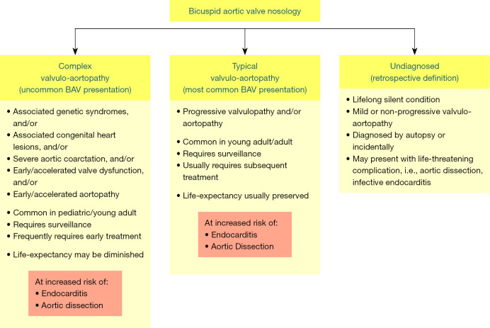
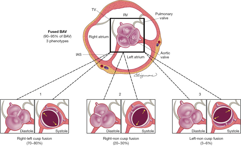
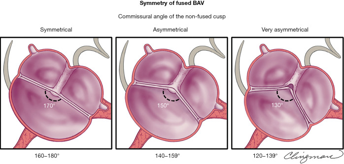
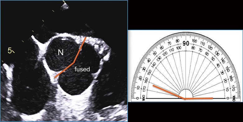
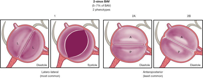
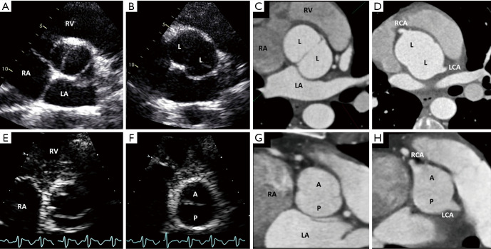
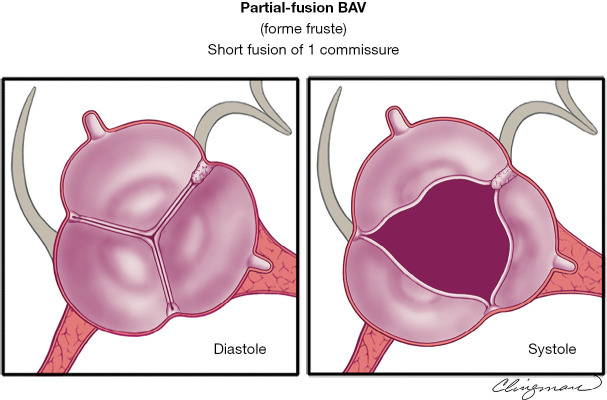
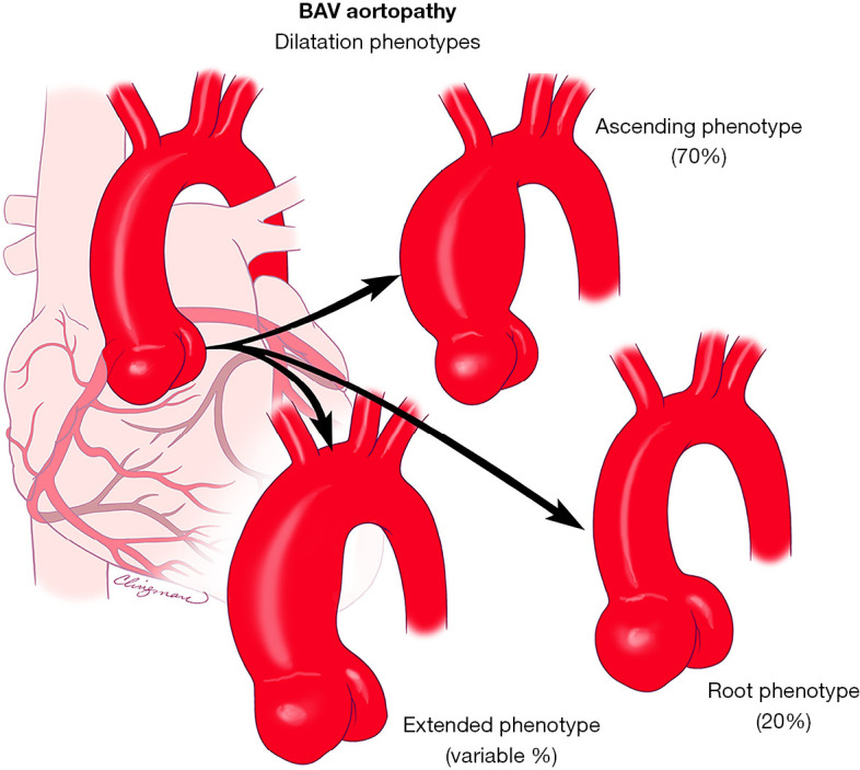
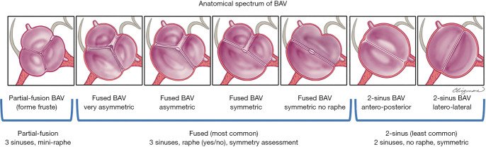
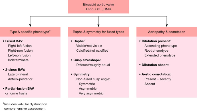
The congenital bicuspid aortic valve (BAV) condition is a valvulo-aortopathy with heterogeneous phenotypic expressions and clinical outcomes. A multitude of pre-existing classification systems, some extensive and some succinct, utilize combinations of numbers and/or letters to describe the condition. These diverse nomenclature systems are used according to the random preference of clinicians and researchers, generating confusion in clinical practice and research alike, effectively creating a barrier of communication at multiple levels. Based on imaging, pathology, surgery and clinical history evidence-based principles, and in utilizing the English language, the international nomenclature and classification consensus on the congenital BAV and its aortopathy offers a simple nosologic and phenotypic format that covers all possible phenotypes and clinical presentations of the bicuspid valvulo-aortopathy. From the nosology perspective, the valvulo-aortopathy has three major clinical presentations: (I) typical valvulo-aortopathy; (II) complex valvulo-aortopathy; and (III) undiagnosed or uncomplicated valvulo-aortopathy. From the valvular perspective, the congenital BAV has three major phenotypic expressions: (I) the fused BAV; (II) the 2-sinus BAV; and (III) the partial fusion (forme fruste) BAV. From the aortopathy perspective, the condition has three major phenotypic expressions: (I) ascending phenotype; (II) root phenotype; and (III) extended phenotypes. The international consensus is intended for universal use by Clinicians (pediatric and adult), echocardiography sonographers and physicians, cardiovascular advanced-imaging specialists, interventional cardiologists, cardiovascular surgeons, pathologists, geneticists and researchers encompassing clinical and basic research areas. When new landmark research is available, this international consensus may be subject to change in accordance with evidence-based data.

Keywords: Bicuspid aortic valve (BAV); aortopathy; classification; nomenclature.


Conflict of interest statement

Conflicts of Interest: Artivion (Consulting fees, speaker), and Mayo co-PI of PROACT Xa trial, Biostable Science and Engineering, Inc. (Consulting fees, speaker). The disclosures are unrelated to the keynote lecture.

Figures

Figure 1
Figure 1
Nosology of the congenital BAV condition. Left: anatomically and prognostically complex presentations of the BAV valvulo-aortopathy are those associated with syndromes, left-sided obstructions, significant aortic coarctation, early/accelerated valve dysfunction (stenosis or regurgitation) and/or early aortopathy. Middle: the anatomically and prognostically typical valvulo-aortopathy is usually diagnosed in young and middle-aged adults although it may be diagnosed in children as well and comprises various degrees of progressive valvular dysfunction (mostly high incidence of aortic stenosis), with a high cumulative incidence of aortopathy over the long-term, manifested as thoracic aortic dilatation, without other major associated conditions. Right: the undiagnosed form is diagnosed retrospectively (without any BAV-related complications, some are diagnosed post-mortem), or may present for the first time with a life-threatening complication such as aortic dissection or infective endocarditis. Modified from Michelena et al. (1-4) with permission. BAV, bicuspid aortic valve.
Figure 2
Figure 2
Diagnosis of congenital BAV by transthoracic echocardiography and pathology. (A) Parasternal short-axis aortic valve systolic still image demonstrating the existence of only 2 commissures (asterisks) delimiting only 2 cusps. (B) Parasternal long-axis still shows systolic doming of the fused (conjoined) cusp (arrow), common for right-left coronary cusp fusion. (C) Pathological congenital BAV specimen shows the area of the raphe (dashed line) from the left ventricular perspective, forming an obtuse angle between the fused cusps. (D) Ventricular side of a tricuspid aortic valve with acquired rheumatic fusion shows the cleavage plane with acute angle (yellow arrow). From Michelena et al. (1-4) with permission. LV, left ventricle; BAV, bicuspid aortic valve.
Figure 3
Figure 3
The aortic root complex. (A) Schematic drawing of the aortic root: the blue line indicates the virtual basal-ring (aortic annulus); the yellow line depicts the ventriculo-aortic junction (whose non-planar nature is schematically emphasized) (33); the red lines show the crown-shaped attachments of the cusps to the wall of the aortic sinuses [note the different height of the underdeveloped commissure (pseudocommissure, asterisk) compared to that of the other 2 true commissures]; the brown line depicts the STJ. (B) All the above boundaries and structures are reported (same colors as above) in an anatomical specimen of a normal aortic root and tricuspid aortic valve. (C) Echocardiographic view of the aortic root: the levels of the aortic annulus, ventriculo-aortic junction and STJ are shown (same colors as above). It is important to recognize that it is the measurement of the virtual annulus, sinuses and STJ that have clinical and practical implications for the patient with BAV. RCO: blue pin and arrow; LCO: green pin and arrow. From Michelena et al. (1-4) with permission. STJ, sinotubular junction; BAV, bicuspid aortic valve; RCO, right coronary orifice; LCO, left coronary orifice.
Figure 4
Figure 4
Schematic of fused BAV phenotypes as seen by parasternal short-axis transthoracic echocardiography. Applicable to similar tomographic views by cardiac computed tomography and cardiovascular magnetic resonance, the figure demonstrates the 3 fused BAV phenotypes as zoomed views of the base of the heart (black square contains a tricuspid aortic valve for reference) for anatomical landmark correlation. Note that all fused BAVs have 3 distinguishable aortic sinuses. Note the oval (American football shape) systolic opening of these 3 valves, as opposed to the triangular opening of a tricuspid aortic valve. 1: Right-left cusp fusion (most common) with visible raphe, different size/shape functional cusps [the non-fused cusp (non-coronary) is commonly a bit larger than the others]. 2: Right-non cusp fusion with visible raphe, different size/shape functional cusps [the non-fused cusp (left) is larger than the others]. 3: Left-non cusp fusion with visible raphe (least common), different size/shape functional cusps [the non-fused cusp (right) is larger than the others]. These 3 fused phenotypes may not have a visible raphe and may also be symmetrical (Figure 5). From Michelena et al. (1-4) with permission. BAV, bicuspid aortic valve; RC, right cusp; LC, left cusp; NC, noncoronary cusp; RV, right ventricle; TV, tricuspid valve; IAS, interatrial septum.
Figure 5
Figure 5
Schematic of the transthoracic echocardiographic evaluation of fused BAV symmetry in the parasternal short-axis. Applicable to similar tomographic views by cardiac computed tomography and cardiovascular magnetic resonance, the figure demonstrates different commissural angles of the non-fused cusps (applicable to the 3 fused BAV phenotypes, only right-left cusp fusion is shown), which define symmetry. Left panel: symmetrical (angle 160–180°) right-left cusp fusion BAV with a raphe, where the 2 functional cusps are almost same size/shape (the non-fused cusp is a little larger), and the commissural angle of the non-fused cusp is about 170°. Middle panel: asymmetrical (angle 140–159°) right-left fusion BAV with a raphe, and the commissural angle of the non-fused cusp is about 150°. Right panel: very asymmetrical (angle 120–139°) right-left fusion BAV shows retraction of the conjoined cusp at the raphal area and the commissural angle of the non-fused cusp is about 130°. Note that retraction is more prominent as the angle decreases, which may cause aortic regurgitation. From Michelena et al. (1-4) with permission. BAV, bicuspid aortic valve.
Figure 6
Figure 6
Transoesophageal echocardiographic measurement of the commissural angle of the non-fused cusp prior to valve repair. Applicable to similar tomographic views by cardiac computed tomography and cardiovascular magnetic resonance, after careful visualization of systolic and diastolic motion of this regurgitant fused-type right-left cusp fusion BAV, the non-fused commissures are identified, and a line is drawn from the position of the commissures to the center of the valve in diastole (left). The angle of the non-fused cusp (N) is then carefully measured at approximately 162° on the protractor to the right, suggesting a good repair chance. From Michelena et al. (1-4) with permission. BAV, bicuspid aortic valve.
Figure 7
Figure 7
Schematic of the 2-sinus BAV phenotypes as seen by the transthoracic echocardiogram parasternal short-axis. Applicable to similar tomographic views by cardiac computed tomography and cardiovascular magnetic resonance, the figure demonstrates 2-sinus BAV phenotypes as zoomed views of the base of the heart for anatomical landmark correlation. 1: 2-sinus laterolateral BAV with only 2 distinguishable aortic sinuses and 2 roughly equal size/shape cusps each occupying 180° of the circumference, with a 180° angle of the commissures. The 2-sinus BAV laterolateral phenotype has 1 coronary artery arising from each sinus. 2A: 2-sinus anteroposterior BAV with only 2 distinguishable aortic sinuses and 2 similar size/shape cusps each occupying 180° of the circumference, with 180° angle of the commissures; 1 coronary arising from each sinus. 2B: 2-sinus anteroposterior BAV, which resembles a fused right-left fusion without a raphe, with only 2 distinguishable aortic sinuses and 2 similar size/shape cusps each occupying 180° of the circumference; with coronaries arising from the same sinus. From Michelena et al. (1-4) with permission. BAV, bicuspid aortic valve.
Figure 8
Figure 8
Diastolic and systolic still images of the 2-sinus BAV phenotypes obtained from transthoracic echocardiogram and diastolic still images by ECG-gated cardiac CT. (A) 2-sinus laterolateral BAV in systole, with only 2 distinguishable aortic sinuses in diastole (B) and roughly equal size/shape cusps occupying 180° of the circumference, reproducible on an equivalent tomographic cut as seen with CT (C). Note the coronary arteries arising 1 from each sinus (D). (E) 2-sinus anteroposterior BAV in systole, with only 2 distinguishable aortic sinuses and roughly equal size/shape cusps occupying 180° of the circumference (F, diastolic still frame), reproducible on an equivalent tomographic cut as seen with CT (G). Note coronary arteries arising 1 from each sinus in this particular example (H). From Michelena et al. (1-4) with permission. BAV, bicuspid aortic valve; ECG, electrocardiographic; CT, computed tomography; RA, right atrium; RV, right ventricle; LA, left atrium; A, anterior cusp; P, posterior cusp; L, lateral cusp; RCA, right coronary artery; LCA, left coronary artery.
Figure 9
Figure 9
Schematic of the partial-fusion BAV phenotype as seen by the transthoracic echocardiographic parasternal short axis. Left panel: The imaging appearance in diastole of the partial-fusion or forme fruste BAV is that of a tricuspid aortic valve. Right panel: The imaging diagnosis is usually made in systole. Although the opening appears triangular, there is a small fusion of the right and left cusps with a “mini-raphe”. These can be suspected by looking at the transoesophageal echocardiogram and sometimes confirmed by a 3D transoesophageal echocardiogram, cardiovascular magnetic resonance or cardiac computed tomography. Definitive confirmation is usually obtained by surgical inspection or pathology. From Michelena et al. (1-4) with permission. BAV, bicuspid aortic valve.
Figure 10
Figure 10
BAV aortopathy phenotypes. On the left is a normal aorta. Top: the most common phenotype, the ascending phenotype (approximately 70%), is preferential dilatation of the tubular ascending aorta. Middle: the root phenotype involves preferential dilatation of the root, seen in approximately 20% of patients with BAVs with aortopathy. Bottom: the extended phenotype shows dilatation of the root, the ascending aorta and the arch. The most common extended phenotypes are root plus ascending aorta and ascending aorta plus arch. From Michelena et al. (1-4) with permission. BAV, bicuspid aortic valve.
Figure 11
Figure 11
Schematic of the BAV anatomical spectrum using the most common right-left cusp fusion as an example. From left to right, note the partial-fusion BAV resembling a tricuspid aortic valve likely associated with a mild embryologic defect, then spanning a continuum of fused BAV with increasing commissural angles and increasing cusp size/shape similarity, ending with the 2-sinus BAV phenotypes that represent almost perfect “bicuspidity” and are likely associated with the most severe embryologic defects. From Michelena et al. (1-4) with permission. BAV, bicuspid aortic valve.
Figure 12
Figure 12
Critical imaging evaluation of the congenital BAV condition. BAV, bicuspid aortic valve; CCT, cardiac computed tomography; CMR, cardiac magnetic resonance.
Video
Video
Speaking a common language: the international consensus on bicuspid aortic valve nomenclature and classification.

Similar articles

Cited by

References

    1. Michelena HI, Corte AD, Evangelista A, et al. International Consensus Statement on Nomenclature and Classification of the Congenital Bicuspid Aortic Valve and Its Aortopathy, for Clinical, Surgical, Interventional and Research Purposes. Radiol Cardiothorac Imaging 2021;3:e200496. 10.1148/ryct.2021200496 - DOI - PMC - PubMed
    1. Michelena HI, Della Corte A, Evangelista A, et al. International consensus statement on nomenclature and classification of the congenital bicuspid aortic valve and its aortopathy, for clinical, surgical, interventional and research purposes. J Thorac Cardiovasc Surg 2021;162:e383-414. 10.1016/j.jtcvs.2021.06.019 - DOI - PubMed
    1. Michelena HI, Della Corte A, Evangelista A, et al. International Consensus Statement on Nomenclature and Classification of the Congenital Bicuspid Aortic Valve and Its Aortopathy, for Clinical, Surgical, Interventional and Research Purposes. Ann Thorac Surg 2021;112:e203-35. 10.1016/j.athoracsur.2020.08.119 - DOI - PubMed
    1. Michelena HI, Della Corte A, Evangelista A, et al. International consensus statement on nomenclature and classification of the congenital bicuspid aortic valve and its aortopathy, for clinical, surgical, interventional and research purposes. Eur J Cardiothorac Surg 2021;60:448-76. 10.1093/ejcts/ezab038 - DOI - PubMed
    1. Michelena HI, Della Corte A, Evangelista A, et al. Speaking a common language: Introduction to a standard terminology for the bicuspid aortic valve and its aortopathy. Prog Cardiovasc Dis 2020;63:419-24. 10.1016/j.pcad.2020.06.006 - DOI - PubMed