. 2022 Jul 25:13:962925.
doi: 10.3389/fimmu.2022.962925. eCollection 2022.

Role of human metapneumovirus glycoprotein G in modulation of immune responses

Affiliations

Role of human metapneumovirus glycoprotein G in modulation of immune responses

Thangam Sudha Velayutham et al. Front Immunol. .

Abstract

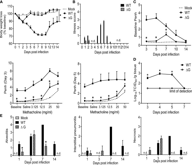
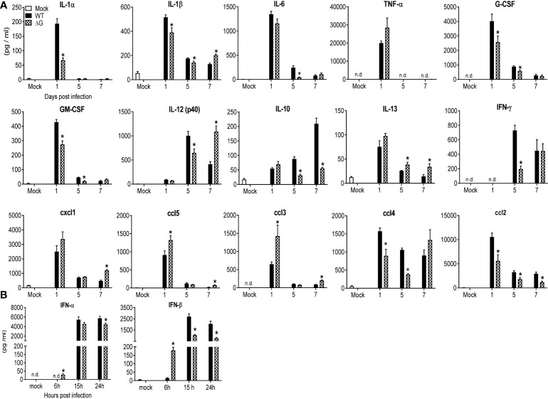
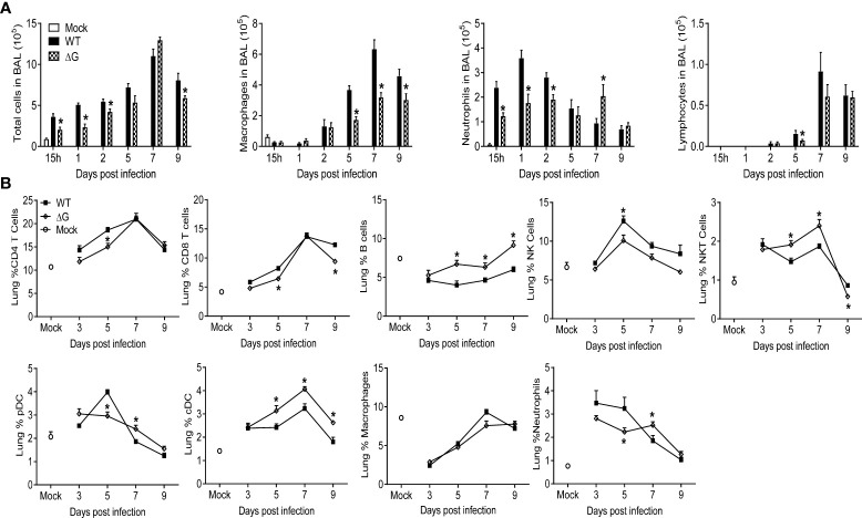
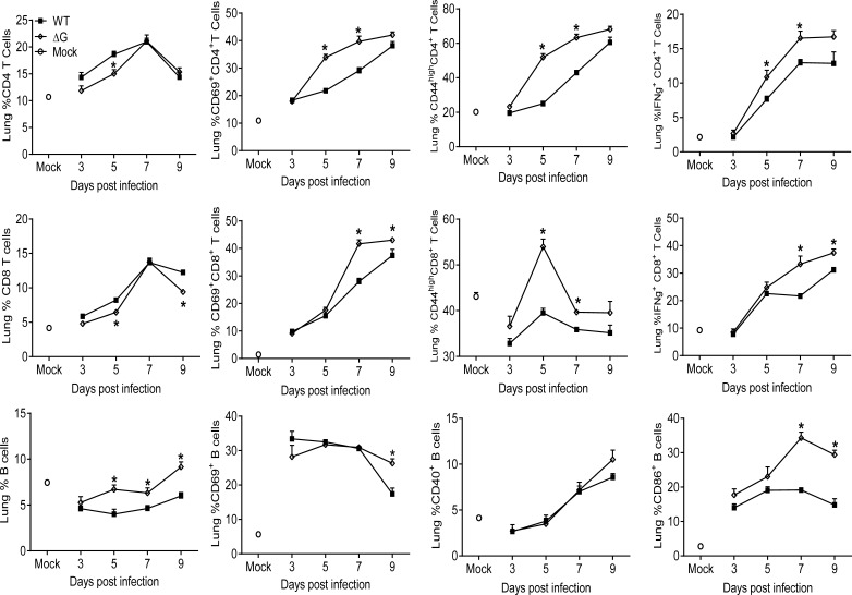
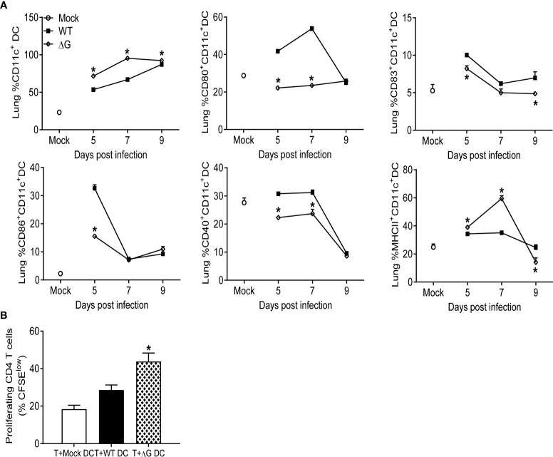
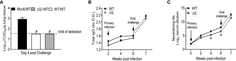
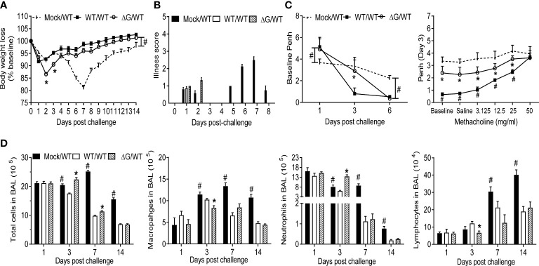
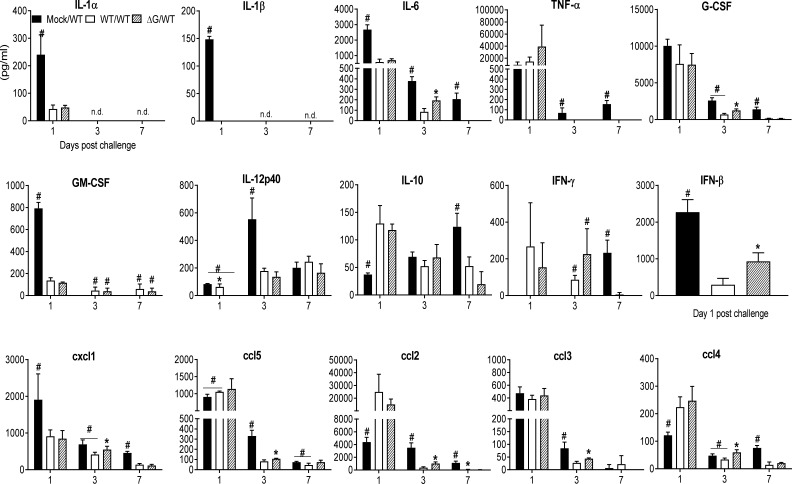
Human metapneumovirus (hMPV) is an important pathogen responsible for acute respiratory tract infections in children, the elderly, and immunocompromised patients, with no effective treatment or vaccine currently available. Knowledge of virus- and host-specific mechanisms contributing to the pathogenesis of hMPV infection is still limited. Studies have shown that hMPV surface glycoprotein G is an important virulence factor, by inhibiting innate immune signaling in airway epithelial cells and immune cells. In this study, we investigated the role of G protein in modulating innate and adaptive immune responses in mice infected with a recombinant virus with deletion of G protein (rhMPV-ΔG). Results show that rhMPV-ΔG was strongly attenuated, as it did not induce significant clinical disease, airway obstruction and airway hyperresponsiveness (AHR), compared to infection with a control strain (rhMPV-WT). By analysis of cells in bronchoalveolar fluid and lung tissue, as well as cytokine production, we found that G protein mediates aspects of both innate and adaptive immune responses, including neutrophils, dendritic cells, natural killer cells and B cells. Lung T cells recruited in response to rhMPV-ΔG had a significantly higher activated phenotype compared to those present after rhMPV-WT infection. Despite highly attenuation characterized by low levels of replication in the lung, rhMPV-ΔG was able to induce neutralizing antibodies and to protect mice from a secondary hMPV challenge. However, challenged mice that had received rhMPV-ΔG as primary infection showed some signs of lung disease at the earliest time points, which were less evident in mice that had received the rhMPV-WT strain as primary infection. These results demonstrate some of the mechanisms by which G protein could contribute to airway disease and modulate immune response to hMPV infection.

Keywords: RNA virus; hMPV G protein; immune response; lung disease; mouse model.


Conflict of interest statement

The authors declare that the research was conducted in the absence of any commercial or financial relationships that could be construed as a potential conflict of interest.

Figures

Figure 1
Figure 1
Clinical disease and virus replication. 6-8 wk old female BALB/c mice were either infected with rhMPV (-WT or -ΔG) or mock infected. (A) Change in body weight was measured over a period of 14 days. Body weight is expressed as percentage of baseline weight. (B) Illness score for infected mice infected was assessed daily by two observers using a grading scale from 0 to 5. (C) Baseline airway obstruction (left panel) and obstruction post-methacholine challenge (two right panels) was measured over a period of 14 days by unrestrained plethysmography. Day 3 and 5 p.i. are shown. (D) Infected mice were sacrificed at days 3, 4, 5 and 7 p.i. to determine viral titers by TCID50 assay. The lower limit of detection of this assay is 1.5 log10/gram of tissue. (E) Lungs were formalin-fixed for slide preparation, and hematoxylin and eosin stained. Alveolitis, interstitial pneumonitis and necrosis were scored on lung sections by a pathologist. Data are expressed as mean ± SEM of four to six animals/group and is representative of one of three independent experiments. *P<0.05 when comparing rhMPV -ΔG to -WT infected mice. rhMPV -ΔG =ΔG; rhMPV -WT = WT. n.d., not detected.
Figure 2
Figure 2
BAL fluid levels of pro-inflammatory cytokines, chemokines and type I IFNs. Mice were either infected with rhMPV-WT or -ΔG or mock infected and sacrificed at various time points p.i. to collect BAL fluid. (A) Levels of cytokines and chemokines in BAL fluid were measured by Bio-Plex. (B) Levels of IFN-α/β were measured by ELISA. Data are expressed as mean ± SEM of four to six animals/group and is representative of one of three independent experiments. *P<0.05 when comparing rhMPV- ΔG infected mice to rhMPV- WT infected mice. rhMPV -ΔG =ΔG; rhMPV -WT = WT. n.d., not detected.
Figure 3
Figure 3
Cell recruitment in BAL and lung tissue. Mice were either infected with rhMPV-WT or rhMPV- ΔG/WT or mock-infected and sacrificed at various time points p.i. to collect BAL fluid and lungs. (A) BAL fluid was subjected to cytospin and analyzed for total and differential cell counts. (B) Cells were isolated from lungs and stained for cell-type specific CD markers and live cells analyzed by flow cytometry. Data are expressed as mean ± SEM of four mice/group and is representative of one of three independent experiments. * P<0.05 when comparing rhMPV- ΔG infected mice to rhMPV- WT infected mice. rhMPV -ΔG =ΔG; rhMPV -WT = WT.
Figure 4
Figure 4
Lymphocyte recruitment and activation. Mice were either infected with rhMPV-WT or -ΔG or mock-infected and sacrificed at various time points p.i. to collect lungs. Single cell suspension of lungs was obtained and stained for CD4 (CD3+CD4+) and CD8 (CD3+CD8+) T cells and B cells (CD19+B220+). Lymphocytes were further stained for expression of various activation markers (CD69, CD44, CD40, CD86). IFN-γ produced by T cells was detected by intracellular staining upon stimulation with PMA/ionomycin and analyzed by flow cytometry. * P<0.05 when comparing rhMPV- ΔG infected mice to rhMPV- WT infected mice. rhMPV -ΔG =ΔG; rhMPV -WT = WT.
Figure 5
Figure 5
Dendritic cells recruitment to the lung, activation, and proliferation. Mice were either infected with rhMPV-WT or -ΔG or mock-infected and sacrificed at various time points p.i. to collect lungs. Single cell suspension of lungs was obtained and stained for DC markers. (A) CD11c+DCs population was enriched from the lung cells and analyzed by flow cytometry for the expression of activation markers (CD80, CD83, CD86, CD40, MHC class II). (B) CD11c+ cells isolated from lungs of infected mice at day 7 p.i. were loaded with 10 µg/mL of OVA peptide for 2h prior to co-culture with T cells. CD4+ T cells isolated from spleen of DO11.10 mice were labeled with CFSE and co-cultured with DCs at a ratio of 1∶2 (DC:T). T cell proliferation was measured by CFSE dilution. Cultures without antigen served as controls. The bar graph shows the percentage of proliferating (CFSE low) T cells among the total CD4+ T cell population. * P<0.05 when comparing rhMPV- ΔG infected mice to rhMPV- WT infected mice. rhMPV -ΔG =ΔG; rhMPV -WT = WT.
Figure 6
Figure 6
Virus replication and antibody titer in response to virus challenge. Mock and previously rhMPV-WT or -ΔG infected mice were challenged with rhMPV-WT virus 6 weeks after primary infection. (A) Mice were sacrificed on day 4 post challenge (peak of viral replication), and lungs were harvested for determining viral titers by TCID50 assay. (B) Total hMPV-specific IgG titers in serum were determined by ELISA. (C) hMPV-specific neutralizing antibody titer was determined by plaque neutralization assay. Data are expressed as mean ± SEM of four animals/group and is representative of one of two independent experiments. *P<0.05 when comparing rhMPV-WT and -ΔG; #P<0.05 when comparing rhMPV-WT and -ΔG and mock- mice challenged with rhMPV -WT. rhMPV -ΔG =ΔG; rhMPV -WT = WT; Mock/rhMPV -WT = Mock/W; rhMPV -ΔG/rhMPV –WT =ΔG/WT; rhMPV -WT/rhMPV -WT = WT/WT.
Figure 7
Figure 7
Clinical disease, cellular immune responses in response to virus challenge. Mock and with rhMPV-WT or -ΔG infected mice were challenged with rhMPV -WT virus 6 weeks after primary infection and sacrificed at various time points p.i. to collect BAL fluid. Change in body weight (A) and illness score (B) were measured over a period of 14 days. Data are expressed as mean ± SEM of four animals/group and the figure represents cumulative data from two independent experiments. (C) Penh values were measured at baseline (airway obstruction) and post-methacholine challenge by unrestrained plethysmography. Data are expressed as mean ± SEM of four animals/group and is representative of one of two independent experiments. (D) BAL fluid was subjected to cytospin and analyzed for total and differential cell counts. *P<0.05 when comparing rhMPV -WT and -ΔG mice challenged with rhMPV -WT.; #P<0.05 when comparing rhMPV (-WT and -ΔG) challenged with rhMPV -WT and mock- mice challenged with rhMPV -WT. Mock/rhMPV -WT = Mock/WT; rhMPV -ΔG/rhMPV –WT =ΔG/WT; rhMPV -WT/rhMPV -WT = WT/WT.
Figure 8
Figure 8
BAL fluid levels of pro-inflammatory cytokines, chemokines and type I IFNs in response to virus challenge. Mock and previously rhMPV-WT or -ΔG infected mice were challenged with rhMPV-WT virus as previously described in Material and Methods. Levels of cytokines and chemokines in BAL fluid were measured by Bio-Plex. Levels of IFN-β were measured by ELISA. Data are expressed as mean ± SEM of four to six animals/group and is representative of one of three independent experiments. *P<0.05 when comparing rhMPV -WT and -ΔG mice challenged with rhMPV -WT.; #P<0.05 when comparing rhMPV (-WT and -ΔG) challenged with rhMPV -WT and mock- mice challenged with rhMPV -WT. Mock/rhMPV -WT = Mock/WT; rhMPV -ΔG/rhMPV –WT =ΔG/WT; rhMPV -WT/rhMPV -WT = WT/WT. n.d., not detected.

Similar articles

Cited by

References

    1. Afonso CL, Amarasinghe GK, Banyai K, Bao Y, Basler CF, Bavari S, et al. . Taxonomy of the order mononegavirales: Update 2016. Arch Virol (2016) 161(8):2351–60. doi: 10.1007/s00705-016-2880-1 - DOI - PMC - PubMed
    1. Kahn JS. Epidemiology of human metapneumovirus. Clin Microbiol Rev (2006) 19(3):546–57. doi: 10.1128/CMR.00014-06 - DOI - PMC - PubMed
    1. Williams JV, Wang CK, Yang CF, Tollefson SJ, House FS, Heck JM, et al. . The role of human metapneumovirus in upper respiratory tract infections in children: A 20-year experience. J Infect Dis (2006) 193(3):387–95. doi: 10.1086/499274 - DOI - PMC - PubMed
    1. van den Hoogen BG, de Jong JC, Groen J, Kuiken T, de Groot R, Fouchier RA, et al. . A newly discovered human pneumovirus isolated from young children with respiratory tract disease. Nat Med (2001) 7(6):719–24. doi: 10.1038/89098 - DOI - PMC - PubMed
    1. Boivin G, Mackay I, Sloots TP, Madhi S, Freymuth F, Wolf D, et al. . Global genetic diversity of human metapneumovirus fusion gene. Emerg Infect Dis (2004) 10(6):1154–7. doi: 10.3201/eid1006.031097 - DOI - PMC - PubMed

Publication types

LinkOut - more resources