

Effect of low-dose rituximab treatment on autoimmune nodopathy with anti-contactin 1 antibody
- PMID: 35958552
- PMCID: PMC9362773
- DOI: 10.3389/fimmu.2022.939062
Effect of low-dose rituximab treatment on autoimmune nodopathy with anti-contactin 1 antibody
Abstract
Background: Autoimmune nodopathy with anti-contactin-1 (CNTN1) responds well to rituximab instead of traditional therapies. Although a low-dose rituximab regimen was administered to patients with other autoimmune diseases, such as myasthenia gravis and neuromyelitis optica spectrum disorders, and satisfactory outcomes were obtained, this low-dose rituximab regimen has not been trialed in anti-CNTN1-positive patients.
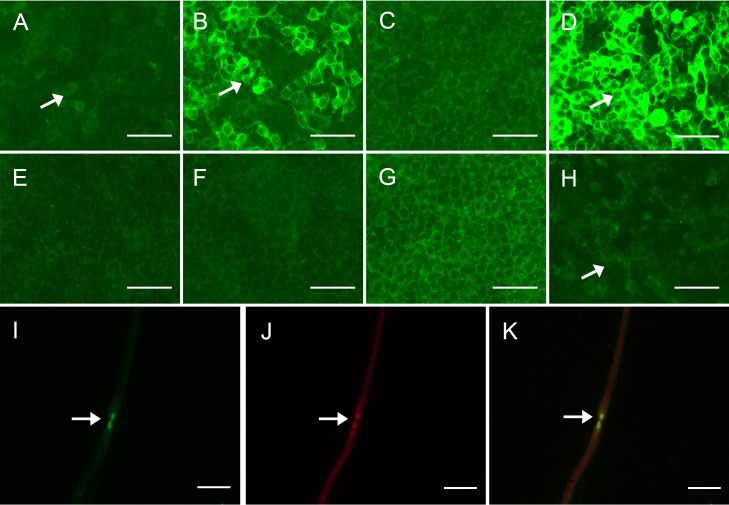
Methods: Anti-CNTN1 nodopathy patients were enrolled in this prospective, open-label, self-controlled pilot study. A cell-based assay was used to detect anti-CNTN1 antibodies and their subclasses in both serum and cerebrospinal fluid. Clinical features were evaluated at baseline, 2 days, 14 days, and 6 months after single low-dose rituximab treatment (600 mg). The titers of the subclasses of anti-CNTN1 antibody and peripheral B cells were also evaluated at baseline, 2 days, and 6 months after the rituximab regimen.
Results: Two patients with anti-CNTN1 antibodies were enrolled. Both patients had neurological symptoms including muscle weakness, tremor, sensory ataxia, numbness and mild nephrotic symptoms. In the field of neurological symptoms, sensory ataxia markedly improved, and the titer of anti-CNTN1 antibody as well as CD19+ B cells decreased only two days following low-dose rituximab treatment. Other neurological symptoms improved within two weeks of rituximab treatment. At the 6-month follow-up, all neurological symptoms steadily improved with steroid reduction, and both the anti-CNTN1 antibody titer and CD19+ B cells steadily decreased. No adverse events were observed after this single low-dose rituximab treatment.
Conclusions: We confirmed the clinical efficacy of low-dose rituximab by B cell depletion in autoimmune nodopathy with anti-CNTN1 antibody. This rapid and long-lasting response suggests that low-dose rituximab is a promising option for anti-CNTN1 nodopathy.
Keywords: anti-CNTN1 antibodies; antibody titer; autoimmune nodopathy; low-dose rituximab; treatment outcome.
Copyright © 2022 Hou, Zhang, Yu, Wang, Zhang, Bai, Yan, Ma, Li, Ji, Cao and Wang.
Conflict of interest statement
The authors declare that the research was conducted in the absence of any commercial or financial relationships that could be construed as potential conflict of interest.
Figures
Similar articles
-
Anti-rituximab antibodies in patients with refractory autoimmune nodopathy with anti-neurofascin-155 antibody.Front Immunol. 2023 Mar 28;14:1121705. doi: 10.3389/fimmu.2023.1121705. eCollection 2023. Front Immunol. 2023. PMID: 37056784 Free PMC article.
-
Autoimmune nodopathy with anti-contactin 1 antibody characterized by cerebellar dysarthria: a case report and literature review.Front Immunol. 2024 Mar 8;15:1308068. doi: 10.3389/fimmu.2024.1308068. eCollection 2024. Front Immunol. 2024. PMID: 38524138 Free PMC article. Review.
-
Recurrent CNTN1 antibody-positive nodopathy: a case report and literature review.Front Immunol. 2024 May 23;15:1368487. doi: 10.3389/fimmu.2024.1368487. eCollection 2024. Front Immunol. 2024. PMID: 38846936 Free PMC article. Review.
-
Long-Term Follow Up in Anti-Contactin-1 Autoimmune Nodopathy.Ann Neurol. 2025 Mar;97(3):529-541. doi: 10.1002/ana.27142. Epub 2024 Nov 27. Ann Neurol. 2025. PMID: 39601182
-
Case report: target antigen and subclass switch in a patient with autoimmune nodopathy.Front Immunol. 2024 Oct 7;15:1475478. doi: 10.3389/fimmu.2024.1475478. eCollection 2024. Front Immunol. 2024. PMID: 39434877 Free PMC article.
Cited by
-
Anti-rituximab antibodies in patients with refractory autoimmune nodopathy with anti-neurofascin-155 antibody.Front Immunol. 2023 Mar 28;14:1121705. doi: 10.3389/fimmu.2023.1121705. eCollection 2023. Front Immunol. 2023. PMID: 37056784 Free PMC article.
-
Autoimmune central nervous system disorders: Antibody testing and its clinical utility.Clin Biochem. 2024 Apr;126:110746. doi: 10.1016/j.clinbiochem.2024.110746. Epub 2024 Mar 8. Clin Biochem. 2024. PMID: 38462203 Free PMC article. Review.
-
Growing Spectrum of Autoimmune Nodopathies.Curr Neurol Neurosci Rep. 2023 May;23(5):201-212. doi: 10.1007/s11910-023-01264-4. Epub 2023 Apr 4. Curr Neurol Neurosci Rep. 2023. PMID: 37014546 Review.
-
Autoimmune nodopathy with anti-contactin 1 antibody characterized by cerebellar dysarthria: a case report and literature review.Front Immunol. 2024 Mar 8;15:1308068. doi: 10.3389/fimmu.2024.1308068. eCollection 2024. Front Immunol. 2024. PMID: 38524138 Free PMC article. Review.
-
Recurrent CNTN1 antibody-positive nodopathy: a case report and literature review.Front Immunol. 2024 May 23;15:1368487. doi: 10.3389/fimmu.2024.1368487. eCollection 2024. Front Immunol. 2024. PMID: 38846936 Free PMC article. Review.
References
-
- Van den Bergh PYK, van Doorn PA, Hadden RDM, Avau B, Vankrunkelsven P, Allen JA, et al. . European Academy of Neurology/Peripheral nerve society guideline on diagnosis and treatment of chronic inflammatory demyelinating polyradiculoneuropathy: Report of a joint task force-second revision. Eur J Neurol (2021) 28(11):3556–83. doi: 10.1111/ene.14959 - DOI - PubMed
-
- Delmont E, Brodovitch A, Kouton L, Allou T, Beltran S, Brisset M, et al. . Antibodies against the node of ranvier: a real-life evaluation of incidence, clinical features and response to treatment based on a prospective analysis of 1500 sera. J Neurol (2020) 267(12):3664–72. doi: 10.1007/s00415-020-10041-z - DOI - PubMed
Publication types
MeSH terms
Substances
LinkOut - more resources
Full Text Sources
Miscellaneous