

Anaphylatoxins spark the flame in early autoimmunity
- PMID: 35958588
- PMCID: PMC9358992
- DOI: 10.3389/fimmu.2022.958392
Anaphylatoxins spark the flame in early autoimmunity
Abstract
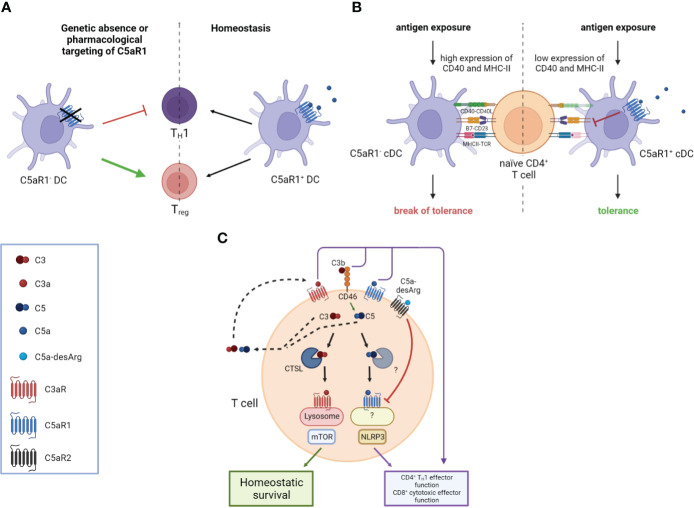
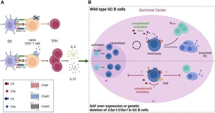
The complement system (CS) is an ancient and highly conserved part of the innate immune system with important functions in immune defense. The multiple fragments bind to specific receptors on innate and adaptive immune cells, the activation of which translates the initial humoral innate immune response (IR) into cellular innate and adaptive immunity. Dysregulation of the CS has been associated with the development of several autoimmune disorders such as systemic lupus erythematosus (SLE), rheumatoid arthritis (RA), ANCA-associated vasculitis, and autoimmune bullous dermatoses (AIBDs), where complement drives the inflammatory response in the effector phase. The role of the CS in autoimmunity is complex. On the one hand, complement deficiencies were identified as risk factors to develop autoimmune disorders. On the other hand, activation of complement can drive autoimmune responses. The anaphylatoxins C3a and C5a are potent mediators and regulators of inflammation during the effector phase of autoimmunity through engagement of specific anaphylatoxin receptors, i.e., C3aR, C5aR1, and C5aR2 either on or in immune cells. In addition to their role in innate IRs, anaphylatoxins regulate humoral and cellular adaptive IRs including B-cell and T-cell activation, differentiation, and survival. They regulate B- and T-lymphocyte responses either directly or indirectly through the activation of anaphylatoxin receptors via dendritic cells that modulate lymphocyte function. Here, we will briefly review our current understanding of the complex roles of anaphylatoxins in the regulation of immunologic tolerance and the early events driving autoimmunity and the implications of such regulation for therapeutic approaches that target the CS.
Keywords: C3a; C5a; anaphylatoxins; break of tolerance; complement; early autoimmunity.
Copyright © 2022 Schanzenbacher, Köhl and Karsten.
Conflict of interest statement
The authors declare that the research was conducted in the absence of any commercial or financial relationships that could be construed as a potential conflict of interest.
Figures
Similar articles
-
Revealing the signaling of complement receptors C3aR and C5aR1 by anaphylatoxins.Nat Chem Biol. 2023 Nov;19(11):1351-1360. doi: 10.1038/s41589-023-01339-w. Epub 2023 May 11. Nat Chem Biol. 2023. PMID: 37169960
-
The role of anaphylatoxins C3a and C5a in regulating innate and adaptive immune responses.Inflamm Allergy Drug Targets. 2009 Jul;8(3):236-46. doi: 10.2174/187152809788681038. Inflamm Allergy Drug Targets. 2009. PMID: 19601884 Review.
-
The anaphylatoxins bridge innate and adaptive immune responses in allergic asthma.Mol Immunol. 2004 Jun;41(2-3):123-31. doi: 10.1016/j.molimm.2004.03.019. Mol Immunol. 2004. PMID: 15159057 Review.
-
The anaphylatoxin C5a: Structure, function, signaling, physiology, disease, and therapeutics.Int Immunopharmacol. 2023 May;118:110081. doi: 10.1016/j.intimp.2023.110081. Epub 2023 Mar 28. Int Immunopharmacol. 2023. PMID: 36989901 Review.
-
The role of C5a receptors in autoimmunity.Immunobiology. 2023 Sep;228(5):152413. doi: 10.1016/j.imbio.2023.152413. Epub 2023 Jun 20. Immunobiology. 2023. PMID: 37598588
Cited by
-
The complement system and human autoimmune diseases.J Autoimmun. 2023 May;137:102979. doi: 10.1016/j.jaut.2022.102979. Epub 2022 Dec 18. J Autoimmun. 2023. PMID: 36535812 Free PMC article. Review.
-
Th2 predominance and decreased NK cells in patients with hereditary angioedema.Front Immunol. 2025 May 14;16:1536128. doi: 10.3389/fimmu.2025.1536128. eCollection 2025. Front Immunol. 2025. PMID: 40438097 Free PMC article.
-
Decrease in multiple complement proteins associated with development of islet autoimmunity and type 1 diabetes.iScience. 2023 Dec 20;27(2):108769. doi: 10.1016/j.isci.2023.108769. eCollection 2024 Feb 16. iScience. 2023. PMID: 38303689 Free PMC article.
-
Association of Complement Factors With Disability Progression in Primary Progressive Multiple Sclerosis.Neurol Neuroimmunol Neuroinflamm. 2024 Jul;11(4):e200270. doi: 10.1212/NXI.0000000000200270. Epub 2024 Jun 21. Neurol Neuroimmunol Neuroinflamm. 2024. PMID: 38912898 Free PMC article.
-
Inhibition of Alternative and Terminal Complement Pathway Components Modulate the Immune Response Against Bacteria and Fungi in Whole Blood.Scand J Immunol. 2025 May;101(5):e70030. doi: 10.1111/sji.70030. Scand J Immunol. 2025. PMID: 40387159 Free PMC article.
References
Publication types
MeSH terms
Substances
LinkOut - more resources
Full Text Sources
Medical