

Combining segments 9 and 10 in DNA and recombinant protein vaccines conferred superior protection against tilapia lake virus in hybrid red tilapia (oreochromis sp.) compared to single segment vaccines
- PMID: 35958595
- PMCID: PMC9359061
- DOI: 10.3389/fimmu.2022.935480
Combining segments 9 and 10 in DNA and recombinant protein vaccines conferred superior protection against tilapia lake virus in hybrid red tilapia (oreochromis sp.) compared to single segment vaccines
Abstract
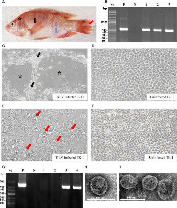
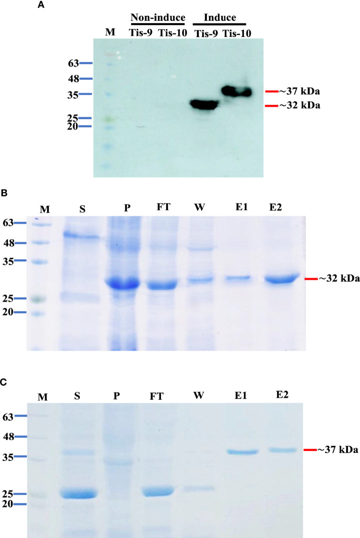
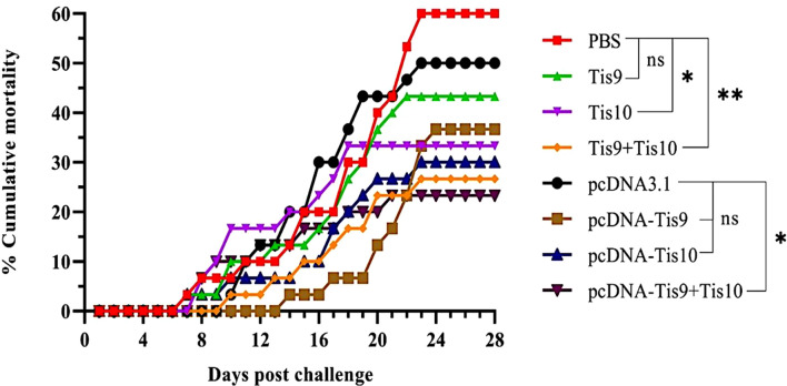
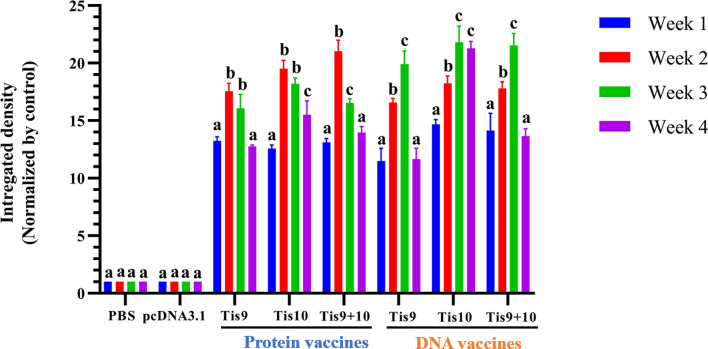
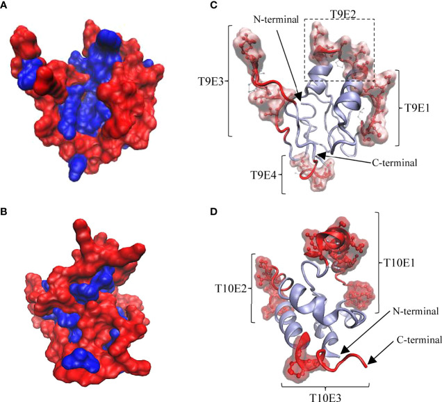
Tilapia lake virus (TiLV) now affects Nile tilapia culture worldwide, with no available commercial vaccine for disease prevention. DNA and recombinant protein-based vaccines were developed and tested following viral isolation and characterization. The viral strain isolated from diseased hybrid red tilapia (Oreochromis sp.) shared high levels of morphological and genomic similarity (95.49-99.52%) with other TiLV isolates in the GenBank database. TiLV segment 9 (Tis9) and segment 10 (Tis10) DNA vaccines (pcDNA-Tis9 and pcDNA-Tis10) and recombinant protein vaccines (Tis9 and Tis10) were prepared and tested for their efficacy in juvenile hybrid red tilapia. Fish were immunized with either single vaccines (pcDNA-Tis9, pcDNA-Tis10, Tis9 and Tis10) or combined vaccines (pcDNA-Tis9 + pcDNA-Tis10 and Tis9 + Tis10) by intramuscular injection and intraperitoneal injection for DNA and protein vaccines, respectively. Negative controls were injected with PBS or a naked pcDNA3.1 vector in the same manner. An experimental challenge with TiLV was carried out at 4 weeks post-vaccination (wpv) by intraperitoneal injection with a dose of 1 × 10⁵ TCID50 per fish. Relative percent survival (RPS) ranged from 16.67 ± 00.00 to 61.11 ± 9.62%. The Tis10 and pcDNA-Tis10 vaccines conferred better protection compared to Tis9 and pcDNA-Tis9. Highest levels of protection were observed in pcDNA-Tis9 + pcDNA-Tis10 (61.11 ± 9.62%) and Tis9 + Tis10 (55.56 ± 9.62%) groups. Specific antibody was detected in all vaccinated groups at 1-4 wpv by Dot Blot method, with the highest integrated density at 2 and 3 wpv. In silico analysis of Tis9 and Tis10 revealed a number of B-cell epitopes in their coil structure, possibly reflecting their immunogenicity. Findings suggested that the combination of Tis9 and Tis10 in DNA and recombinant protein vaccine showed high efficacy for the prevention of TiLV disease in hybrid red tilapia.
Keywords: DNA vaccine; TiLV; TiLV ORF10; TiLV ORF9; recombinant protein vaccine; tilapia (fish).
Copyright © 2022 Chamtim, Suwan, Dong, Sirisuay, Areechon, Wangkahart, Hirono, Mavichak and Unajak.
Conflict of interest statement
Author RM is employed by Charoen Pokphand Foods Public Co., Ltd. The remaining authors declare that this research was conducted in the absence of any commercial or financial relationships that could be construed as a potential conflict of interest.
Figures
Similar articles
-
Tilapia Lake Virus Vaccine Development: A Review on the Recent Advances.Vaccines (Basel). 2023 Jan 23;11(2):251. doi: 10.3390/vaccines11020251. Vaccines (Basel). 2023. PMID: 36851129 Free PMC article. Review.
-
Efficacy of heat-killed and formalin-killed vaccines against Tilapia tilapinevirus in juvenile Nile tilapia (Oreochromis niloticus).J Fish Dis. 2021 Dec;44(12):2097-2109. doi: 10.1111/jfd.13523. Epub 2021 Sep 3. J Fish Dis. 2021. PMID: 34477227 Free PMC article.
-
Expression and purification of S5196-272 and S6200-317 proteins from Tilapia Lake Virus (TiLV) and their potential use as vaccines.Protein Expr Purif. 2022 Feb;190:106013. doi: 10.1016/j.pep.2021.106013. Epub 2021 Nov 6. Protein Expr Purif. 2022. PMID: 34752859
-
Combining Phage Display Technology with In Silico-Designed Epitope Vaccine to Elicit Robust Antibody Responses against Emerging Pathogen Tilapia Lake Virus.J Virol. 2023 Apr 27;97(4):e0005023. doi: 10.1128/jvi.00050-23. Epub 2023 Mar 28. J Virol. 2023. PMID: 36975794 Free PMC article.
-
Susceptibilities of ten fish cell lines to infection with Tilapia lake virus.Microb Pathog. 2022 May;166:105510. doi: 10.1016/j.micpath.2022.105510. Epub 2022 Apr 11. Microb Pathog. 2022. PMID: 35421555 Review.
Cited by
-
Computational design of novel chimeric multiepitope vaccine against bacterial and viral disease in tilapia (Oreochromis sp.).Sci Rep. 2024 Jun 18;14(1):14048. doi: 10.1038/s41598-024-64383-z. Sci Rep. 2024. PMID: 38890454 Free PMC article.
-
Tilapia Lake Virus Vaccine Development: A Review on the Recent Advances.Vaccines (Basel). 2023 Jan 23;11(2):251. doi: 10.3390/vaccines11020251. Vaccines (Basel). 2023. PMID: 36851129 Free PMC article. Review.
-
Tilapia lake virus: A structured phylogenetic approach.Front Genet. 2023 Apr 18;14:1069300. doi: 10.3389/fgene.2023.1069300. eCollection 2023. Front Genet. 2023. PMID: 37144122 Free PMC article.
-
Expression and purification of recombinant tilapia lake virus segment 4 protein and its in-vitro biological activity for potential use in vaccine development.Sci Rep. 2024 Dec 28;14(1):31529. doi: 10.1038/s41598-024-83293-8. Sci Rep. 2024. PMID: 39733177 Free PMC article.
-
Biosecurity and Vaccines for Emerging Aquatic Animal RNA Viruses.Viruses. 2025 May 28;17(6):768. doi: 10.3390/v17060768. Viruses. 2025. PMID: 40573359 Free PMC article. Review.
References
-
- Senapin S, Shyam KU, Meemetta W, Rattanarojpong T, Dong HT. Inapparent infection cases of tilapia lake virus (TiLV) in farmed tilapia. Aquaculture (2018) 487:51–5. doi: 10.1016/J.AQUACULTURE.2018.01.007 - DOI
-
- Jansen MD, Dong HT, Mohan CV. Tilapia lake virus: a threat to the global tilapia industry? Rev Aquacult (2019) 11:725–39. doi: 10.1111/raq.12254 - DOI
-
- Liamnimitr P, Thammatorn W, U-thoomporn S, Tattiyapong P, Surachetpong W. Non-lethal sampling for tilapia lake virus detection by RT-qPCR and cell culture. Aquaculture (2018) 486:75–80. doi: 10.1016/j.aquaculture.2017.12.015 - DOI
Publication types
MeSH terms
Substances
LinkOut - more resources
Full Text Sources